Thursday, November 30, 2017

MRI-Compatible Robot Could Lead to Individualized Treatments for Stroke Survivors

It is true that no two stroke survivors are alike but good stroke medical persons would find the commonality behind the stroke damage and thus not have to create 10 million  separate recovery plans every year.

MRI-Compatible Robot Could Lead to Individualized Treatments for Stroke Survivors

No two stroke survivors are alike. While many do have lingering brain damage that makes motor tasks difficult, the rate at which each person heals, and what techniques best help them to heal, varies vastly amongst patients.
Despite these differences, the current method for assessing brain damage is the same for each patient.
Motor performance is typically assessed with simple tests—such as how well the patient can pick up a pen and put it down again—but these indicators aren’t sensitive enough to detect short-term neural reorganization, or rewiring and recovery in the brain. 
Fabrizio Sergi, PhD, an assistant professor of biomedical engineering at the University of Delaware, is taking a new approach, combining cutting-edge imaging and robotics technology to create a treatment paradigm for stroke survivors that is patient-specific and patient-adaptive. 
In order to assess brain activity in response to a changing set of rehabilitation exercises, Sergi is using functional MRI (fMRI), a non-invasive brain imaging technique, to see how patients’ brains respond to exercises they perform with a wrist-controlled, MRI-compatible robot. 
How it works
Operating the wrist-controlled robot is similar to using a video game joystick— except that the robot interacts physically with the subject, applying forces to either assist or to challenge the player during the video game.
“As you can imagine, throughout the observation process the subject gets better and improves motor function for a specific task,” explained Sergi in an interview with R&D Magazine. “The way to continue engaging the patient in the therapeutic process is to make the task more complicated. In that case, the robot can apply forces that distort the natural movement, so the patient has to learn to deal with a new environment. This process is called motor adaptation, and this is a classic paradigm that has been studied in neuroscience for several decades.”
As the patient is engaging in motor adaptation exercises with the robot, they are also undergoing fMRI to measure their brain activity, as well as shortly before and after.
This is done in order to observe neural reorganization, especially in the brain’s cortico-thalamic-cerebellar pathway.
“What we are trying to do now is study the process of motor adaptation from a neural level and look at which brain areas are responsible for the capability of subjects to adapt to a new environment,” said Sergi. “This could lead to localized and individualized treatments.”
By using fMRI to measure this process of neural reorganization directly, Sergi will then be able to program the robot to engage the patient in actions that specifically target the areas of the brain known to improve motor function. Sergi is also comparing the MRI results with the motor improvements visible to the naked eye when patients do the exercises and using these insights to predict subsequent gains in motor function.
As the patient improves, the actions the robots are doing with them are adjusted based on what is learned from the fMRI.
Robots are an ideal way to engage this patient population, said Sergi.
“Sometimes subjects are heavily impaired and they cannot move on their own or do basis motor functions on their own,” he explained. “With robot-aided rehabilitation they could, for example, stand without having to use all their energy to support gravity, because the machine can help them do that. Then the robot can apply the forces that are needed for that motor task.”
What they’ve learned so far
Much of Sergi’s research so far has been focused on designing a robot that can work with these patients effectively while still operating during an MRI.  The next step is to take a closer look at exactly what is happening in each patient’s brain and notice trends.
“One interesting aspect that we are starting to observe now is related to motor memory information,” said Sergi. “This is an ongoing concept, but it turns out if you expose subjects to a motor learning paradigm for 15 to 30 minutes, what happens is that the brain activity is motivated not only during that activity time frame, but also immediately after.”
These findings could serve as a predictor of how subjects will do with repeated exposure to the same training.  
“This would have a high clinical potential if we could predict the outcome of a longer intervention and assign subjects to different groups in a more efficient way,” said Sergi.

Virtual reality rehabilitation for stroke patients: Recent review and research issues

Your doctor was correct in not reading or bringing in all those older research trials on VR. No point in being a leader when you can just wait for a review like this and bring in the best. Except that your doctor probably won't read this review either. You'll have to take on the challenge of training your doctor of the finer and newer points of stroke rehab. Good luck with that, hope your doctor is trainable. And all those earlier stroke patients that could have been helped by bringing in VR earlier. Well, so what?
http://aip.scitation.org/doi/abs/10.1063/1.5012226

AIP Conference Proceedings 1905, 050007 (2017); https://doi.org/10.1063/1.5012226
Stroke is one of the main causes of disability in the world. In order for stroke survivors to reduce their disability, they need to go through a rehabilitation process to regain back their independence and improve their quality of life. To guide patients in their rehabilitation process and improve their receptiveness in performing repetitive exercises, a new rehabilitation training program using Virtual Reality (VR) technology has been introduced. This has attracted many researchers to explore more on VR technology as a new tool for stroke patient’s rehabilitation. This paper presents a review on existing VR systems that have been developed for stroke rehabilitation. First, recent VR systems utilized for rehabilitation after stroke are delineated and categorized. Each of these categories concludes with a discussion on limitations and any issues that arise from it. Finally, a concise summary with significant findings and future possibilities in VR rehabilitation research is presented in table format.

Wednesday, November 29, 2017

Stroke in Childhood: Clinical guideline for diagnosis, management and rehabilitation (2017) | RCPCH

I really dislike these types of guidelines when all they talk about is 'care'. Not how to get them 100% recovered. Survivors don't care about 'care', they want recovery.  GET THEM THERE!
Stroke in Childhood: Clinical guideline for diagnosis, management and rehabilitation (2017) | RCPCH

Stroke in Childhood 2017 is a nationally developed evidence-based clinical guideline for all UK paediatricians and healthcare professionals involved in the regulation or practice of the care of children and young people who have had or are suspected of having a stroke.

Join the conversation on Twitter at #childhealthmatters, #stroke, #makemaypurple to improve child health and stroke awareness.

Quick links:

Full clinical guideline
Guideline summary
Key recommendations
Parent/carer guideline
Additional resources

About

The updated Stroke in Childhood evidence-based clinical and parent guidelines were published in May 2017.
Funded by the Stroke Association, the clinical guideline was prepared by the Royal College of Paediatric and Child Health’s (RCPCH) multi-professional Stroke in Childhood Guideline Development Group (GDG), chaired by Dr Vijeya Ganesan. The GDG was represented by 27 stakeholder organisations and included lay representation from three parents of affected children and young people.
The clinical guideline is accompanied by a parent guideline, which aims to ensure that families understand what a stroke is, why they occur, and how children and young people affected by stroke should be treated and cared for.
Watch Stroke in Childhood: My Story, below, where a parent from the Stroke in Childhood Guideline Development Group talks of her experiences.


Background

The first clinical guideline on stroke in childhood was published by the Royal College of Physicians (RCP) in 2004. These guidelines may no longer reflect the best and most up-to-date clinical practice, and as such required urgent updating to ensure utilisation of current evidence.
This 2017 iteration of the clinical guideline delivers an update and scope extension, and provides guidance on the identification, diagnosis, management and rehabilitation of children and young people (aged 29 days to 18 years at time of presentation) with arterial ischaemic stroke (AIS) and haemorrhagic stroke (HS).

Full clinical guideline 

This clinical guideline is the most comprehensive and up-to-date guidance on how stroke care should be provided, covering the whole pathway from identification, diagnosis and management of children and young people with AIS and HS until their transition to adult care.
The 2017 iteration is intended for use by all UK paediatricians and other healthcare professionals involved in the regulation or practice of the care of children and young people who have had or are suspected of having a stroke, as well as non-healthcare professionals involved with educational/social services. While sections may also be relevant to education and social care professionals, it is intended to inform clinical decision making.
RCPCH notes:
On 01.06.2017 an amendment was made to the recommendations under the acute diagnosis (clinical presentation) section on page 24 of the full clinical guideline. The third recommendation relating to urgent brain imaging was revised to read 'Reduced level of consciousness (age-appropriate Glasgow Coma Scale (GCS) less than 12 or AVPU (‘Alert, Voice, Pain, Unresponsive’) less than V) at presentation'.
Full clinical guideline (PDF, 197 pages, 15MB)
Appendices (PDF, 357 pages, 27MB)

Guidelines summary

Working with the Guidelines Team this summary provides a concise overview for GPs on the management of stroke in childhood.
Guidelines summary (weblink)

Key recommendations

The concise key recommendations guide contains 83 key recommendations identified by the RCPCH Stroke in Childhood GDG, which, if followed, will enhance the quality of stroke care in children and young people.
These recommendations have been extracted from the clinical guideline, which contains over 250 individual recommendations covering the diagnosis, management and rehabilitation of stroke in children and young people.
It is recommended that the concise key recommendations guide should not be read in isolation, and individuals should always consider the guideline in full.
Key recommendations (PDF, 9 pages, 114KB)
Full recommendations (PDF, 25 pages, 220KB)

Parent/carer guideline 

This lay version of the guideline provides information to parents, carers, and families of children and young people affected by stroke. The information is based on detailed clinical guidelines produced for healthcare professionals who are involved in the care of children and young people affected by stroke.
The information is designed to help the reader understand what a stroke is, why strokes occur, and how children and young people affected by stroke should be treated and cared for. It describes the usual journey from diagnosis to rehabilitation that a child or young person will follow after a stroke and will outline what you should expect at each stage of treatment and rehabilitation.
Parent/carer guideline (PDF, 30 pages, 4.2MB)
Welsh version (PDF, 31 pages, 4.1MB)

Additional resources

Posters

Want to raise awareness that stroke happens to children and young people?
Poster 1 (PDF, 1 page, 1.6MB)
Poster 2 (PDF, 1 page, 1MB)
Poster 3 (PDF, 1 page, 2.3MB)

Contact

If you have any questions in relation to the development of this guideline, please contact us on clinical.standards@rcpch.ac.uk. For media queries, please contact us on press.office@rcpch.ac.uk and see the press release.

Technology and Stroke Infographic - National Stroke Association

Totally useless, generic crap with no links to any protocols on how to use or what the criteria is for use.  But great conscience laundering, thinking you are helping but actually doing absolutely fucking nothing. Contact me for ideas, I dare you to actually listen to your greatest critic.
Technology and Stroke Infographic

A model of stroke care enhanced by IT

What a pile of shit! 'CARE' not RESULTS. Is everyone in stroke that fucking lazy and incompetent?
https://twitter.com/IntJStroke?t=1&cn=ZmxleGlibGVfcmVjcw%3D%3D&refsrc=email&iid=ffc5473c57dc49c1a9ecffb97c159876&fl=4&uid=625967110&nid=244+273285133


Prof Martin Dennis - a future model of stroke care utilising IT to deliver quality care every time

Effect of EMG-triggered neuromuscular electrical stimulation with bilateral arm training on hemiplegic shoulder pain and arm function after stroke: a randomized controlled trial

Where is the publicly available protocol for this? Of course there is none since why would you publish useful information to stroke patients to bring to their doctors and therapists? Stroke patients know nothing and are just waiting for their medical providers to solve everything for them. Scream at your doctor for their obtuseness and lack of solutions.
https://jneuroengrehab.biomedcentral.com/articles/10.1186/s12984-017-0332-0

  • Li-Ling Chuang,
  • You-Lin Chen,
  • Chih-Chung Chen,
  • Yen-Chen Li,
  • Alice May-Kuen Wong,
  • An-Lun HsuEmail author and
  • Ya-Ju ChangEmail author
Contributed equally
Journal of NeuroEngineering and Rehabilitation201714:122
Received: 13 July 2017
Accepted: 1 November 2017
Published: 28 November 2017

Abstract

Background

Hemiplegic shoulder pain is a frequent complication after stroke, leading to limited use of the affected arm. Neuromuscular electrical stimulation (NMES) and transcutaneous electrical nerve stimulation (TENS) are two widely used interventions to reduce pain, but the comparative efficacy of these two modalities remains uncertain. The purpose of this research was to compare the immediate and retained effects of EMG-triggered NMES and TENS, both in combination with bilateral arm training, on hemiplegic shoulder pain and arm function of stroke patients.

Methods

A single-blind, randomized controlled trial was conducted at two medical centers. Thirty-eight patients (25 males and 13 females, 60.75 ± 10.84 years old, post stroke duration 32.68 ± 53.07 months) who had experienced a stroke more than 3 months ago at the time of recruitment and hemiplegic shoulder pain were randomized to EMG-triggered NMES or TENS. Both groups received electrical stimulation followed by bilateral arm training 3 times a week for 4 weeks. The primary outcome measures included a vertical Numerical Rating Scale supplemented with a Faces Rating Scale, and the short form of the Brief Pain Inventory. The secondary outcome measures were the upper-limb subscale of the Fugl-Meyer Assessment, and pain-free passive shoulder range of motion. All outcomes were measured pretreatment, post-treatment, and at 1-month after post-treatment. Two-way mixed repeated measures ANOVAs were used to examine treatment effects.

Results

Compared to TENS with bilateral arm training, the EMG-triggered NMES with bilateral arm training was associated with lower pain intensity during active and passive shoulder movement (P =0.007, P =0.008), lower worst pain intensity (P = 0.003), and greater pain-free passive shoulder abduction (P =0.001) and internal rotation (P =0.004) at follow-up. Both groups improved in pain at rest (P =0.02), pain interference with daily activities, the Fugl-Meyer Assessment, and pain-free passive shoulder flexion and external rotation post-treatment (P < 0.001) and maintained the improvement at follow-up (P < 0.001), except for resting pain (P =0.08).

Conclusions

EMG-triggered NMES with bilateral arm training exhibited greater immediate and retained effects than TENS with bilateral arm training with respect to pain and shoulder impairment for chronic and subacute stroke patients with hemiplegic shoulder pain.

Trial registration

Gel Could Help Regrow Heart Muscles After Heart Attack

Look at that, a solution for heart attacks. Nothing for stroke because our stroke teams have NO strategy and NO leadership to solve anything in stroke. Complete fucking incompetence displayed from the get go.

Gel Could Help Regrow Heart Muscles After Heart Attack

Wed, 11/29/2017 - 3:30pm
by Kenny Walter - Digital Reporter -
Scientists may have found a way to regenerate heart muscles following a heart attack.
Researchers from the University of Pennsylvania created an injectable gel that that slowly releases short gene sequences called microRNAs into the heart muscle, restarting replication in existing cardiomyocytes—contractile cells in the heart.
The microRNAs targeted signaling pathways are related to cell proliferation and were able to inhibit some of the inherent stop signals that keep cardiomyocytes from replicating, resulting in cardiomyocytes reactivating their proliferative potential.
The researchers found that with more heart cells dividing and reproducing, mice treated with the gel after a heart attack showed improved recovery in key clinically relevant categories.
“Biologic drugs turn over very fast,” Edward Morrisey, a professor in Medicine at Penn, a member of the Cell and Molecular Biology graduate group and scientific director of the Penn Institute for Regenerative Medicine in Penn Medicine, said in a statement. “The microRNAs that we used last less than eight hours in the bloodstream, so having a high local concentration has strong advantages.”
The short lifespan indicates that if patients were treated systemically they would need to be injected frequently with large doses to ensure that a sufficient amount of microRNAs reaches their target in the heart. 
The cells that contract the heart muscle and enable it to beat do not regenerate in mammals after an injury. Following a heart attack there is a dramatic loss of the heart muscle cells, which those that survive cannot effectively replicate.
Fewer of the cardiomyocytes means the heart pumps less blood with each beat, leading to the increased mortality associated with heart disease.
“We want to design the right material for a specific drug and application,” Jason Burdick, a professor in Bioengineering at Penn, said in a statement. “The most important traits of this gel are that it's shear-thinning and self-healing.
“Shear-thinning means it has bonds that can be broken under mechanical stress, making it more fluid and allowing it to flow through a syringe or catheter,” he added. “Self-healing means that when that stress is removed, the gel's bonds re-form, allowing it to stay in place within the heart muscle.”
The gel features attachment sites that keep the microRNAs in place and as the gel breaks down, it loses its grip on the microRNAs, which can slip out of the gel and into the cardiomyocytes.
While encapsulated, the microRNAs are also protected from degradation, maximizing the period that they can be effective without the risk of them invading off-target cells.
“There's likely a time window that the cardiomyocytes are susceptible to this stimulus—maybe a week or two after injury,” Morrisey said. “We want to promote proliferation for a short period and then stop.”
The researchers tested the gel with normal, healthy mice, genetically engineered mice that have individual cardiomyocytes that randomly express one of four different fluorescent proteins and mice in which heart attacks were induced so that clinically relevant outcomes of the treatment could be studied.
Within a few days after injecting the gel, the heart tissue of the healthy mice showed increased biomarkers of cardiomyocyte proliferation.
For the mice expressing different fluorescent proteins, after inducing heart attacks and introducing the gel, the researchers saw that single red, yellow or green cardiomyocytes had become clusters, ranging from two to eight cells of the same color.
The third group of mice showed improved recovery as compared to controls, including higher ejection fraction—more blood pumped with each beat—and smaller increases in heart size.
The researchers will now test the gel on human heart cells in vitro and conduct physiological experiments in animals with more human-like hearts, including pigs.
The study was published in Nature Biomedical Engineering.

Study finds gadolinium retention causes no neurologic harm

So you shouldn't have to worry about those MRI scans. Of course I was never told that gadolinium was present in my MRI scans.
http://www.auntminnie.com/index.aspx?sec=rca&sub=rsna_2017&pag=dis&ItemID=119283
By Wayne Forrest, AuntMinnie.com staff writerNovember 29, 2017

CHICAGO - Gadolinium accumulation in the brain after an MRI scan does not degrade a person's overall neurologic or neurocognitive performance, nor does it significantly affect the natural progression of cognitive decline, according to a new study presented on Wednesday at RSNA 2017.
The researchers reviewed more than 1,000 patients who received at least one dose of the linear gadolinium-based contrast agent (GBCA) gadodiamide (Omniscan, GE Healthcare) and compared their cognitive capabilities with those of a group of matched control subjects. They found no significant decrease in neuropsychological performance, no diminished motor function, and no rise in dementia related to GBCA exposure.
"Right now there is concern over the safety of gadolinium-based contrast agents, particularly relating to gadolinium retention in the brain and other tissues," said Dr. Robert McDonald, PhD, a neuroradiologist at the Mayo Clinic in Rochester, MN. "This study provides useful data that at the reasonable doses 95% of the population is likely to receive in their lifetime, there is no evidence at this point that gadolinium retention in the brain is associated with adverse clinical outcomes."
McDonald's contributions

Dr. Robert McDonald, PhD, from the Mayo Clinic.
Some 400 million doses of gadolinium have been administered for MRI scans since 1988, McDonald said. Today, GBCAs are used in 40% to 50% of MRI exams.
McDonald has been a leading researcher on the issue of gadolinium retention and how it may or may not affect patients years after they undergo an MRI scan.
In a June 2017 study, he and his colleagues found that gadolinium retention in the brain may be more widespread than previous studies have shown and may not be limited to patients with brain abnormalities such as a tumor or infection. The findings challenged previous theories regarding the permeability and role of the blood-brain barrier in the accumulation of gadolinium within the neural tissues.
In a March 2015 study, McDonald et al provided evidence of traces of gadolinium in the dentate nuclei, pons, globus pallidus, and thalamus from autopsies of 13 deceased patients who received GBCAs between 2000 and 2014. The deposits appeared to occur in all patients exposed to gadolinium and were detectable with as few as four doses. What's more, the patients had relatively normal renal function at the time of their MRI exams.
For the current study, McDonald and colleagues set out to identify the potential influence of neurotoxicity from intracranial gadolinium deposition following the intravenous administration of GBCAs for MR imaging.
The researchers used the Mayo Clinic Study of Aging (MCSA), the world's largest prospective population-based cohort on aging, to study the effects of gadolinium exposure on neurologic and neurocognitive function. All MCSA participants underwent extensive neurologic evaluation and neuropsychological testing at baseline and at 15-month follow-up intervals.
With and without GBCAs
The researchers looked at 4,261 cognitively normal MCSA study participants who had a mean age of 71.9 years (range, 50-90 years). Among those subjects, 1,092 (25%) received one or more doses of a GBCA, with a median of two doses (range, 1-28 doses). Their GBCA-enhanced MRI scans were unrelated to their participation in the MCSA. The median time since their first gadolinium exposure was 5.6 years.
McDonald and colleagues also compared the neurologic and neurocognitive scores of subjects who had undergone GBCA-enhanced MRI scans with scores from MCSA participants with no history of gadolinium exposure. Progression from normal cognitive status to mild cognitive impairment and dementia was assessed using multistate Markov model analysis.
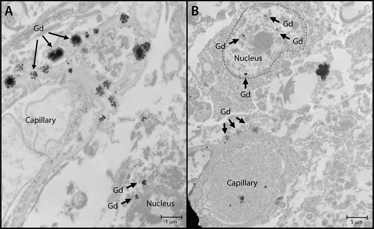
Nuclear localization of gadolinium deposits
Nuclear localization of gadolinium deposits is shown in transmission electron microscopy images. Cellular localization of gadolinium is evident in the dentate nuclei tissue samples in one gadolinium-exposed patient (A) and another gadolinium-exposed patient (B) at a 2,000- to 10,000-fold magnification. X-ray spectra were collected for selected electron-dense foci (arrows) to verify their identity. Images courtesy of RSNA.
After adjusting for age, sex, education level, baseline neurocognitive performance, and other factors, the researchers found that GBCA exposure was not a significant predictor of cognitive decline, based on comparisons between the healthy control subjects and those who were exposed to gadolinium.
Changes in clinical dementia rating (p = 0.48), dementia scale (p = 0.68), and mental status exam score (p = 0.55) showed no statistical significance, along with diminished neuropsychological performance (p = 0.13) or diminished motor performance (p = 0.43). Finally, gadolinium exposure was not an independent risk factor in the rate of cognitive decline from normal cognitive status to dementia (p = 0.91).
"I think right now there is a lot of smoke [and] no fire in terms of whether or not there is any evidence of harm," McDonald told RSNA attendees during his talk, adding that he hopes more studies from other centers provide data on the safety of GBCAs.
"Importantly, these studies do a good job of assessing the risk primarily on the 95% of patients who get very low doses of gadolinium in their lifetime -- not those [patients] who get 20 or 30 doses," he said. "The risk-benefit equation for these patients is entirely different than for those who only get one or two doses. We need to focus on real-world questions and real-world scenarios with our patients."
The researchers plan to reanalyze the current data and follow additional patients as the Mayo Clinic transitions to the macrocyclic GBCA gadobutrol (Gadovist, Bayer HealthCare).
This study by McDonald and colleagues received the RSNA's Kuo York Chynn Neuroradiology Research Award for the annual meeting's best neuroradiology paper. With the award comes a check for $3,000.

Tuesday, November 28, 2017

Effect of Action Observation Training with Auditory Feedback for Gait Function of Stroke Patients with Hemiparesis

Earlier articles which these researchers should know about and compare their research to. Because we don't have a database of stroke research and protocols we get incomplete research like this because the mentors and senior researchers are not up-to-date on previous research.

Metronome Cueing of Walking Reduces Gait Variability after a Cerebellar Stroke July 2016

Rhythmic Auditory Cueing in Motor Rehabilitation for Stroke Patients: Systematic Review and Meta-Analysis April 2016


http://www.papersearch.net/thesis/article.asp?key=3556006

( Hyeong Min Kim ) , ( Sung Min Son )
- 발행기관 : 대한물리치료학회
- 발행년도 : 2017
- 간행물 : 대한물리치료학회지(JKPT), 29권 5호
- 페이지 : pp.246-254 ( 총 9 페이지 )

학술발표대회집, 워크숍 자료집 중 1,2 페이지 논문은 ‘요약’만 제공되는 경우가 있으니,
구매 전에 간행물명, 페이지 수 확인 부탁 드립니다.
4,000

논문제목
초록(외국어)
Purpose: Previous studies have reported that action observation training has beneficial effects on enhancing the motor task, such as balance and gait functions. On the other hand, there have been few studies combined with action observation training and auditory feedback. The purpose of this study was to determine the effects of action observation training with auditory feed-back on the gait function in stroke patients with hemiparesis
Methods: A total of 24 inpatients with post-stroke hemiparesis were assigned randomly to either an experimental group 1 (EG 1, n=8), experiment group 2 (EG 2, n=8), control group (CG, n=8, EG 1). The EG 2 and CG watched video clip demonstrating three functional walking tasks with auditory feedback, without auditory feedback, and showing a landscape image, respectively. The exercise program consisted of 30 minutes, five times a week, for four weeks. The participants were measured to 10MWT (10 m walk test), 6MWT (6 min-utes walking distance test), TUG (timed up and go test), DGI (dynamic gait index), time and steps of F8WT (figure-of-8 walk test).
Results: In the intra-group comparison after the intervention, EG 1 and EG 2 showed a significantly different gait function (10MWT, 6MWT, DGI, TUG, F8WT)  (p<0.05). In the inter-group comparison after intervention, EG 1 showed significant improvements in the entire gait parameters and EG 2 only showed significant improvement in DGI and TUG compared to CG (p<0.05). Conclusion: These findings show that action observation training with auditory feedback may be used beneficially for improving the gait function of stroke patients with hemiparesis.

Twilight zone: Human head successfully transplanted on a corpse; professor claims the next operation will be on a live human

The definition of success for this type of operation is so low as to be non-existent. I could use crazy glue and call it a success.
https://www.naturalnews.com/2017-11-26-twilight-zone-human-head-successfully-transplanted-on-a-corpse.html

Twilight zone: Human head successfully transplanted on a corpse; professor claims the next operation will be on a live human


Image: Twilight zone: Human head successfully transplanted on a corpse; professor claims the next operation will be on a live human
(Natural News) On the morning of Nov. 17, 2017, Italian neurosurgeon Sergio Canavero made a shocking announcement at a press conference in Vienna. The professor claimed that the world’s first human head transplant was performed on a corpse in China. Led by Chinese orthopedic surgeon Xiaoping Ren, the team at Harbin Medical University managed to successfully reconnect the spine, nerves, and blood vessels of a severed head onto a dead body.
“The first human head transplant, in the human mode, has been realized,” said Canavero at the conference. “The surgery lasted 18 hours. The paper will be released in a few days. Everyone said it was impossible, but the surgery was successful.
Though no evidence to back his claims was shown at the press conference, Canavero stated that the details of the procedure would be laid out in a scientific paper due to be released in the coming days. Speaking further, he remarked: “For too long nature has dictated her rules to us. We’re born, we grow, we age, and we die. For millions of years humans have evolved and 100 billion humans have died. That’s genocide on a mass scale.
“We have entered an age where we will take our destiny back in our hands. It will change everything. It will change you at every level.”
Following the operation on human cadavers, Canavero added that the next step for him and his team is to carry out a complete head swap between brain dead organ donors. This surgical procedure, which he said is “imminent”, would be for medical purposes. “It will be for a medical, neurological condition, not for life-extension,” Canavero noted.
Though his bold proclamation has attracted a flurry of excitement from many people, the medical community at large has lobbied the doctor with a barrage of criticism. Many experts have pointed out that spinal cord fusion is an impossibility with the technology of today, calling his plan all but implausible. Others have raised concerns on the morality and legality of the operation.
Speaking to DailyMail.co.uk, Dr. Jan Schupp, professor of neuroscience at the University of Oxford, commented: “I find it inconceivable that ethics committees in any reputable research or clinical institutions would give a green light to living human head transplants in the foreseeable future. Indeed, attempting such a thing given the current state of the art would be nothing short of criminal.”
Dr. James Fildes, NHS principal research scientist at the University Hospital of South Manchester’s Transplant Centre, chimed in with: “Perhaps far more worryingly, this endeavor appears to revolve around immortality, but in each case a body is needed for the transplant, and therefore a human needs to die as part of the process.
“Where does Canavero propose to get the donor body from if the goal is to tackle the laws of nature? Has Canavero considered how he will tackle acute rejection of the constituent parts of the head? What will rejection of the skin, muscles, eyes, and brain manifest as? I hope this is not just egotistical pseudoscience.” (Related: Organ transplant industry pushes for legal right to buy and sell body parts on the global market.)
For his part, Canavero was described as remaining “defiant of his critics” by NewsWeek.com. In a statement to the publication, he estimated the success of the up-and-coming operation to be about 90 percent, calling it “long and tedious, but absolutely feasible”, then closed with: “One thing is certain. Spinal cord fusion is a reality and head transplants will happen.”

Fast facts on the head transplant

  • Canavero’s first subject for the head transplant was Valery Spiridonov, a 30-year-old Russian computer scientist with Werdnig-Hoffman disease, also known as spinal muscular atrophy. With the venue being moved to China, Spiridonov has since accepted that he would no longer be able to attain a healthy new body. Instead, he would opt for conventional treatments by way of crowdfunding.
  • On the cost of the procedure, Canavero has speculated that it would cost $11,922,550 and need 150 doctors and nurses.

Reconsidering Neuroprotection in the Reperfusion Era

So WHOM  is going to create a strategy to solve the neuronal cascade of death(neuroprotection) problem? And lead that strategy to a successful conclusion?
http://stroke.ahajournals.org/content/48/12/3413?etoc=

Sean I. Savitz, Jean-Claude Baron, Midori A. Yenari, Nerses Sanossian, Marc Fisher
This article requires a subscription to view the full text. If you have a subscription you may use the login form below to view the article. Access to this article can also be purchased.


Neuroprotection to prevent infarct progression as a potential treatment for acute ischemic stroke carries a long and disappointing history. The failures of these prior neuroprotection trials have many potential explanations that encompass both problems with the preclinical assessment of these drugs and the design/implementation of clinical trials.1 Prior development of neuroprotection has focused primarily on its use as a monotherapy, and no clinical trial was designed to determine whether neuroprotection could extend the time window for successful reperfusion or ameliorate the consequences of reperfusion. Acute stroke therapy has now entered the era of highly effective reperfusion with the recent publication of 5 positive thrombectomy trials.2 Combining neuroprotection with intravenous or intra-arterial reperfusion therapy is now an important next step in the development of acute stroke therapies. One approach to neuroprotection would be to initiate therapy early after ischemic stroke onset, either in the ambulance or at a primary stroke center/community hospital to potentially extend the time window for intravenous or intra-arterial therapy. A second approach would be to use neuroprotection during or after partial or complete reperfusion to reduce the consequences of reperfusion injury. Both future neuroprotective approaches will require preclinical studies that anticipate novel clinical trial designs and trials that will be organized and evaluated differently than past monotherapy neuroprotection trials.

Ischemic Penumbra and Core

Acute ischemic stroke therapy is designed to reduce infarction of hypoperfused brain tissue and limit the expansion of already irreversibly injured tissue when treatment is initiated, leading to smaller infarction and improved clinical outcomes.3,4 Severely hypoperfused but still potentially viable ischemic brain represents the ischemic penumbra, whereas irreversibly injured tissue is the ischemic core.5 Early after stroke onset, most patients with a large vessel occlusion (LVO) have an extensive ischemic penumbra that can be salvaged by timely reperfusion. This concept was proven …
View Full Text

Impact on Prehospital Delay of a Stroke Preparedness Campaign

More blame the patient for not recognizing a stroke fast enough. Deal with the real problem. You as doctors and stroke hospitals have no protocols to get everyone 100% recovered regardless of when they arrive. But it is so much easier to blame the patient than actually work on and solve the BHAGs(Big Hairy Audacious Goals) in stroke. 
http://stroke.ahajournals.org/content/48/12/3316?etoc= 

A SW-RCT (Stepped-Wedge Cluster Randomized Controlled Trial)

Licia Denti, Caterina Caminiti, Umberto Scoditti, Andrea Zini, Giovanni Malferrari, Maria Luisa Zedde, Donata Guidetti, Mario Baratti, Luca Vaghi, Enrico Montanari, Barbara Marcomini, Silvia Riva, Elisa Iezzi, Paola Castellini, Silvia Olivato, Filippo Barbi, Eva Perticaroli, Daniela Monaco, Ilaria Iafelice, Guido Bigliardi, Laura Vandelli, Angelica Guareschi, Andrea Artoni, Carla Zanferrari, Peter J. Schulz
This article requires a subscription to view the full text. If you have a subscription you may use the login form below to view the article. Access to this article can also be purchased.

Abstract

Background and Purpose—Public campaigns to increase stroke preparedness have been tested in different contexts, showing contradictory results. We evaluated the effectiveness of a stroke campaign, designed specifically for the Italian population in reducing prehospital delay.
Methods—According to an SW-RCT (Stepped-Wedge Cluster Randomized Controlled Trial) design, the campaign was launched in 4 provinces in the northern part of the region Emilia Romagna at 3-month intervals in randomized sequence. The units of analysis were the patients admitted to hospital, with stroke and transient ischemic attack, over a time period of 15 months, beginning 3 months before the intervention was launched in the first province to allow for baseline data collection. The proportion of early arrivals (within 2 hours of symptom onset) was the primary outcome. Thrombolysis rate and some behavioral end points were the secondary outcomes. Data were analyzed using a fixed-effect model, adjusting for cluster and time trends.
Results—We enrolled 1622 patients, 912 exposed and 710 nonexposed to the campaign. The proportion of early access was nonsignificantly lower in exposed patients (354 [38.8%] versus 315 [44.4%]; adjusted odds ratio, 0.81; 95% confidence interval, 0.60–1.08; P=0.15). As for secondary end points, an increase was found for stroke recognition, which approximated but did not reach statistical significance (P=0.07).
Conclusions—Our campaign was not effective in reducing prehospital delay. Even if some limitations of the intervention, mainly in terms of duration, are taken into account, our study demonstrates that new communication strategies should be tested before large-scale implementation.
Clinical Trial Registration—URL: http://www.clinicaltrials.gov. Unique identifier: NCT01881152.

Development and Validation of a Predictive Model for Functional Outcome After Stroke Rehabilitation

You fucking bastards are once again wasting time and money on predictions rather than solving all the problems in stroke. Your mentors and senior researchers need to be keel-hauled.
http://stroke.ahajournals.org/content/48/12/3308?etoc=

The Maugeri Model

Domenico Scrutinio, Bernardo Lanzillo, Pietro Guida, Filippo Mastropasqua, Vincenzo Monitillo, Monica Pusineri, Roberto Formica, Giovanna Russo, Caterina Guarnaschelli, Chiara Ferretti, Gianluigi Calabrese
This article requires a subscription to view the full text. If you have a subscription you may use the login form below to view the article. Access to this article can also be purchased.

Abstract

Background and Purpose—Prediction of outcome after stroke rehabilitation may help clinicians in decision-making and planning rehabilitation care. We developed and validated a predictive tool to estimate the probability of achieving improvement in physical functioning (model 1) and a level of independence requiring no more than supervision (model 2) after stroke rehabilitation.
Methods—The models were derived from 717 patients admitted for stroke rehabilitation. We used multivariable logistic regression analysis to build each model. Then, each model was prospectively validated in 875 patients.
Results—Model 1 included age, time from stroke occurrence to rehabilitation admission, admission motor and cognitive Functional Independence Measure scores, and neglect. Model 2 included age, male gender, time since stroke onset, and admission motor and cognitive Functional Independence Measure score. Both models demonstrated excellent discrimination. In the derivation cohort, the area under the curve was 0.883 (95% confidence intervals, 0.858–0.910) for model 1 and 0.913 (95% confidence intervals, 0.884–0.942) for model 2. The Hosmer–Lemeshow χ2 was 4.12 (P=0.249) and 1.20 (P=0.754), respectively. In the validation cohort, the area under the curve was 0.866 (95% confidence intervals, 0.840–0.892) for model 1 and 0.850 (95% confidence intervals, 0.815–0.885) for model 2. The Hosmer–Lemeshow χ2 was 8.86 (P=0.115) and 34.50 (P=0.001), respectively. Both improvement in physical functioning (hazard ratios, 0.43; 0.25–0.71; P=0.001) and a level of independence requiring no more than supervision (hazard ratios, 0.32; 0.14–0.68; P=0.004) were independently associated with improved 4-year survival. A calculator is freely available for download at https://goo.gl/fEAp81.
Conclusions—This study provides researchers and clinicians with an easy-to-use, accurate, and validated predictive tool for potential application in rehabilitation research and stroke management.