Friday, December 31, 2021

‘We think it’s cognitive engagement’—a study finds that delaying retirement may help ward off dementia

I totally disagree about the keep working part, been retired for almost 4 years now. Am cognitively engaged trying to solve stroke, even though no one in charge is listening to me.  When I was working I would fly out Monday morning, fly back Thursday night, wash clothes on Friday, put those clothes back into the suitcase and do it all over again. It was destroying my health.

‘We think it’s cognitive engagement’—a study finds that delaying retirement may help ward off dementia

The science behind staying busy to keep your mind sharp

It’s not necessarily paid work, but cognitive engagement that protects against dementia, the researchers say.

This article is reprinted by permission from NextAvenue.org.

We’ve all heard someone say it: they’ve decided to keep working beyond retirement age to “keep their mind sharp.” Now, that widely held notion has some science behind it.

Three researchers at the Max Planck Institute for Demographic Research in Germany have released a study showing measurable differences in cognitive decline between those who bow out of the workforce earlier versus later in life. Some of the differences were stark.

“Oh, it’s absolutely substantial,” says Jo Mhairi Hale, a sociologist at the University of St. Andrews in Scotland and the lead author on the study.

Hale’s team drew its data from the massive Health and Retirement Study, an extensive ongoing trove of information of 20,000 Americans maintained by the University of Michigan.

What the study found about cognitive decline

The authors didn’t try to pinpoint an optimal retirement age — that would depend heavily on individual circumstances — but their results do suggest that generally speaking, sticking it out until age 67 (vs. retiring between age 55 and 66) can ward off the type of cognitive decline suffered by people with Alzheimer’s disease.

Subjects in the study averaged a one-third reduction in typical cognitive declines observed in people aged 61 to 67. What’s more, the positive effects can be enduring, say the authors, lasting from age 67 at least through age 74.

Hale says one surprising finding was that it doesn’t appear to matter what kind of work you do — whether it’s highly brain-intensive or nearly mindless. It all helps. In fact, the cognitive benefits may not be related to paid employment at all.

“The three of us who wrote the paper are not suggesting that it’s paid work per se that is protective against cognitive decline,” Hale told Next Avenue. “We think it’s cognitive engagement.”

That idea is borne out by some of the specific findings of Hale’s team, including that just having a life partner offers some protection against decline.

“What if you retire at age 60 but you’re a grandparent and part of your daily activity becomes grandparenting?” muses Hale. “Or you’re an active volunteer. Or you work part time as a museum docent or whatever. Does that provide the same sort of protective effects against cognitive decline? I would guess that it does.”

The thing that matters, Hale said, “is cognitive engagement, not that you get a paycheck for your cognitive engagement.”

Also see: Planning to retire early? Watch out for these tax traps

A 75-year-old finding ways to challenge herself

Case in point: Beverly Farr of Richmond, Calif. At 75, she’s been retired from her work as an educational researcher for more than five years, but that doesn’t mean she’s slowed down. On the contrary.

“I just liked being active and I wanted to stay active,” says Farr. “And I think a little part of that was the idea of keeping your mind active and, you know, just being active in general.”

That she has done. Today, Farr juggles church activities with being a court-appointed advocate for foster youth. She’s also taken on the daunting task of presiding over the homeowners’ association of her 488-unit condo complex. As if that weren’t enough, when her brother, Roger, died in 2019, Farr took over managing his boutique research firm part-time.

She especially credits the often thankless homeowners’ association work for keeping her on her toes.

“It just energized me,” Farr says. “It just gave me a lot of energy and a lot of excitement and drive about getting things done.”

Hale notes this kind of engagement appears to benefit men and women alike, even though prior studies have shown that men tend to be more dependent on their jobs for their personal identity and social networks. It also applies across racial and ethnic differences.

“I wasn’t surprised at all,” says the Milken Institute Center for the Future of Aging’s Paul Irving, after reviewing the study’s findings. (He’s also a Next Avenue Influencer in Aging.)

Check out: Want to share your life experiences and help others? Here’s how to do it well

The importance of purpose

“Meeting other people and engaging with other people is stimulating,” Irving says. “Work can be challenging and can provide opportunities for learning. A changing environment requires adaptability and flexibility. I think that has consequences for brain health.”

Irving says the Max Planck results help affirm earlier research, including one 2018 study that found that “positive age beliefs” can be a buffer against dementia, even in those genetically predisposed to age-related cognitive loss.

Irving points to other research that suggests factors such as purpose, connection and lifelong learning are on a par with body-mass index, smoking and exercise as determinants of longevity.

“Having some sense of challenge, having a reason to wake up in the morning — best defined as purpose,” says Irving, whose 2014 book, “The Upside of Aging,” emphasized the importance of the “P-word.”

“This notion of purpose throughout life, but particularly the realization of purpose later in life, just couldn’t be more important,” says Irving. “And that can be achieved in many ways. We all define our own purpose, and it might be family or community activity or volunteering. But work can very much be part of that.”

People who feel a sense of purpose, are engaged and continue to involve themselves in the world, Irving adds, tend to be happier and healthier.

“And the corollary benefit is that they’re continuing to provide value — their wisdom and judgment and experience — which benefits all of us, young people as well,” notes Irving.

You might like: Why my retirement won’t include a ‘bucket list’

Hale’s major takeaway from her team’s results: “I think what we could say is that you need to stay cognitively engaged, like basically forever, as long as you can. And honestly, that’s not about doing crossword puzzles. I mean, do your crossword puzzles, but cognitive engagement needs to be seen much more broadly. Our paper suggests that full-time work is one way to do that.”


Morphological Subtypes of Intracranial Internal Carotid Artery Arteriosclerosis and the Risk of Stroke


My doctors never found the 80% blockage in my right carotid artery even though they told me that artery had dissected and thrown a clot into my brain. So they left me with a risk of further strokes until 3 years later when that artery completely closed up.

Morphological Subtypes of Intracranial Internal Carotid Artery Arteriosclerosis and the Risk of Stroke

Originally publishedhttps://doi.org/10.1161/STROKEAHA.121.036213Stroke. 2021;0:STROKEAHA.121.036213

Background and Purpose:

Accumulating evidence highlights the existence of distinct morphological subtypes of intracranial carotid arteriosclerosis. So far, little is known on the prevalence of these subtypes and subsequent stroke risk in the general population. We determined the prevalence of morphological subtypes of intracranial arteriosclerosis and assessed the risk of stroke associated with these subtypes.

Methods:

Between 2003 and 2006, 2391 stroke-free participants (mean age 69.6, 51.7% women) from the population-based Rotterdam Study underwent noncontrast computed tomography to visualize calcification in the intracranial carotid arteries as a proxy for intracranial arteriosclerosis. Calcification morphology was evaluated according to a validated grading scale and categorized into intimal, internal elastic lamina (IEL), or mixed subtype. Follow-up for stroke was complete until January 1, 2016. We used multivariable Cox regression to assess associations of each subtype with incident stroke.

Results:

The prevalence of calcification was 82% of which 39% had the intimal subtype, 48% IEL subtype, and 13% a mixed subtype. During a median follow-up of 10.4 years, 155 participants had a stroke. All 3 subtypes were associated with a higher risk of stroke (adjusted hazard ratio [95% CI] for intimal: 2.11 [1.07–4.13], IEL: 2.66 [1.39–5.11], and mixed subtype 2.57 [1.18–5.61]). The association of the IEL subtype with stroke was strongest among older participants. The association of the intimal subtype with stroke was noticeably stronger in women than in men.

Conclusions:

Calcification of the IEL was the most prevalent subtype of intracranial arteriosclerosis. All 3 subtypes were associated with an increased risk of stroke, with noticeable age and sex-specific differences.


Thursday, December 30, 2021

Plémont Ward ‘not adequate’ for stroke care; Jersey UK

  As long as any stroke hospital talks about 'care' it means they are not providing anything even approaching 100% recovery.  Any hospital talking about 'care' I consider a total failure.

Oops, I'm not playing by the polite rules of Dale Carnegie,  'How to Win Friends and Influence People'. 

Telling stroke medical persons they know nothing about stroke is a no-no even if it is true. 

Politeness will never solve anything in stroke. Yes, I'm a bomb thrower and proud of it. Someday a stroke 'leader' will try to ream me out for making them look bad by being truthful , I look forward to that day.

Plémont Ward ‘not adequate’ for stroke care; Jersey UK


REHABILITATION facilities at the Hospital’s Plémont Ward are not adequate for stroke survivors and full services need to be reinstated at Overdale’s Samarès Ward, according to a charity.

Jacqui Cuthbert, from the Stroke Association Jersey, said the current offerings at the General Hospital ‘lack space and privacy’ for patients to have conversations with therapists and family members.

The charity’s calls come after Health Minister Richard Renouf said there was no need to reconsider moving the Island’s rehabilitation services back to Samarès Ward. He said that transferring the service back to the facility at Overdale would cause more ‘disruption’ and only be ‘temporary’ due to the buildings being demolished to make way for the new hospital.

Earlier this month, Senator Steve Pallett said he would be lodging a proposition to immediately reinstate the Island’s rehabilitation services at Samarès Ward. He also called for a ‘temporary purpose-built’ facility to be constructed, while the ward at Overdale was demolished to make way for the new hospital.

A petition launched by Jean Lelliott which called for the new hospital to have the same equipment and specialist staff that used to be based at what she called the ‘world-class’ Samarès Ward has received more than 1,400 signatures.

The facility had 28 beds for people needing physical rehabilitation, but last year the Health Department closed the ward and opened 12 beds in Plémont Ward with shorter stays provided. Mrs Cuthbert said that while some stroke survivors received support at home, others required therapy and dedicated support in ‘bedded rehab settings’.

‘The current facilities are not adequate, they lack space and privacy for stroke survivors to have conversations with therapists or family members on deeply personal matters,’ she said.

‘Staff on the stroke ward do an amazing job but are then faced with the pressure of making the difficult decision to send people home when they really need continuing care in a hospital environment.

‘In some cases people are going back to homes which are unsuitable for their disabilities, where the lack of therapy means they are more likely to develop psychological problems, and where their ability to make the best possible recovery from their stroke is reduced.

‘As a charity dedicated to stroke, we want to work closely with the Health Minister and his team to look at how we can improve the whole stroke pathway from prevention through to rehab and life after stroke to ensure that people get the right support for the long-term effects of living with stroke.’

Malcolm Ferey, chief executive of brain injury charity Headway Jersey, said: ‘We are really concerned about the situation, particularly for those who have suffered a stroke or brain injury and currently use the Island’s rehabilitation services. The closure of Samarès Ward has put a strain on other areas of the service and after speaking with people who have been impacted by the move to Plémont Ward, we would advocate for a move back to Samarès.’


Transcranial Direct-Current Stimulation and Behavioral Training, a Promising Tool for a Tailor-Made Post-stroke Aphasia Rehabilitation: A Review

So just a review and NOTHING ON CREATING PROTOCOLS FOR APHASIA! Useless.

Transcranial Direct-Current Stimulation and Behavioral Training, a Promising Tool for a Tailor-Made Post-stroke Aphasia Rehabilitation: A Review

Zettin, MarinaBondesan, CaterinaNada, GiuliaVarini, MatteoDimitri, Danilo. Frontiers in Human Neuroscience; Lausanne (Dec 20, 2021). DOI:10.3389/fnhum.2021.742136

Full text

Introduction

Aphasia is an acquired language disorder resulting from damage to the portions of the brain which are responsible for language comprehension and formulation. The most common causes of this disorder include vascular lesions, encephalic traumatic injury, and brain tumors (Marangolo and Caltagirone, 2014), with a prevalence of 250,000 cases in the United Kingdom and 1 million in the United States (Crinion, 2016). Aphasia may also be associated with other degenerative, inflammatory, autoimmune or parasitic disorders. About 0.7 to 3% of people presenting with multiple sclerosis also show aphasic symptoms (Naro et al., 2021).

Although damages to specific brain areas and their connections mainly occur in the left hemisphere, functional magnetic resonance imaging (fMRI) studies such as the one carried out by Thompson and den Ouden (2008) showed that in some cases the dominant language areas can be located in the right hemisphere.

Aphasia can involve different levels of language processing with impairments in both oral and written comprehension and production. Most patients who experience aphasia show some degree of spontaneous recovery within the first two to three months, due to a functional neural reactivation and reorganization. The most important factors that determine recovery are the lesion size and location, the type and severity of aphasia, the treatment received and, to some extent, the nature of early hemodynamic response (Watila and Balarabe, 2015).

From the second half of the twentieth century, different rehabilitation perspectives and therapeutic interventions for aphasia rehabilitation have been developed. The most recommended treatment for this disorder is Speech and Language Therapy (SLT). However, it is argued that SLT would lead to moderate effects, even when administered at high intensity. For this reason, over the last few years, new strategies have been implemented to enhance the effects of traditional rehabilitation.

These effects depend on the metaplasticity, which “refers to activity-dependent changes in neural functions that modulate subsequent synaptic plasticity such as long-term potentiation (LTP) and long-term depression (LTD)” (Abraham and Philpot, 2009). Changes in the pathomechanisms underlying psychiatric and neurological disorders are possible by acting on metaplasticity (Cantone et al., 2021). Non-Invasive Brain Stimulation Techniques (NIBS) can be a beneficial tool to transiently modulate cortical excitability and lead to lasting changes after the stimulation time (Fisicaro et al., 2020). Non-invasive brain stimulation techniques facilitate the activation of single brain areas, or the inhibition of other ones whose hyperactivation could have a maladaptive effect on cognitive recovery (Simonetta-Moreau, 2014). One of the main neuromodulation tools is Transcranial Magnetic Stimulation (TMS), which generates magnetic field pulses under the scalp. A single impulse leads to a short-term effect, while a sequence of stimulation on the same region of interest can generate long-term effects. These can either be inhibiting or excitatory, depending on the stimulation frequency (Fisicaro et al., 2020). For instance, repetitive TMS (rTMS) can successfully treat both motor and non-motor symptoms in stroke patients, including depression, which often affects the rehabilitation process after a stroke (Fisicaro et al., 2019). Both for safety and cost issues, tDCS is often preferred over TMS.

The main strength of tDCS is its feasibility associated with relatively minor side effects, if safely and properly administered. Another strength is that it shows promise as an effective and versatile neurostimulation tool. It has the potential to be a treatment for several conditions characterized by an alteration of the cerebral cortex activation. Indeed, it has been proven to have beneficial effects on both neuropsychiatric and neurological disorders, such as mood disorders, substance abuse, Alzheimer’s and Parkinson’s disease, multiple sclerosis, as well as post-stroke motor and cognitive impairment (Lefaucheur et al., 2017). Additionally, tDCS can be applied in sham mode, making it easier to carry out a single-blind study (Nitsche et al., 2003). Therefore, tDCS represents one of the most promising tools for the treatment of aphasia (Biou et al., 2019). It does not directly induce an action potential, but it delivers a continuous current flow at a low intensity (1/2 mA) instead. Transcranial Direct-Current Stimulation requires two electrodes, an anode and a cathode, which are generally placed on the scalp. Depending on the polarity and the consequent positioning of the electrodes, the experimenter can obtain a depolarizing effect, thus favoring neuronal firing (anodic tDCS), or a hyperpolarized effect by decreasing the discharge rate (cathodic tDCS) (Liebetanz et al., 2002). Because of the electrode size, tDCS allows the stimulation of large cortical areas, with a consequent reduction of stimulation focality. The effects of tDCS are variable and depend on the stimulation duration, the current density, the characteristics of the neuronal tissue involved and the current flow direction, which can move from the anode to the cathode or vice versa (Chase et al., 2020). Non-invasive brain stimulation is an important resource in neuropsychological rehabilitation, however, its application is not risk-free, as most non-invasive current induction tools. The ultimate goal of applying tDCS in rehabilitation is to re-establish an interhemispheric balance by promoting functional brain reorganization and facilitating relearning (Simonetta-Moreau, 2014).

To date, scientific literature offers a comprehensive overview of the several therapeutic treatments used for the rehabilitation of aphasia. Unfortunately, these many specific training methods lead to moderate effects. Therefore, a number of techniques have been implemented over the years in support of speech therapy and neuropsychological rehabilitation to promote a faster and more effective recovery. As tDCS is the most widely used method in rehabilitation, this review aims at examining those studies which associate rehabilitation with tDCS, and investigate its effectiveness (Marangolo, 2017).

Summary of Findings

Several articles were selected and analyzed for this review. PubMed, PsycInfo and Cochrane were consulted for the systematic search of the relevant articles. As for keywords, different combinations of the terms “aphasia,” “speech impairment,” “Broca’s aphasia,” “non-fluent aphasia,” “tDCS,” “transcranial direct current stimulation,” “non-invasive brain stimulation,” “cognitive rehabilitation,” “neurorehabilitation,” “aphasia training,” “cognitive training,” “language recovery” were used. The whole research process started from an accurate analysis of the most quoted and detailed reviews available on this topic. Out of the 37 most relevant reviews and meta-analysis, only 33 specifically analyzed tDCS studies on aphasic patients with acquired cerebral lesions (Figure 1). The experimental studies were subsequently extracted from these reviews. From a total of 93 studies analyzed, 46 were considered the most relevant. The main exclusion criterion was the type of aphasia: only tDCS studies conducted on a sample of aphasic patients at the chronic phase with an acquired cerebral lesion were included. Studies on other types of aphasia (e.g., primary progressive aphasia) or carried out earlier than six months from the damage were excluded. Moreover, it was decided to include only studies enrolling a minimum of three subjects, thus removing single-case studies from the total count of papers. The conclusions drawn from those studies could have been weak and not significant enough for the purpose of this study. All experimental studies using TMS or other brain stimulation techniques other than tDCS were not included as well. Out of the 79 articles included in this review, 26 were reviews, 7 meta-analysis and 46 were experimental studies (Supplementary Table 1).

More at link.


Clinical assessment and screening of stroke patients with aphasia: a best practices implementation project

Oh for fucks sake, this is totally useless, not one survivor gives a damn about your assessments. I'd have your asses fired in no time.   The only goal in stroke is 100% recovery, this does nothing for that.

Give us EXACT STROKE PROTOCOLS PRODUCING EXACT RESULTS.


Clinical assessment and screening of stroke patients with aphasia: a best practices implementation project

JBI Evid Implement. 2021 Nov 11. doi: 10.1097/XEB.0000000000000300. Online ahead of print.

ABSTRACT

INTRODUCTION AND AIMS: As a critical form of stroke damage, aphasia negatively impacts stroke patients’ return to society. Speech and language intervention has been found to assist in optimizing poststroke aphasia patient outcomes; consequently, early identification and diagnosis are vital for poststroke aphasia to ensure that patients receive the rehabilitation they require. This project aimed to promote evidence-based practice (EBP) in the assessment and screening of stroke patients with aphasia and to improve the clinical outcomes of patients who suffer from poststroke aphasia in a large tertiary hospital.

METHODS: The current evidence implementation project was conducted in the neurology and rehabilitation departments of a tertiary hospital in China. Six audit criteria were developed for the baseline and follow-up audits. The project used the Joanna Briggs Institute’s (JBI) PACES software, as well as JBI’s Getting Research into Practice audit and feedback tool, to foster evidence-based healthcare in practice.

RESULTS: Although the performance of all evidence-based criteria during the baseline audit was poor, barriers were identified through baseline, and the project team carried out and implemented developed strategies following Getting Research into Practice resources. All the criteria improved from baseline after the follow-up cycle, with four out of six criteria achieving a compliance rate of 100%, and two evidence-based criteria recorded at 73 and 80% compliance, respectively.

CONCLUSION: The current project successfully increased EBP for the assessment and screening of stroke patients with aphasia. Further studies are needed to ensure the project’s long-term sustainability.

PMID:34772826 | DOI:10.1097/XEB.0000000000000300


Effect of robotic-assisted gait training on objective biomechanical measures of gait in persons post-stroke: a systematic review and meta-analysis


This meta-analysis should never have to occur, that database of all stroke protocols and research should be updated every time new research comes. That way survivors could look at that and see the best way to get recovered.

Hell, research on robotic gait training goes back to 2009. So your hospital has been incompetent since then in not providing it.

Effect of robotic-assisted gait training on objective biomechanical measures of gait in persons post-stroke: a systematic review and meta-analysis

Abstract

Background

Robotic-Assisted Gait Training (RAGT) may enable high-intensive and task-specific gait training post-stroke. The effect of RAGT on gait movement patterns has however not been comprehensively reviewed. The purpose of this review was to summarize the evidence for potentially superior effects of RAGT on biomechanical measures of gait post-stroke when compared with non-robotic gait training alone.

Methods

Nine databases were searched using database-specific search terms from their inception until January 2021. We included randomized controlled trials investigating the effects of RAGT (e.g., using exoskeletons or end-effectors) on spatiotemporal, kinematic and kinetic parameters among adults suffering from any stage of stroke. Screening, data extraction and judgement of risk of bias (using the Cochrane Risk of bias 2 tool) were performed by 2–3 independent reviewers. The Grading of Recommendations Assessment Development and Evaluation (GRADE) criteria were used to evaluate the certainty of evidence for the biomechanical gait measures of interest.

Results

Thirteen studies including a total of 412 individuals (mean age: 52–69 years; 264 males) met eligibility criteria and were included. RAGT was employed either as monotherapy or in combination with other therapies in a subacute or chronic phase post-stroke. The included studies showed a high risk of bias (n = 6), some concerns (n = 6) or a low risk of bias (n = 1). Meta-analyses using a random-effects model for gait speed, cadence, step length (non-affected side) and spatial asymmetry revealed no significant differences between the RAGT and comparator groups, while stride length (mean difference [MD] 2.86 cm), step length (affected side; MD 2.67 cm) and temporal asymmetry calculated in ratio-values (MD 0.09) improved slightly more in the RAGT groups. There were serious weaknesses with almost all GRADE domains (risk of bias, consistency, directness, or precision of the findings) for the included outcome measures (spatiotemporal and kinematic gait parameters). Kinetic parameters were not reported at all.

Conclusion

There were few relevant studies and the review synthesis revealed a very low certainty in current evidence for employing RAGT to improve gait biomechanics post-stroke. Further high-quality, robust clinical trials on RAGT that complement clinical data with biomechanical data are thus warranted to disentangle the potential effects of such interventions on gait biomechanics post-stroke.

Background

Technology-assisted interventions to enhance gait rehabilitation post-stroke are highly interesting from a clinical perspective. Robotic-assisted gait training (RAGT) employs electromechanical devices that assist stepping cycles by supporting body weight while automatizing the gait process through support and facilitation of movement in one or several lower limb joints. RAGT is suggested to be less energy-consuming and cardiorespiratory demanding when compared with walking without a robot [1]. Implementing RAGT may thus enable higher intensities and longer, task-specific training sessions when compared with non-robotic gait training.

Various forms of robotic devices are commercially available and they are commonly categorized according to the support they apply [2]. Treadmill-based RAGT (t-RAGT) is most commonly used in combination with body weight support [3]. This is either performed with end-effector robots that drive two footplates, simulating the phases of the gait, or with exoskeleton orthoses that move the lower body extremity joints in coordination with the phases of gait. Overground RAGT (o-RAGT) is provided by wearable powered exoskeletons that allow a person to walk overground on hard and flat surfaces [4], supposedly enabling the user to experience increased proprioceptive input when compared with the stationary treadmill training [5].

Earlier reviews revealed that RAGT, together with conventional physiotherapy, might have a slightly better or similar positive effect on gait speed and ambulation when compared with conventional gait training alone [6,7,8,9,10,11,12,13,14,15,16]. However, the need for a broadened perspective in the evaluation of gait ability after RAGT post-stroke has been highlighted [13, 15, 17, 18]. The International Classification of Functioning, Disability and Health (ICF), advocated by the World Health Organization, is a classification system widely used in clinical practice [19]. It is a foundation for understanding the patient’s personal and environmental resources and limitations, hence also used when evaluating rehabilitation effects from different perspectives. The classification system identifies three domains of a health condition: (1) body function (physiological and psychological) and structure (related to organs, limbs, etc.), (2) activity (related to the execution of a task, and (3) participation (related to involvement in a real-life situation). Although the domains are interrelated, measurements of all domains and contextual factors are necessary to describe a person’s condition from a holistic point of view. In a 2013 review, Geroin and colleagues [20] emphasize that a comprehensive post-intervention evaluation of RAGT, such as that of any other intervention, should use outcome measures that include all domains of the ICF. In general, tests that evaluate walking ability post-stroke address activity limitations alone (6 min Walk Test, Timed Up and Go, Functional Ambulation Category). These tests might fail to identify restrictions related to the domain of body function and structure since they do not investigate specific gait characteristics, such as coordination, muscle power, joint mobility or extremity positions during gait. In persons post-stroke, gains in walking ability following rehabilitation may be considered a result of the restitution of underlying impairments. However, improvements in activity measures could also partly be explained by an adaptation of non-optimal movement strategies that compensate for existing deficits [21, 22]. A paradigm shift has occurred in the research area of gait rehabilitation post-stroke [23], claiming that rehabilitation methods that stimulate the nervous system's ability to recover a normalized movement pattern should be preferred before those encouraging compensation for impaired mobility, motor control, and balance. In line with this, the quantitative evaluation of gait quality and movement pattern may allow for differentiation of recovery mechanisms and foster a deeper understanding of the effects of different gait rehabilitation interventions post-stroke [18, 23, 24]. To manage this, various biomechanical variables of temporal (related to time) or spatial (related to distance) information have been applied. These are derived from kinematic (parameters of registered position, motion and/or marker trajectories of interest to describe the locomotion pattern) or kinetic (registered forces that act on the body during movement) measures of gait [24]. A gait-assisting robot aims to replicate a movement pattern that is as close to normal as possible with regards to temporal and spatial parameters. It is also believed to generate more repetitions with regards to the number of steps during one training session as compared with non-robotic gait training. RAGT could thus be assumed to improve gait quality to a greater extent than training without a robot by normalizing the movement pattern and increasing training volume with a carryover effect to when the person is walking without the assisting robot. This review aims to summarize the level of evidence for any potential superior effects of RAGT (with or without a combination of non-robotic training) compared with non-robotic training alone on post-stroke gait movement pattern quantified with objective biomechanical measures.

More at link.


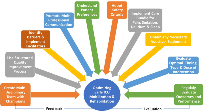
Ten strategies to optimize early mobilization and rehabilitation in intensive care

YOU have to make sure  you completely state your only preference is 100% recovery. You'll have to scream at your doctors because they incompetently have not been solving that problem for decades. 

Ten strategies to optimize early mobilization and rehabilitation in intensive care

Introduction

In the last decade, there have been more than 40 randomized trials evaluating early mobilization and rehabilitation in intensive care units (ICU) [1]. Such trials generally aim to reduce the incidence of ICU-acquired weakness (ICUAW) which is associated with poor long-term survival, physical functioning, and quality of life [2]. At least eight international guidelines have recommended ICU early mobilization and rehabilitation [3].

Despite supporting evidence and guidelines, implementation of ICU mobilization and rehabilitation is highly variable[4]. Hence, we report on 10 steps to help ICU clinicians in optimizing early mobilization and rehabilitation.

Create multidisciplinary team with designated champions

Early mobilization and rehabilitation is more successful in ICUs with a culture that prioritizes and values this intervention [5]. Mobility champions can help develop this culture using leadership and communication skills to educate, train, coordinate, and promote patient mobilization [3, 4, 6]. They support staff with an emphasis on safety and practical skills to improve the team’s confidence and capabilities [6].

Use structured quality improvement (QI) processes

A structured QI approach can greatly enhance successful implementation of early mobilization and rehabilitation [7]. One approach to QI includes four steps: (1) summarizing the evidence; (2) identifying barriers (e.g., sedation or lack of equipment); (3) establishing performance measures (e.g., sedation targets, frequency, and level of patient mobilization); and (4) ensuring all eligible patients receive the intervention (via appropriate engagement, education, execution, and evaluation) [6, 7].

Identify barriers and facilitators

A systematic review identified 28 unique barriers to early mobilization and rehabilitation, including patient-related barriers (e.g., physiological instability and medical devices), structural barriers (e.g., limited staff and equipment), procedural barriers (e.g., lack of coordination and delayed screening for eligibility), and cultural barriers (e.g., prior staff experience and ICU priorities for patient care) [4]. There are many strategies to effectively overcome barriers, including implementation of safety guidelines; use of mobility protocols; interprofessional training, education, and rounds; and inclusion of physician champions [4].

Promote multi-professional communication

The multi-professional team effort required for early mobilization and rehabilitation program depends on optimal communication. We recommend that interprofessional communication is facilitated using a structure adapted to the individual ICU that allows (algorithm-based) mobilization goals, including an opportunity for all team members to raise concerns and ensure flow of information regarding mobility goals and achievement across staff and over time [8].

Understand patient preferences

And the one and only preferences is 100% recovery. Don't allow your stroke staff to downgrade that goal because they haven't set up the proper protocols to get there.

ICU patients’ experience with early mobilization and rehabilitation is variable. It may be tiring, uncomfortable and difficult, while at other times be motivating and rewarding for patients [9]. With improving cognitive status, patients may be shocked by the severity of their muscle weakness. In the early stages of critical illness, patients may prefer to focus on short-time goals (e.g., sitting in a chair) set by the multidisciplinary team [9]. As patients progress, they may become more engaged in goal setting and longer-term rehabilitation planning (e.g., walking longer distances, sitting outside) (Fig. 1).

Fig. 1
figure1

Ten strategies to optimize early mobilization and rehabilitation in ICU

Adopt safety criteria

Meta-analyses have demonstrated the safety of in-bed and out-of-bed ICU mobilization, with rare occurrence of serious events [10]. One method of assessing safety is a traffic light system that provides specific criteria, across respiratory, hemodynamic, neurological, and other body systems, to be considered in mobilizing individual patients [11]. In this system, “red light” criteria indicate an increased potential for a serious safety event during mobilization requiring experienced decision-making, “yellow light” indicates potential risk that should be evaluated in terms of benefits versus risks, and “green light” indicates that mobilization is generally safe [11].

Implement care bundles for pain, sedation, delirium, and sleep

Patients’ sedation and delirium status is a common barrier to early mobilization and rehabilitation [4]. More broadly, pain, sedation, delirium, sleep, and early mobilization and rehabilitation are closely inter-related, as considered in clinical guidelines[3]. Assessment and management of these issues, via existing evidence-based practices (as synthesized in the guidelines), are important to maximize patients’ ability to participate in rehabilitation.

Obtain any necessary equipment

If this doesn't include lever or rowing wheelchairs you don't have the right equipment. 


Barriers to early mobilization and rehabilitation may include ICUAW, impaired physical functioning, traumatic injuries, and obesity [6]. Equipment can expand treatment options, increase patient mobility and activity levels, and reduce risk of injury to staff [12]. Selecting rehabilitation equipment may be challenging, with important considerations including the equipment cost/availability, ability to share equipment between units or patients (including infection control considerations), and the physical space available for patient mobilization and for convenient storage of equipment. Evidence supporting use of specific equipment is still evolving, including evaluation of neuromuscular electrical stimulation (NMES), in-bed cycle ergometry, tilt tables, and other devices [12, 13].

Evaluate optimal timing, type, and dose of intervention

Important knowledge gaps exist regarding exercise, including the timing, type, and dose of interventions. There is some evidence suggesting that starting rehabilitation within 2 or 3 days of ICU admission may be superior to later initiation [3]. Types of interventions to be considered include active functional mobilization, in-bed cycle ergometry, electrical muscle stimulation (with or without passive/active exercises), tilt tables, and use of various rehabilitation equipment. In addition, the intensity, duration, and frequency of each intervention type are important considerations [14]. Additional research is needed to further understand potential benefit or harm. Until that time, clinician judgement will play an important role and must be tailored to individual patients and to the dynamic nature of critical illness.

Assess outcomes and performance

Mobility and rehabilitation-related measures, appropriate to the ICU setting and integrated into clinical care, are needed to set patient goals and track their progress, allocate scarce rehabilitation resources to those patients who may benefit the most, and conduct evaluations of structured quality improvement programs [15]. Understanding patients’ functioning prior to critical illness, and their own goals, are also important considerations.

Conclusion

Evidence is still evolving about early mobilization in ICU with ongoing large, multi-center trials. Further research is needed to understand the optimal timing, type and dose of interventions, and their effect on long-term patient outcomes. These 10 strategies provide guidance for implementing early mobilization and rehabilitation in the ICU with the goal of optimizing safety and effectiveness to improve patients’ experiences and outcomes.

References at link,

Dr. Sukhdeo-Singh named medical director of UM SRH Primary Stroke Center at Easton, Maryland

So contact her NOW, before your stroke to ensure she has set in process plans for 100% recovery and implementing research into interventions. YOUR RESPONSIBILITY. You can't assume that the board of directors has set proper goals for her to achieve.  Since 'care' is referred to multiple times the mindset of this stroke center is completely wrong. YOU ARE RESPONSIBLE FOR CHANGING THAT TO RECOVERY AND RESULTS!

Dr. Sukhdeo-Singh named medical director of UM SRH Primary Stroke Center at Easton, Maryland


Dr. Rena Sukhdeo-Singh

Dr. Rena Sukhdeo-Singh has been named Medical Director of the Primary Stroke Center at the University of Maryland Shore Medical Center at Easton.

EASTON — Dr. Rena Sukhdeo-Singh has been named Medical Director of the Primary Stroke Center at University of Maryland Shore Medical Center at Easton.

A member of the UM Shore Regional Health medical staff since August 2021, Sukhdeo-Singh sees patients at UM Shore Medical Group-Neurology and Sleep Medicine as well as in the Primary Stroke Center at Easton.

“I am excited to lead the outstanding care(This already means the wrong goals have been set. Results is the proper goal.)  and treatment offered in our Primary Stroke Center,” Sukhdeo-Singh said. “Our community is fortunate that our Stroke Center has an outstanding team of physicians, nurses, therapists and techs who achieved the level of excellence required for Primary Stroke Center designation and who work so well with our hospitalists, emergency department physicians, staff, stroke coordinator and rehabilitation specialists. I am passionate about acute stroke care, outcomes and rehabilitation. I am looking forward to continuing to advance quality stroke care for our region.”

Dr. M. Walid Kamsheh, the center’s former medical director, left in late October to focus on his work as Medical Director at UM Shore Medical Group-Neurology and Sleep Medicine.

A board-certified neurologist, Sukhdeo-Singh most recently completed a Fellowship in Vascular Neurology (Stroke) at the National Institutes of Health in Bethesda. She previously served as Chief Neurology Resident at the University of Tennessee Health Science Center in Memphis, Tennessee, and was an Associate Investigator for the Natural History Study and White Matter Study at the National Institute of Neurological Disorders and Stroke, Stroke Branch. She also has co-authored multiple articles and presentations in the field of neurology and stroke, with a recent book chapter publication on post-stroke depression.

“I feel my training as a Vascular Neurology Fellow at the NIH has prepared me well for this opportunity,” she said. “Our hospital’s designation as a Primary Stroke Center by the Maryland Institute for Emergency Medical Services Systems shows that we are committed to a higher level of stroke care. It demonstrates that we are following standardized practices to deliver the best outcomes for our patients and are dedicated to their continued care.”

Often the first line of defense during a stroke are the emergency department teams, Sukhdeo-Singh said. “UM Shore Regional Health at UM Shore Medical Center at Cambridge, Chestertown and Easton, and UM Shore Emergency Center at Queenstown are excellent at triaging acute strokes.”

Besides ensuring UM Shore Regional Health continues to meet national benchmarks for brain imaging and reaching goal treatment times, Sukhdeo-Singh said she is committed to following patients’ recovery periods.

“After the acute phase, there is usually a long recovery period that takes a lot of patience and adjustments,” she said. “I always want my patients to feel like they are being heard and are getting the best possible care.”

The Primary Stroke Center at UM Shore Medical Center at Easton has received multiple awards from the American Heart Association and the American Stroke Association’s Get with the Guidelines Stroke Recognition program, which recognizes hospitals for providing the most effective stroke treatment according to nationally acknowledged, research-based guidelines.

Most recently, University of Maryland Shore Medical Center at Easton’s Primary Stroke Center was recognized by the American Heart Association/American Stroke Association by receiving the Get With The Guidelines®-Stroke Gold Plus quality achievement award with Target Type 2 Diabetes Honor Roll.

According to the ASA, hospitals earning the Gold Plus Quality Achievement Award have reached an aggressive goal of treating patients with 85 percent or higher adherence to all Get With The Guidelines-Stroke achievement indicators for two or more consecutive 12-month periods and have achieved 75 percent or higher compliance with five of eight Get With The Guidelines-Stroke Quality measures.

“We follow each treatment case to look for opportunities for improvement,” Sukhdeo-Singh said. “As Medical Director, I also plan to engage in public education about strokes, collaborate with our local emergency medical services and the Maryland Institute for Emergency Medical Services Systems counterparts, and ensure that from the first 911 call to discharge, patients are getting the best care for their stroke.”

Kamsheh has been the Primary Stroke Center’s Medical Director since 2017, and has been practicing medicine as a neurologist on Maryland’s Eastern Shore for more than 30 years. This year, he celebrated 10 years with the University of Maryland Medical System (UMMS).

“For the past few years I have had the honor and privilege to be the Medical Director of the Stroke Center,” Kamsheh said. “We should be proud, as such a small hospital, to be certified as a Primary Stroke Center. To achieve recognition, it takes a special, dedicated team on the ground that includes the ER staff, physicians, nurses, techs and support from UMMS, as well as the support of local emergency crews. Now it is time for me to pass this directorship to another great physician. She is stroke-trained at NIH and will lead us into the future of stroke care.”

Kamsheh continues to see patients at UM SMG-Neurology and Sleep Medicine’s Easton office. This growing practice specializes in the study and treatment of brain, spinal cord and nerve disorders, as well as sleep-related disorders.

UM Shore Regional Health also offers a Stroke Support Group, facilitated by Nicole Leonard, UM SRH’s Stroke Coordinator. This virtual group takes place on the first Thursday of each month, from noon to 1 p.m. Patients, caregivers and loved ones are invited to participate. For more information or to receive login credentials for the Zoom group, please call Leonard at 410-822-1000, extension 5068.

UM SMG-Neurology and Sleep Medicine offers appointments in-person and via telemedicine. For more information or to schedule an appointment, speak to your primary care physician, call 410-770-5250 or visit umshoreregional.org.