Wednesday, May 31, 2023

F82 The 10th Annual Upper Limb Neurorehabilitation Course

I see nothing in either day that remotely suggests they are providing EXACT PROTOCOLS FOR RECOVERY!  Useless blathering amongst themselves, nothing that will concretely help survivors. Prove me wrong, I dare you.

F82 The 10th Annual Upper Limb Neurorehabilitation Course

Event Information

The 10th Annual Upper Limb Neurorehabilitation Course
Dates of Event
29th June 2023 – 30th June 2023
Last Booking Date for this Event
28th June 2023

Description

UCL Centre for Neurorehabilitation and the National Hospital for Neurology and Neurosurgery present:


The 10th Annual Upper Limb Neurorehabilitation Course

Treating patients with upper limb deficit: Integrating research into practice


Thursday 29th June and Friday 30th June 2023


This two-day course provides an up-to-date overview of current research in treatment and rehabilitation options for the neurological patient with upper limb deficit. The course looks at the practical, real-life translation of scientific evidence into clinical practice and discusses the ingredients that make an upper limb therapy effective. Delegates will have the opportunity to trial novel devices and robotic technology. Course lecturers are leading clinicians and researchers in the field of upper limb rehabilitation in neurological patients. Full programme can be found under ‘more info’ tab.


Standard fee: £250 (includes catering)

Student fee: £125 (includes catering)

Enquiries to: cnr@ucl.ca.uk


Attendee CategoryCost   

CLCH Remaining Payment.£62.50[Read More]

Standard.£250.00[Read More]

Student Rate.£125.00[Read More]

More Information

DAY 1

Time

Title

Speaker

08:55-09:00

Introduction

Nick Ward

09.00-09.45 

Upper limb recoverythe state of play (1)           

Nick Ward

09:45-10:30

Integrated approach to UL rehabilitation              

Fran Brander & Kate Kelly

10.30-11.00 

Coffee break & Exhibitors          


11.00-11:45 

Recognising and treating the painful shoulder    

Ben Beare & Zeina Ghanem

11:45-12:15

Arm preparation – dealing with spasticity             

Amanda Strawson & Hannah Martin

12:15-13.00

Assessing Motor control

Steve Scott

13.00-14.00 

Lunch & Exhibitors & Demonstrations 


14.00-14:45 

Case Study #1  

Amanda Strawson & Helen Stancombe

14.45-15.15 

The role of priming in a rehabilitation service      

Fran Brander & Kate Kelly            

15.15-15:45 

Coffee break & Exhibitors


15.45-16.30  

Severe arm – keeping it in the game 

Emma Playfair & Hannah Griffiths            

16.30-17.00

Discussion


17.00-19.00

Wine & Chat & Exhibitors


DAY 2

Time

Title

Speaker

09.00-09.45 

The role of self-management in recovery

Fiona Jones & Scott Ballard-Ridley

09:45-10:30

Upper limb recoverythe state of play (1)           

Nick Ward

10.30-11.00 

Coffee break & Exhibitors          


11.00-11:45 

Don’t forget about sensation

Kathleen O’Keeffe & Mags Bean

11:45-12:30

The role of electrical stimulation 

Georgina Masters & Emma Playfair

12.30-13.30 

Lunch & Exhibitors & Demonstrations 


13.30-14:15

Creative splinting for function                                    

Elisha O’Brien & Amanda Strawson

14.15-15.00

Case Study #2 

Jess Steel & Mags Bean

15.00-15:30 

Coffee break & Exhibitors


15.30-16.15  

Gaming your way to recovery

Mireia Coll

16.15-17.00

Case Study #3

Zeina Ghanem & Emma Playfair

17:00-17:30

Closing discussion


17.30-19.00

Wine & Chat & Exhibitors


National Stroke Awareness Month: Vagus Nerve Stimulation Post-Stroke

 She may be certified and much more professionally trained than me but still has NO protocol written up on this. Survivors demand better!

National Stroke Awareness Month: Vagus Nerve Stimulation Post-Stroke

Written By: Liza Meiksins PT, DPT
Board Certified Specialist in Neurologic Physical Therapy
Certified Lymphedema Therapist


Many patients and therapists would likely agree that one of the most challenging aspects of stroke rehab is improving upper extremity function and recovery. As a former patient once said, “I would be happy with my recovery if this arm just wasn’t so stubborn!” But with few options to promote upper extremity recovery, there has been a lot of talk recently about the use of vagus nerve stimulation. So, what exactly is vagus nerve stimulation and how does it impact upper extremity recovery post-stroke?

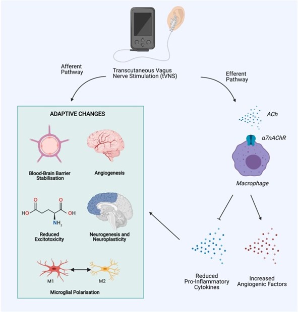
Invasive vagus nerve stimulation (i-VNS) has been widely accepted to improve upper extremity function post-stroke, but this requires a surgical procedure and is not appropriate nor encouraged for many patients1. However, in recent years, transcutaneous vagus nerve stimulation (t-VNS) has been shown to activate similar vagal nerve projections as i-VNS and is generally safe and well-tolerated1. t-VNS can be carried out through auricular VNS in the ear or cervical branch VNS in the neck and has been shown to elicit the following responses:


Physiologic Changes with t-VNS1,2:

  • Cholinergic & monoaminergic modulation of motor cortex neurons
  • Reduced systemic inflammation
  • Reduced blood-brain barrier breakdown
  • Increased angiogenesis
  • Reduced excitotoxicity
  • Improved axon regeneration & reorganization
 

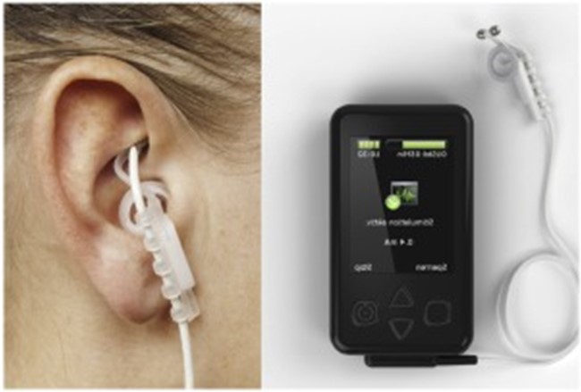
Example of auricular transcutaneous VNS device4

Functional Outcomes with t-VNS1,2,3:

  • Significant improvement in immediate and long-term upper extremity outcomes when t-VNS was applied in conjunction with intensive, task-specific rehabilitation (quantified using the UE Fugl-Meyer scale, Wolf Motor Function Test, Stroke Impact Scale, and Motor Activity Log)
  • Most research has been done on patients with moderate-severe UE impairment
  • Results consistent in both sub-acute and chronic strokes
  • Most studies have examined the use of t-VNS immediately prior to rehab interventions and used auricular VNS as the method of delivery


Further research(EXACTLY WHOM is doing that? So we can follow up and make sure it gets done.) continues to be carried out to expand our understanding of dosing, optimal patient selection, laterality, and auricular vs cervical stimulation, among other variables to optimize outcomes. There is also the potential to utilize t-VNS for other impairments besides motor function, including: dysphagia, dysphasia, cognition, and visual field dysfunction1.

While many questions surrounding transcutaneous vagus nerve stimulation still abound, it remains an extremely promising intervention to potentiate rehabilitation-driven neuroplasticity and improve functional outcomes for our patients post-stroke!


Explore online continuing education courses from Liza below:

Clinical Application of Neuromuscular Electrical Stimulation (NMES) for Neurologic Conditions

High-Intensity Gait Training for Stroke and Spinal Cord Injury Patients


Visit summit-education.com for more information.


References:

Baig SS, Kamarova M, Ali A et al. Transcutaneous vagus nerve stimulation (tVNS) in stroke: the evidence, challenges and future directions. J Autonom Neurosci. 2022;237.

Dawson J, Liu CY, Francisco GE, et al. Vagus nerve stimulation paired with rehabilitation for upper limb motor function after ischaemic stroke (VNS-REHAB): a randomised, blinded, pivotal, device trial. The Lancet. 2021;397(10284):1545-1553.

Hamer HM, Bauer S. Lessons learned from transcutaneous vagus nerve stimulation (tVNS). Epilepsy Research. 2019;153:83-84.

Lui Y, Zhang L, Zhang X, et al. Effect of Combined Vagus Nerve Stimulation on Recovery of Upper Extremity Function in Patients with Stroke: A Systematic Review and Meta-Analysis. J Stroke Cerebrovas Dis. 2022;31(6)

Study found long-term brain damage associated with COVID-19, not vaccine | Fact check by USA Todaay

In case you are getting bamboozled by vaccine claims. I'm much more worried about brain damage from COVID-19 than the vaccine. I already have enough brain damage, I'm preventing more.

 Study found long-term brain damage associated with COVID-19, not vaccine | Fact check

A May 12 Instagram post (direct link, archive link) shows a screenshot of a now-deleted tweet.

"BREAKING: A European study has found COVID vaccines could be causing 'long-term brain damage,'" reads the tweet.

The post garnered more than 200 likes in five days. Similar versions of the claim have been shared on Instagram and Twitter.

Follow us on Facebook! Like our page to get updates throughout the day on our latest debunks

Our rating: False

The study explored the long-term neurological effects of being infected with the COVID-19 virus, not the vaccine.

Study focuses on virus, not vaccine

The viral claim appears to stem from a May 9 article published by The People's Voice, formerly known as NewsPunch. The website has a lengthy history of publishing misinformation.

The article references a pre-print study published in April that explored the long-term neurological effects observed in those who had been infected with COVID-19. It claims the study revealed that "spike proteins from mRNA jabs infest the brain tissue of vaccinated people."

But Dr. Ali Ertürk, a co-author of the paper and director of the Institute of Tissue Engineering and Regenerative Medicine in Munich, told USA TODAY the study did not examine the COVID-19 vaccine of its side effects.

"We have done zero experiments using the vaccine, and we have shown and claim zero side effects of the vaccine," said Ertürk in an email. "Our work reports the presence of the spike protein in the skull of deceased individuals long after their COVID-19 infection, suggesting that the spike's persistence may contribute to long-term neurological symptoms."

Fact check: FDA still recommends COVID-19 vaccine, contrary to viral claim

None of the COVID-19 vaccines authorized in the U.S. contain the live virus, according to the Centers for Disease Control and Prevention. Instead, the mRNA in the vaccine teaches the body's cells to make copies of the COVID-19 spike protein so they can later recognize and fight off the virus if they become infected.

"The issue is that during infection, there is an enormous amount of viral replication and spike protein production, which impacts many organs including the brain," Ertürk said.

The COVID-19 vaccine is safe and effective, according to the CDC.

USA TODAY reached out to the users who shared the post for comment but did not immediately receive a response.

The claim has also been debunked by the Associated Press and PolitiFact.


$4.5 million grant to fund research exploring link between exercise and slowing Alzheimer's progression

Your risk of dementia, has your doctor told you of this?  Your doctor is responsible for preventing this! Which means your doctor has to get you 100% recovered to be able to do these aerobic exercises.

1. A documented 33% dementia chance post-stroke from an Australian study?   May 2012.

2. Then this study came out and seems to have a range from 17-66%. December 2013.`    

3. A 20% chance in this research.   July 2013.

4. Dementia Risk Doubled in Patients Following Stroke September 2018 

The latest here:


$4.5 million grant to fund research exploring link between exercise and slowing Alzheimer's progression

A $4.5 million groundbreaking grant will fund research to explore a promising link between aerobic exercise and slowing the progression of Alzheimer's disease in a study led by an Arizona State University researcher.

An estimated 6.7 million Americans age 65 and older are living with Alzheimer's disease, according to the Alzheimer's Association's 2023 report. A $4.5 million groundbreaking grant from the National Institute on Aging will fund research exploring a promising link between aerobic exercise and slowing the progression of Alzheimer's.

Alzheimer's is a complex disease with many factors contributing to it, which is part of the reason we haven't found a single cure. Recently though, we've found that modifying lifestyle factors may contribute to slowing the progression of the disease."

Professor Fang Yu, Edson Chair in Dementia Translational Nursing Science at the Edson College of Nursing and Health Innovation

The grant comes from the National Institute on Aging, a division of the National Institutes of Health. Yu and her team will conduct the first-ever sequential, multiple assignment, randomized trial, or SMART, for Alzheimer's disease.

"I think this grant shows what Professor Yu and her team are doing is really cutting edge. This research could impact millions of people, potentially giving them some hope of relief from a terrible disease that steals their very essence. I'm looking forward to the findings and seeing how they develop into real-world interventions," said Edson College Dean Judith Karshmer.

We recommend Management innovation of Chang’e-5 project Jizhong LIU et al., Frontiers of Engineering Management, 2021
  • Blockchain-based smart contract for smart payment in construction: A focus on the payment freezing and disbursement cycle
    Liupengfei WU et al., Frontiers of Engineering Management, 2022
    1. Fiber-reinforced composites in milling and grinding: machining bottlenecks and advanced strategies
      Teng GAO et al., Frontiers of Mechanical Engineering, 2022
    2. Quantifying the Contribution of Track Changes to Interannual Variations of North Atlantic Intense Hurricanes
      Jun Lu, Liguang Wu & Shunwu Zhou, Special Issue on Climate Change and Variability of Tropical Cyclone Activity, 2022

    This area of research builds on Yu's previous work, which found that a six-month exercise program significantly reduced cognitive decline in comparison to the natural course of changes for Alzheimer's dementia. Specifically, aerobic exercise is effective in reducing white matter hyperintensities progression, which is associated with cognitive decline and the development of dementia.

    The new Phase 2 clinical trial will examine the best exercises to improve aerobic fitness in older adults with early Alzheimer's disease and examine how those exercises impact their memory.

    "The most exciting part of this is trying to reduce the variations in responses to exercise among participants. To this point, we've seen some people improve, while some decline. So, what we're trying to do is to reduce the variability to help people respond positively in some way so we can truly understand if exercise has an impact and for who," Yu said.

    The team is planning to recruit more than 100 participants in Arizona to enroll in the study. They're looking for people who are 65 and older, who have noticed changes in their memory and have someone who knows them to serve as their study partner.

    Arizona State University

    Ear acupuncture with reduced food intake linked to weight loss for adults

    You have got to be kidding:

    Impossible to have effects except as a placebo. Energy meridians have never been proven to exist.
    No mechanism of action is possible. 

    But if you believe, have at it, recognizing these possible side effects;

    acupuncture side effects


    Ear acupuncture with reduced food intake linked to weight loss for adults 

    Key takeaways:

    • Men from Japan who underwent auricular acupuncture stimulation followed by a diet lost 8.61 kg of body weight at 3 months.
    • Women who received acupuncture lost a similar amount of weight as men.

    An ear acupuncture intervention using metal beads may help induce weight loss for adults, according to findings from two studies presented at the European Congress on Obesity.

    In data from 81 Japanese men who underwent an auricular acupuncture stimulation, researchers observed decreases in body weight, BMI, waist circumference and body fat. Similar reductions were observed in a study of 81 Japanese women, though women had a greater decrease in body fat and men had a higher increase in muscle mass.

    Auricular acupuncture linked to lower muscle mass and body fat percentage at 3 months.
    Data were derived from Fujimoto T, et al. PO4.085. Presented at: European Congress on Obesity; May 17-20, 2023; Dublin.

    “By stimulating the auricular acupoints using beads instead of traditional acupuncture needles, we have found that it is possible to support weight-loss treatments,” Takahiro Fujimoto, MD, PhD, of Clinic F in Tokyo, told Healio. “We propose this as a convenient means of supporting weight-loss treatments beyond dietary and exercise therapies.”

    Fujimoto presented data from two studies of an auricular acupuncture stimulation-based weight-loss program. In the first study, researchers enrolled 81 men to receive acupuncture stimulation using metal beads on six points of the ear. Participants received dietary guidance to reduce food intake in half and nutritional supplements to compensate for any nutritional deficiencies. Clinic visits took place twice a week for bead sticking and diet progress monitoring. Body composition measurements were collected over 3 months.

    From baseline to 3 months, the men had an 8.61 kg reduction in body weight, a 2.88 kg/m2 reduction in BMI, a 10.43 cm decrease in waist circumference, a 3.98% decrease in body fat and a 2.11% increase in muscle mass. The cohort also had increases in muscle-to-fat ratio and leg muscle mass and decreases in basal metabolic rate and visceral fat level.

    In the second study, researchers obtained data from 81 women in Japan aged 23 to 74 years from a dataset of participants who underwent the same acupuncture and diet weight-loss program as the men in the first study. Women were matched with men from the first study 1:1, based on age and BMI values at baseline. Body composition measurement changes from baseline to 3 months were compared between men and women.

    From baseline to 3 months, men and women had similar reductions in weight loss, BMI and waist circumference. Men had a greater decrease in muscle mass compared with women (mean change, –5% vs. –2.8%; P < .001) while women had a greater decrease in body fat percentage than men (mean change, –4.5% vs. –4%; P = .04). Basal metabolic rate dropped more in men vs. women (mean change, –101 kcal vs. –66 kcal; P < .001). The ratio of muscle-to-fat also increase more in men vs. women (mean change, 0.26 vs. 0.15; P < .001).

    Fujimoto said future research is needed to examine the mechanisms behind how acupuncture contributes to weight loss in adults.

    “It is likely that the stimulation of the parasympathetic nervous system is involved in the production of leptin, but we would like to conduct further research to gather supporting data,” Fujimoto said.

    Vitamins can make your brain 3 years younger, study says

    But where do we get genuine vitamins?  And this still leaves your doctor to come up with interventions to recover the extra two years.

    The supplements in the US have zero guarantee of purity or efficacy due to the fucking stupidity of the US Congress passing the Dietary Supplement Health and Education Act of 1994 (DSHEA).

    Your doctor should already have interventions to recover your lost 5 cognitive years from your stroke  if they are competent at all.


    Vitamins can make your brain 3 years younger, study says

    Peer-reviewed scientific study tracked 3,500 seniors over a three-year span

    Regular vitamins and walks can slow or even reverse the effects of cognitive decline. Getty Images/iStockphoto

    If you’re getting more forgetful as you age — and who among us isn’t? — there are two things you can do about it this Memorial Day weekend.

    Take a multivitamin. And go for a 30-minute walk.

    Then keep both of those practices up.

    So reports a peer-reviewed scientific study out last week, which found that regular vitamins and walks can slow or even reverse the effects of cognitive decline on the aging brain.

    The average effect on the aging brain of a daily multivitamin is the equivalent of being a full three years younger, according to a study conducted by researchers at Columbia and Harvard medical schools and the New York State Psychiatric Institute. “We estimate that the effect of the multivitamin intervention improved memory performance above placebo by the equivalent of 3.1 years of age-related memory change,” the researchers report in the latest issue of the American Journal of Clinical Nutrition.

    This was based on a study of more than 3,500 senior citizens over three years. The participants were randomly assigned either to take a Centrum Silver multivitamin — Centrum is a Pfizer-owned PFE, -1.34% brand — or a placebo every day. They were subjected to various standard brain tests every year, such as tests requiring people to recall as many words as possible from a random list.

    Researchers say the benefits were most pronounced in those with underlying heart disease.

    The benefits of the daily vitamin showed up as early as the first annual exam, and persisted in the years afterward, the researchers said.


    These findings confirm similar findings in a parallel study published last year, which found that a daily multivitamin benefited the whole brain, not just the memory. That study, too, found the effects were most pronounced among those with underlying heart disease.

    Meanwhile another independent study, conducted at the University of Maryland, found that walking for 30 minutes three or four times a week also has a significant beneficial effect on the brains of older people.

    The study involved 33 participants aged between the ages of 71 and 85 who exercised on a treadmill under supervision over a 12-week span. Verbal memory tests and MRI scans showed brain and memory benefits, even that quickly.

    There are so many scientific studies coming out these days — on age-related cognitive decline and more generally — that it’s easy to become inured to them. But cognitive impairment and full-blown dementia are already pandemics way more extensive than COVID-19 even at its most acute stage. Alzheimer’s is currently killing over 6 million Americans (six times as many Americans as died with the coronavirus-borne disease), and the numbers are rising.


    And scientific breakthroughs in terms of medical treatments, let alone cures, are scarce and expensive.

    So it’s good news that there are things we can do on our own. These include not just taking vitamins and walking but eating the right foods, avoiding the wrong ones, studying, doing crosswords and meditating.

    We can hardly do them all at once. But anything is better than nothing.

    Next challenge for those of us getting older? Remember to take the multivitamin every morning. And remembering where we put them.

    Stroke Recovery Strategies June 16 webinar by Anysia Ensslen-Boggs, Ed.D., M.S., CCC-SLP

    Looking at the objectives and outline there is nothing on recovery protocols to use, so in my opinion, worthless.

    Stroke Recovery Strategies June 16 webinar by Anysia Ensslen-Boggs, Ed.D., M.S., CCC-SLP

     
    • Learning Objectives
    1. Identify various etiologies of cognitive deficits as they relate to the neuroanatomy of strokes.
    2. Define principles of experience-dependent neuroplasticity and its impact on rehabilitation.
    3. Select appropriate screenings and assessments to informally and formally evaluate cognitive deficits.
    4. Explain how various cognitive impairments impact the patient's ability to meet transdisciplinary treatment objectives.
    5. Implement treatment techniques for orientation, short-term memory, safety awareness, and executive functioning forfunctional improvement of ADL's across all therapydisciplines.
    6. Compose measurable goals that allow for documentation of patient outcomes.
    Outline
    1. The Current Landscape of Cognitive Deficits
      1. Stroke as etiology
      2. Neuroanatomy
      3. Orientation and awareness
      4. Attention
      5. Memory
      6. High-level cognitive deficits associated with right CVA
    2. Assessment of Cognitive Deficits Following Stroke
      1. SLUMS
      2. MoCA
      3. Other standardized assessments and screening tools
      4. Criterion-referenced assessments
      5. Setting functional treatment objectives
    3. Leveraging Neuroplasticity for Stroke Recovery
      1. What is neuroplasticity?
      2. 10 principles of experience-dependent neuroplasticity
      3. Neuroplasticity and evidence-based practice
      4. Application to levels of assistance and cueing hierarchy
    4. Intervention Strategies to Improve ADLs
      1. Treatment techniques for improving functional cognition
      2. Utilizing neuroplasticity to achieve functional treatment objectives
      3. Improving completion of ADLs by improving cognition
      4. Executive functioning and application to patient safety and independence
      5. Examples of cognitive interventions
    5. Effectively Communicating Across a Multidisciplinary Team
      1. Targeting cognitive goals in PT and OT
      2. Co-treating/interdisciplinary treatment plans
      3. Family/caregiver/staff education
      4. Discharge plans: Is this patient safe at home?
    6. Accurate Documentation Tips and Tricks
      1. Goal-writing
      2. Patient outcomes
      3. Justifying medical necessity
      4. ICD-10 and Medicare guidelines
    7. Case Studies
      1. Review of case studies in small interdisciplinary groups
      2. Application of knowledge
      3. Discussion
      4. Case Study

    Inhale, Exhale, Remember: Uncovering the Breath-Memory Connection


    What is your doctor's breathing protocol? Doesn't have one, then you don't have a functioning stroke doctor. 

    RUN AWAY!


    What does your doctor think? Should you be doing slow breathing as the book

    'Breath: The New Science of a Lost Art' by James Nestor

    says?

    5 Ways To Improve Your Breathing with James Nestor

    Or should you be doing fast breathing in

    Creation of nitric oxide via Breath of Fire  February 2014 

    And why doesn't your doctor know a damn thing about a breathing protocol?

    The latest here:

    Inhale, Exhale, Remember: Uncovering the Breath-Memory Connection

    Summary: Intricate links between breathing and memory recall have been unraveled by recent scientific research, painting a more complex picture of our cognitive processes.

    Studies reveal that the rhythm of our breathing can influence neural activity, impacting cognitive functions such as emotional processing and memory recall.

    The most compelling evidence highlights that inhalation, particularly through the nose, can improve memory function. As this field of study emerges, these insights could lead to novel therapeutic approaches for cognitive decline and memory-related conditions.

    Key Facts:

    1. The rhythm of our breathing creates electrical activity in the brain, enhancing emotional judgment and memory recall, with this effect being most pronounced during inhalation through the nose.
    2. The amygdala and hippocampus, brain areas linked to emotion and memory, are significantly affected by the rhythm of breathing, suggesting that the act of breathing can modulate the functions of these regions.
    3. Deep, controlled breathing, often used in mindfulness practices, can improve working memory capacity, the kind of memory we use to hold and manipulate information over short periods.

    Source: Neuroscience News

    Breathing: it’s an automatic process we often don’t give a second thought. Yet recent scientific discoveries have begun to shed light on a fascinating relationship between breathing and memory function.

    Our breath influences our neural activity, which in turn, impacts our cognitive functions including attention, memory recall, and emotional processing.

    The rhythm of our breathing creates electrical activity in the brain that contributes to the enhancement of emotional judgments and memory recall.

    In fact, a study led by Christina Zelano at Northwestern University demonstrated that the act of breathing, specifically through the nose, can have a direct impact on cognitive functions such as memory recall.

    Credit: Neuroscience News

    Zelano’s research team carried out a series of experiments involving human subjects and found that memory recall was significantly better during inhalation compared to exhalation. This effect was most pronounced when the subjects were breathing through their noses.

    The study showed that the rhythm of breathing can induce changes in the brain, enhancing the emotional judgment and improving memory recall.

    Furthermore, the amygdala and the hippocampus, two brain regions linked to emotion, memory function, and smell, are significantly affected by the breathing rhythm.

    These areas of the brain are part of the limbic system, which controls emotions and memory. It’s thought that the act of breathing may modulate the functions of these brain regions, thereby influencing memory and emotional processing.

    Moreover, the act of controlled, deep breathing, often utilized in mindfulness and meditation practices, has been shown to enhance memory recall.

    A study published in the Journal of Sport and Exercise Psychology showed that mindfulness-based attention, which involves focusing on one’s breathing, increases the ability to maintain visuospatial information over short periods.

    This suggests that deep, controlled breathing can improve working memory capacity, the kind of memory we use to hold and manipulate information in our minds over short periods.

    While the relationship between breathing and memory remains an emerging field, these findings suggest exciting possibilities for future research and potential therapeutic applications.

    Understanding the impact of breathing on memory could have implications for interventions related to cognitive decline, stress, anxiety, and conditions such as ADHD and Alzheimer’s disease.

    In conclusion, it seems that the simple act of breathing, often taken for granted, can play a significant role in our cognitive functions, specifically memory recall.

    So next time you’re struggling to remember something, take a moment, take a deep breath, and see if it helps. It appears our breath holds more power over our brains than we might think.

    About this neuroscience and memory research news

    Author: Neuroscience News Communications
    Source: Neuroscience News
    Contact: Neuroscience News Communications – Neuroscience News
    Image: The image is credited to Neuroscience News

    Citations:

    Nasal Respiration Entrains Human Limbic Oscillations and Modulates Cognitive Function” by Christina Zelano et al. Journal of Neuroscience

    Mindfulness, Movement Control, and Attentional Focus Strategies: Effects of Mindfulness on a Postural Balance Task” by Kee et al. Journal of Sport and Exercise Psychology

    Respiration modulates olfactory memory consolidation in humans” by Artin Arshamian, Behzad Iravani, Asifa Majid and Johan N. Lundström in Journal of Neuroscience.

    New Ankle Exosuit Helps Post-Stroke Patients Walk Independently

    Can't find a picture of this. Ask you doctor how this compares to all the other walking helpers out there.

    Here is the picture from another article.

    Designed for independent use in community settings, the new exosuit could help stroke survivors improve their gait outside of the lab and during their daily routines
    Designed for independent use in community settings, the new exosuit could help stroke survivors improve their gait outside of the lab and during their daily routines - Biodesign Lab / Harvard SEAS

    New Ankle Exosuit Helps Post-Stroke Patients Walk Independently

    Harvard John A. Paulson School of Engineering and Applied Sciences

    Every 40 seconds, someone in the United States has a stroke. According to the U.S. Centers for Disease Control and Prevention, that totals about 795,000 strokes each year. More than 80 percent of stroke survivors experience gait challenges, often relating to a loss of control over ankle movement. As survivors progress into the chronic stage of stroke, most continue to walk slower and less efficiently.

    An agile, untethered, and easy-to-use ankle exosuit could change that. Designed for independent use in community settings, the new exosuit could help stroke survivors improve their gait outside of the lab and during their daily routines. A proof-of-concept study suggests the community-use ankle exosuit could help stroke survivors improve their walking propulsion and boost their overall walking confidence and ability while ambulating around their own homes, workplaces, and neighborhoods. The work, led by Conor Walsh’s team at the Harvard John A. Paulson School of Engineering and Applied Sciences (SEAS), is published online in Annals of the New York Academy of Sciences.

    Recent studies have proven that post-stroke study participants can improve their walking speed, distance, propulsion, and gait symmetry with the help of an assistive robotic exosuit, but those studies have all occurred in labs or clinical settings.

    “We saw an opportunity to leverage wearable technology to rethink how we approach physical therapy and rehabilitation” says Walsh, senior author on the paper and the Paul A. Maeder Professor of Engineering and Applied Sciences at SEAS. “If we can shift some of these clinical services from the clinic to the home and community, we can improve access, reduce costs and deliver better care. It is exciting to see the fields of engineering and physical therapy come together to make this happen.”

    For over a decade, Walsh’s Biodesign Lab at Harvard has been developing assistive and rehabilitative exosuit technologies for various applications. Some of that technology has already been licensed and commercialized by ReWalk Robotics and been given breakthrough status by the U.S. Food and Drug Administration. To design an ankle exosuit meant for use in the community, Walsh’s team need to simplify the exosuit’s mechanical components and make it easy for wearers to control.

    “In the past, our ankle exosuits had two active actuators – one that helped with dorsiflexion to keep the wearer’s toes up, and another to help with plantarflexion, propelling the foot and body away from the ground,” says Richard Nuckols, a former postdoctoral fellow in Walsh’s lab at SEAS, and co-first author of the paper.

    Instead of an active dorsiflexion actuator, the new exosuit contains a passive material that flexes and performs like a spring, helping the toes stay up during the foot’s swing phase and preventing the wearer from catching their toes on the ground. “By replacing an active actuator with a passive actuator, the exosuit is inherently safer; in the case of an unexpected power loss or controller failure, the default state will keep the users toes up and reduce risk of a trip and fall,” Nuckols says.

    “We also developed a mobile app to enable wearers to easily interact with the device and remotely check in with our team,” says Chih-Kang Chang, a Ph.D. candidate in Walsh’s lab and a co-first author on the paper. “The app allows wearers to turn the device on themselves and tell the exosuit when they want to start walking.”

    In addition, the team incorporated sensors to allow for remote monitoring of the wearer’s progress over time. “We are collecting data while people are walking in the exosuit, and measuring how they improve their gait over time,” Chang says. “Going forward, this information could be a really powerful aspect of using this exosuit for long-term rehabilitation in partnership with a physical therapist.”

    “These sensors – located on the foot, shank, and pelvis – are converted using a machine-learning algorithm into estimates of propulsion, helping us understand how well people are generating proper ankle mechanics and how effectively they are walking,” Nuckols says.

    “Collecting the amount of data needed to train a typical machine learning model from individual wearers is extremely challenging, given the limited ability to walk for extended periods of time post-stroke,” says Daekyum Kim, a postdoctoral fellow in Walsh’s lab, and co-first author of the paper. “The key advantage of our approach is that it leverages walking data gathered from multiple individuals to better tune a machine learning model to each user.”

    To test the community ankle exosuit, Walsh’s team partnered with the labs of Lou Awad and Terry Ellis from Boston University’s Sargent College of Health & Rehabilitation Sciences. They recruited four participants to use the device in their own community settings for four weeks, walking independently three to five times each week. All participants safely completed the study and reported no safety issues. Due to individual variability in response (participants with lower baseline walking propulsion saw more benefit from wearing the exosuit), therapeutic benefit was not observed across the whole group. But two of the participants improved their propulsion by an average of 27 percent. They also walked an average of 4,000 steps further in the week after the study than they had walked in the week before the start of the study.

    “I was 33 when I had my stroke. As a result of the stroke, I have diminished sensitivity on my entire left side,” says Bryant Butler, 51, one of the study’s participants. “Walking is a challenge. I can’t feel my toes very well when I am walking, and I have difficulty bending my leg. I frequently scuff the toe of my shoe, and sometimes I trip.”

    During the study, Butler used the exosuit on Boston’s Commonwealth Avenue Mall, walking 20 to 30 minutes at a time several days a week.

    “The experience of walking with the exosuit was liberating, because I no longer had to expend so much mental energy when going from one place to another,” he says. “The exosuit gradually corrected my gait with every step. I learned how my leg muscles, knee, and toes should feel when I walk without it. [Even] when I wasn’t using [the exosuit], my walking improved, because the device taught me how to better compensate for the shortcomings of my left leg post-stroke.”

    Butler adds: “The device became an extra piece of clothing – [except for the] wires and a battery pack – that I wore for a specific purpose. Most of the time, I forgot it was there. The exosuit nudged me into being a better walker. It inspired me to walk more, and to enjoy it.”

    Additional authors on the paper include Asa Eckert-Erdheim, Dorothy Orzel, Lauren Baker, Teresa Baker, Nicholas C. Wendel, Brendan Quinlivan, Patrick Murphy, Jesse Grupper, and Jacqueline Villalobos.

    Harvard University’s Office of Technology Development is exploring commercial opportunities stemming from this intellectual property.

    This work was supported by the National Institutes of Health (BRG-R01HD088619), the National Science Foundation (CMMI-1925085), a MassTech Collaborative Research and Development Matching Grant, and support from a Blavatnik National Award for Young Scientists.

    /Public Release. This material from the originating organization/author(s) might be of the point-in-time nature, and edited for clarity, style and length. Mirage.News does not take institutional positions or sides, and all views, positions, and conclusions expressed herein are solely those of the author(s).View in full here.

    Prolonged red light disrupts circadian hormones associated with phrenic neuroplasticity

    Way beyond my ability to understand, ask your doctor what it means.

    Prolonged red light disrupts circadian hormones associated with phrenic neuroplasticity


    Neurodegenerative disease and spinal cord injury impair breathing ability, significantly impacting the quality and duration of life. New therapeutic interventions for these conditions aim to improve respiratory function by using intermittent exposure to periods of low oxygen (acute intermittent hypoxia; AIH). AIH elicits one form of respiratory motor plasticity known as phrenic long-term facilitation (pLTF) via competing cellular mechanisms initiated by serotonin and adenosine, respectively. Circadian rhythm and the daily rest/active cycle have recently been shown to modulate the serotonin vs adenosine balance and pLTF. Melatonin and corticosterone are light-sensitive hormones that regulate neuroplasticity and molecules necessary for some forms of pLTF, particularly adenosine. Since the impact of these light-sensitive hormones on pLTF has not been directly investigated, we performed a preparatory investigation concerning the impact of dim red light on daily cycles. We hypothesized that prolonged exposure to low-intensity red light during the active/dark phase (rLEN) impacts light-sensitive hormones. Male Sprague-Dawley rats (3-4 months) were housed in a 12 on/12 off light cycle in 3 different light conditions: 1) normal-cycle (light onset 0700h; n=6); 2) reverse-cycle (light onset 1900h; n=8); or 3) reverse-cycle with rLEN (32 Lux) during the dark phase (light onset 1900h; n=6). Serum samples were collected at mid time-points in the daily rest/active cycle (i.e. 12PM and 12AM). Competitive ELISAs were used to quantify serum melatonin and corticosterone levels; an adenosine assay was used to quantify spinal adenosine. During the midactive phase, melatonin levels were significantly reduced in rLEN rats (63pg/mL; p<0.050) vs rats in normal- (104pg/mL) and reverse- (107pg/mL) cycle housing. Spinal adenosine levels were similar between normal- (15uM) and reverse- (20uM) cycled rats in the midactive phase, but were significantly lower in the rLEN rats (2uM; p<0.001). Melatonin and spinal adenosine levels during midrest were not different between groups. Corticosterone levels during the midactive phase were similar between normal- (46.6ng/mL) and reverse- (34.3ng/mL) cycled rats, but were significantly lower with rLEN (23.5ng/mL; p<0.050). Corticosterone during the midrest phase in the normal- (12ng/mL) and reverse- (12.4ng/mL) cycle were similar, but was significantly elevated in rLEN rats (25ng/mL; p=0.022). We also observed a shift in sleep behavior in rLEN rats. Thus: 1) rats housed in normal and reverse light cycle exhibit appropriate cycles in light-sensitive hormones; but 2) even low intensity rLEN during the active phase disrupts these light-sensitive & stress-related hormones. These data yield striking evidence for the need to limit rLEN exposure to rodent models, and may have important implications for the use of AIH as a therapeutic modality to improve breathing (or non-respiratory motor) function in people with disrupted circadian rhythms.

    Supported by: NIH HL147554, HL148030, T32HL134621-5 (ABM), the American Physiology Society (LRB) and the UF McKnight Brain Institute.

    This is the full abstract presented at the American Physiology Summit 2023 meeting and is only available in HTML format. There are no additional versions or additional content available for this abstract. Physiology was not involved in the peer review process.

    Meditation Diminishes Bias Toward Negative Information

    With all the negative information you're getting from your doctor and therapists, namely nothing positive on how they are going to get you recovered. They should immediately counteract that negative bias by them by introducing meditation. But they won't, you're on your own to figure this out.

    Meditation Diminishes Bias Toward Negative Information

    Summary: A new study reveals that daily mindfulness meditation can help reduce individuals’ tendency to avoid negative information.(I think this sentence is worded completely wrong - should be - to seek out negative information) The practice of regular mindfulness meditation allows people to better process uncomfortable emotions, thus enabling them to handle negative information more objectively.

    The study concluded that meditation could foster better decision-making since meditators are more likely to consider information, even if it could elicit a negative response. This counteracts the cognitive bias that typically leads individuals to disregard potentially unsettling information.

    Key Facts:

    1. The study found that daily mindfulness meditation reduces the tendency to avoid potentially negative information, a common cognitive bias.
    2. Regular meditation not only aids in dealing with negative emotions but also facilitates a more objective processing of unfavorable information.
    3. This study suggests that daily meditation could enhance decision-making by encouraging the consideration of a wider spectrum of information, including potentially negative inputs.

    Source: ETH Zurich

    People who meditate every day are less likely to avoid negative information. This is a finding in a new study by a research team including researchers from ETH Zurich.

    Smart decision makers gather all the pertinent information, and weigh the pros and cons dispassionately. This also includes gathering information that could prove unsettling or unpleasant. At least that’s the theory.

    However, the reality is often quite different. Due to what experts call cognitive bias, people tend to ignore potentially negative information—even when such information is available to them.

    Credit: Neuroscience News

    For example, they don’t want to find out whether an investment is no longer worthwhile, a medical test has confirmed an illness, or a friend has betrayed their trust. The reason is that even just thinking about negative information triggers fear and worry.

    A recent study by Elliott Ash, Professor of Law, Economics, and Data Science at ETH Zurich, shows that people can reduce this tendency towards information avoidance through regular mindfulness meditation.

    Coping better with negative emotions

    The researchers define the practice of mindfulness meditation as sitting still with eyes closed, observing—but not responding to—breathing, physical sensations, thoughts, and emotions.

    Numerous scientific studies have shown that practicing meditation on a regular basis has a positive effect on both body and mind.

    Meditating for just 15 minutes a day helps people relieve stress, increases their ability to concentrate, cuts their risk of depression, and enhances their productivity.

    Like in other studies, Ash and his co-authors were able to show that daily meditation boosts people’s ability to contend with negative emotions.

    Rapid Cognitive Improvement With Noninvasive Brain Stimulation

    What does your doctor think of using this to combat your brain fog immediately post stroke?  

    Do you prefer your  doctor incompetence NOT KNOWING? OR NOT DOING?

    Rapid Cognitive Improvement With Noninvasive Brain Stimulation

    Transcranial alternating current stimulation (tACS) provides moderate cognitive benefits in healthy older adults as well as those with neuropsychiatric disorders, results of the largest and most comprehensive meta-analysis of tACS to date show.

    "tACS has shown great promise at enhancing mental function, but whether this technology can truly fulfill its promise has been a topic of considerable debate in the field of brain stimulation," senior investigator Robert Reinhart, PhD, with the Cognitive & Clinical Neuroscience Laboratory, Boston University, told Medscape Medical News. 

    Given conflicting evidence on tACS for boosting cognition, Reinhart and colleagues leveraged statistical meta-analytic techniques to quantify how consistent the evidence is across several studies.

    The time was right to do this, Reinhart said, as the number of tACS studies has "more than doubled since the previous meta-analysis and tACS designs have rapidly evolved, becoming increasingly sophisticated."

    The study was published online May 24 in Science Translational Medicine.

    Significant, Immediate Improvement

    The analysis included 102 studies of tACS in healthy individuals as well as those with neurological or psychiatric conditions published between 2006 and 2021.

    Together, these studies involved 2893 participants (1290 men and 1603 women) with a mean age of 30 years; 333 participants were older (mean age 67 years).

    A total of 177 participants had a clinical disorder such as major depressive disorder, attention deficit hyperactivity disorder (ADHD), epilepsy, Parkinson's disease, schizophrenia, and mild cognitive impairment.

    When compiling over 300 measures of mental function across all the studies, there was evidence for significant, reliable, and immediate improvement in mental function with tACS, Reinhart told Medscape Medical News.

    When examining specific mental functions separately — such as memory, attention, or intelligence — tACS produced the strongest improvement in executive control, or the ability to adapt behavior in the face of new, surprising, or conflicting information, he noted.

    "We also found improvements in the ability to pay attention, the ability to memorize information for both short and long periods of time, as well as in measures of intelligence. Together, these results suggest that tACS has the ability to particularly improve specific kinds of mental function, at least in the short term," Reinhart said.

    To establish the effect of tACS in people who might most need it, the researchers examined how tACS impacted mental function in two subpopulations which may be particularly vulnerable to brain changes: older adults and clinical populations.

    "In both subpopulations, we found reliable evidence for improvements in mental function with tACS. Interestingly, we also found that in specialized tACS, which can target two brain regions at the same time, manipulating the relationship between the two regions can both enhance or reduce mental function," Reinhart told Medscape Medical News

    This bidirectional regulation of mental function could be particularly useful in the clinic, he noted.

    "For example, conditions like depression may involve reduced reward-processing capabilities while others like bipolar disorder may involve a highly active reward-processing system. With the capability to change mental function in either direction, we may be able to flexibly develop targeted designs to cater to specific clinical needs," Reinhart said.

    The researchers caution that while the analyses suggest immediate enhancements in cognitive function with tACS, they do not speak to the sustainability of these improvements. The durability of cognitive changes is a key question for future studies.

    "Great Potential" but Questions Remain

    Commenting on this research for Medscape Medical News, Shaheen Lakhan, MD, PhD, a neurologist and researcher in Boston, said tACS has shown "great potential" in modulating brain systems and circuits. "However, it is important to note that tACS is still in the early stages of development and requires further refinement."

    "Before tACS can become widely applicable, several crucial aspects need to be addressed. Firstly, we need to determine the specific brain regions that should be targeted for optimal results. Additionally, we must identify which types of individuals would benefit the most from this technology," said Lakhan, who was not involved in the meta-analysis.

    "Moreover, it is crucial to ascertain whether the benefits observed in controlled experiments truly translate into real-life scenarios such as driving a car, work performance, academic achievements, and improved social relationships. It is one thing to witness improvements on brain tests, but it is essential to understand the practical implications of these enhancements," Lakhan commented.

    He said he envisions "a combination of drugs, devices, and applications will work together harmoniously within a closed system to modulate the brain, specifically targeting and alleviating conditions like depression, anxiety, and hyperactivity."

    "However, this advancement raises profound questions about the boundaries between clinical use and neuro-enhancement, challenging our existing social constructs," said Lakhan.

    "Our personalities, to a certain extent, are shaped by the intricate networks within our brains. Remarkably, our brain signatures can reveal aspects of our character, including agreeableness, openness, conscientiousness, neuroticism, and even our political affiliations," he added.

    "As we venture into this exciting realm of brain unlocking and manipulation, it is imperative that we approach it with caution, ethical considerations, and a deep understanding of the potential consequences," Lakhan said.

    "Only through responsible exploration and careful navigation can we fully harness the power of these technologies while respecting the complexities of our individual identities and the broader social implications they entail," he added.

    This work was supported by grants from the National Institutes of Health and a gift from an individual philanthropist. Reinhart and Lakhan report no relevant financial relationships.

    Sci Transl Med. Published online May 24, 2023. Abstract