Wednesday, September 30, 2020

What to do if you test positive for COVID-19

I totally disagree, you see a doctor immediately and get anti-coagulation going. You don't wait until it gets severe before you see a doctor. I'm going to demand heparin.

 Heparin binds to cells at a site adjacent to ACE2, the portal for SARS-CoV-2 infection, and "potently" blocks the virus, which could open up therapy options.

Anticoagulation Again Shown to Improve Survival in COVID-19 Patients;-Mortality risk about 50% lower

Stroke occurs frequently in COVID-19, leads to ‘devastating consequences’ for patients


I'm not medically trained so I know nothing, don't listen to me.

The latest here:


What to do if you test positive for COVID-19

You've gotten tested for COVID-19. What happens if it comes back positive?

Typically, a nurse or another health care professional will call to give you the news. Then they'll walk you through everything you need to know to keep you and those around you safe.

"We know that if someone tests positive, they have a lot of questions, and we want to make sure they have all their questions answered," said Linda Barman, MD, associate director of Stanford Medicine's CROWN Clinic, which was launched in April to care for COVID-19 patients who don't need hospitalization.

Barman provided these tips for weathering COVID-19.

How to quarantine

Each situation is unique and depends on who lives in the house and how vulnerable they are. When an entire family has tested positive at the same time-- which is not uncommon -- they can self-quarantine together.

If just one member of a family is infected, it's important to discuss the situation with a health care provider, who can make recommendations to keep the whole family safe. Sometimes positive patients will stay in a hotel while they are contagious. Other times, a positive patient can self-quarantine in a bedroom and connected bathroom. If that isn't possible, all members of the household should wear masks at all times in shared spaces. With a shared bathroom, everyone should wipe down every surface with disinfectant wipes after every use

Also, since the coronavirus has been detected in stool, "Close the lid on the toilet before flushing," said Barman, who is a clinical assistant professor of medicine and the associate director of Stanford's Express Care Clinics.

Who should prepare food

Infected people should avoid preparing food, if possible. If they must cook, they should prepare food only for themselves and thoroughly wipe down kitchen surfaces afterward.

When to contact a clinician(Wrong, wrong,wrong. See a doctor immediately)

Typically, COVID-19 is at its worst around 8 to 10 days after symptoms start. Patients should call a doctor or another clinician if their breathing gets more difficult or if they experience chest pain, Barman said.

They also should look out for what Barman calls the "shower sign" -- feeling so tired, they can't muster the strength to shower.

 "That's happened so many times with people who ended up getting really sick," she said. "So, if you're so tired you can't take a shower, I need to see you in person."

How to monitor your oxygen level at home

COVID-19 often negatively impacts how well oxygen is transferred into the bloodstream, but a patient doesn't always feel short of breath when their oxygen levels are low. Patients at home can monitor the percentage of oxygen in their blood using a pulse oximeter, a relatively inexpensive device that comfortably clamps onto a finger. Products that are "FDA-approved for home medical use" are best, Barman said.

A reading of 97% or higher is considered healthy, she said. If a patient has a reading of 96% or lower, they should contact a clinician; that may signal that their lungs are not functioning well.

When taking a reading, she said, it can take 30 to 60 seconds to come to a steady reading. The monitors may pick up a signal better on one finger than another -- if it is reading less than 96%, patients should try it on another finger. The highest pulse oximeter number is the correct one -- it could read artificially low, but not artificially high. Many pulse oximeters also record a heart rate or pulse. The instructions should indicate which is which.

How to stay comfortable

To ease breathing, Barman suggested sleeping and lying on the stomach, called proning. She recommended acetaminophen for fever or aches. If possible, patients should get up and move around once an hour, as lying in bed for long periods can cause back pain and stiffness.

How to speed healing

For exercise and vitamin D from sunshine, Barman urged COVID-19 patients to get outside once a day for as little as 15 minutes -- but only if the air quality is good and they can do it safely without going into public spaces. It is also important to stay well-hydrated.

When to end quarantine

Patients who are generally healthy and have mild symptoms should follow the 10-3 Rule, Barman said: self-quarantine for 10 days after symptoms begin and for at least three days following the onset of a fever.

People who are immunosuppressed or are admitted to the hospital should isolate for 20 days following the onset of symptoms, she said. After ending self-isolation, patients should continue to wear masks when out in public and wash hands frequently, Barman said.


'Stroke research that is saving lives'

 Great, but what the fuck are you doing to get survivors to 100% recovery?

'Stroke research that is saving lives' 

For the last twelve years, the Chair of Clinical Neurology has been revolutionising the way New Zealand treats strokes that come into the Emergency Department.Year after year the number of Kiwis affected by stroke has been on the rise. It is estimated that in 2020 11,000 Kiwis will be affected by a stroke. This staggering number proves how desperately we need more research into the treatment and rehabilitation of stroke patients.In 2008 a group of people who believed in what research could accomplish helped establish the Neurological Foundation Chair of Clinical Neurology. This position’s aim was to bridge the gap between neuroscience and neurology, with the theory that this strategy would improve patient outcomes if researchers could work alongside clinicians.In 2011, this theory was proven correct when Professor Alan Barber, Chair of Clinical Neurology and the team of clinicians, nurses, researchers and rehabilitation specialist introduced New Zealand to the revolutionary clot retrieval procedure. This procedure, better known in the medical world as a thrombectomy was known as ‘science fiction’ when Professor Barber first started out. "Thrombectomy used to be science fiction. Now we are routinely going in and pulling out clots. One in every five people who receive the treatment will return home as healthy as they were before the stroke occurred."Since 2011, the work the Chair of Clinical Neurology has conducted has treated over 1,000 Kiwis across Auckland, Wellington and Christchurch, saving more than 200 lives that normally wouldn’t have survived."Currently we’re thrombolysing around 10% percent of stroke cases in New Zealand. In some centres they’re managing to thrombolyse up to 20% of stroke patients. About a third of people are going to do better than if they hadn’t had the drug. A few times a year, I see a miraculous response. "When the Chair was first introduced, Professor Barber only had one stroke nurse specialist. As a team, the nurse and him would visit all newly admitted stroke patients. From there the next step was to establish a stroke unit within the neurology ward. It was only four beds at first, but it was a beginning of something no one could imagine. Now, they not only have an entire stroke ward and research centre dedicated to treating incoming strokes with either the clot busting drug alteplase, (also led by the Chair of Clinical Neurology programme) and the clot retrieval procedure, they also have a rehabilitation specialist and her team working with patients after their stroke.Professor Cathy Stinear and her team developed the PREP2 algorithm for predicting hand and arm function after stroke. This algorithm is used to develop personalised rehabilitation programmes for each patient to optimise their recovery after stroke. PREP2 is now routine clinical care at Auckland Hospital and several other DHBs around the country. Cathy and her team also developed the TWIST algorithm, which predicts whether and when a patient will be able to walk unaided following stroke. The algorithm is being tested at Auckland, North Shore and Waitakere hospitals currently, and hopefully will be used in rehabilitation regimes in the future.Professor Barber says that none of these achievements would have been possible without the ongoing support of the Neurological Foundation, which last year committed $2.2 million to not only fund the Chair of Clinical Neurology role for another five years, but also to take on a new research fellow. This both provides support to the Chair, as well as begins the training of the next generation of neurologists and neuroscientists."Thanks to the Neurological Foundation and their loyal supporters, we have created an environment where neurological researchers can gather data from real-life clinical situations every day."Professor Barber will be speaking via webinar on 14 October, to provide an update on stroke clot retrieval in New Zealand. Free for anyone to attend, registrations can be made at https://ccn-2020.eventbrite.co.nz 
 

Robotics in shoulder rehabilitation

 Ask your doctor what is the present status of robotic technologies? No knowledge, have everyone in that stroke hospital fired.

Robotics in shoulder rehabilitation

Giovanni Merolla
2014, Muscles, Ligaments and Tendons Journal
D. Cervesi Hospital, Cattolica, Italy
Corresponding author:
Giovanni Merolla
Unit of Shoulder and Elbow Surgery
Biomechanics Laboratory “Marco Simoncelli”
D. Cervesi Hospital, AUSL della Romagna Ambito
Territoriale di Rimini
L.V. Beethoven, 5
47841 Cattolica (RN), Italy
phone: +39 0541 966382
fax: +39 0541 966312
E-mail: giovannimerolla@hotmail.com;
giovanni.merolla@auslrn.net

Summary

In the last few decades, several researches have been conducted in the field of robotic rehabilitation to meet the intensive, repetitive and task-oriented training, with the goal to recover the motor function. Up to now, robotic rehabilitation studies of the upper extremity have generally focused on stroke survivors leaving less explored the field of orthopaedic shoulder rehabilitation. In this review we analyse the present status of robotic technologies, in order to understand which are the current indications and which may be the future perspective for their application in both neurological and orthopaedic shoulder rehabilitation.

Introduction

The aim of conventional rehabilitation is to recover the motor function using therapeutic exercises guided by a therapist who moves the patient’s body. An early and repetitive rehabilitation can substantially improve the long-term mobility of the shoulder in both neurological and orthopaedic patients1,2; furthermore, longer and more frequent training sessions have been shown to
have beneficial effect in the short term3-5. Traditional rehabilitation techniques rely on well-established standard exercises, carried out by a therapist during in-patient hospital care and continued at home. As the rehabilitation sessions require involvement of a therapist for each patient this entails human and financial resources. In the last decades, in order to meet the intensive, repetitive and task-oriented rehabilitation, numerous and extensive research programs have been conducted in the field of robotic rehabilitation1-4. These systems can provide external assistive support to the human body, helping patients to experience pre-programmed limb movements and to improve related sensory-motor functions through repetitive practices. This may allow the patient to extend their training sessions providing an objective measure of the repeatability that it is hard to achieve with conventional physiotherapy. Up to date, robotic rehabilitation of the upper extremity have focused on stroke survivors studies1,2,5 without significant applications in orthopaedics. Motor disorders of the upper extremities, following orthopaedic or neurological injuries, include joint and muscular stiffness, muscle weakness, spasms, disturbed muscle timing and reduced ability to selectively activate muscles with abnormal synergistic movement patterns of arm and shoulder girdle. In the rehabilitation field, disabilities, residual motor function and efficacy of treatment cannot be quantified reliably as semi-quantitative evaluation scales are the only established methods to assess motor functions and its changes. Robots could allow quantitative measures of physical properties in a wide range of variation with
levels of speed, accuracy, power and endurance over time that are unachievable by humans. Anyway, robots lack flexibility and adaptability, code-independent communication, high level information processing, detection and responsiveness to weak and otherwise undetected significant sensory inputs that characterize humans6-9. In the current study we describe the modern robotic systems for shoulder rehabilitation, focusing on the indications and other potential technologies that combined with robots can increase the benefits of rehabilitation to restore shoulder function.

Changes in the activation and function of the ankle plantar flexor muscles due to gait retraining in chronic stroke survivors

What the hell did your doctor do with this in the past seven years? NOTHING?

Then get that doctor, the stroke department head, the president and the board of diectors fired. FOR INCOMPETENCE!

Changes in the activation and function of the ankle plantar flexor muscles due to gait retraining in chronic stroke survivors

2013, Journal of NeuroEngineering and Rehabilitation
 Brian A Knarr 1,5*, 
Trisha M Kesar 4, 
Darcy S Reisman 1,2, 
Stuart A Binder-Macleod 1,2
and Jill S Higginson 1,3
1 Biomechanics and Movement Science, University of Delaware, Newark, DE,USA.2 Department of Physical Therapy, University of Delaware, Newark, DE,USA.
3 Department of Mechanical Engineering, University of Delaware,Newark, DE, USA.
4 Department of Rehabilitation Medicine, Division of Physical Therapy, Emory University, Atlanta, GA, USA.
5 University of Delaware,126 Spencer Lab, Newark, DE 19716, USA.

Abstract

Background:
 A common goal of persons post-stroke is to regain community ambulation. The plantar flexor muscles play an important role in propulsion generation and swing initiation as previous musculoskeletal simulations have shown. The purpose of this study was to demonstrate that simulation results quantifying changes in plantar flexor activation and function in individuals post-stroke were consistent with (1) the purpose of an intervention designed to enhance plantar flexor function and (2) expected muscle function during gait based on previous literature.
Methods:
 Three-dimensional, forward dynamic simulations were created to determine the changes in model activation and function of the paretic ankle plantar flexor muscles for eight patients post-stroke after a 12-weeksFastFES gait retraining program.
Results:
 An median increase of 0.07 (Range [−0.01,0.22]) was seen in simulated activation averaged across all plantar flexors during the double support phase of gait from pre- to post-intervention. A concurrent increase in walking speed and plantar flexor induced forward center of mass acceleration by the plantar flexors was seen post-intervention for seven of the eight subject simulations. Additionally, post-training, the plantar flexors had an simulated increase in contribution to knee flexion acceleration during double support.
Conclusions:
 For the first time, muscle-actuated musculoskeletal models were used to simulate the effect of a gait retraining intervention on post-stroke muscle model predicted activation and function. The simulations showed a new pattern of simulated activation for the plantar flexor muscles after training, suggesting that the subjects activated these muscles with more appropriate timing following the intervention. Functionally, simulations calculated that the plantar flexors provided greater contribution to knee flexion acceleration after training, which is important for increasing swing phase knee flexion and foot clearance.
Keywords:
 Gait, Stroke, Musculoskeletal simulation, Plantar flexors, Muscle function
Background
3The degree of locomotor impairment post-stroke can vary greatly [1], but a majority of individuals post stroke have decreased walking speed and abnormal gait kinematics [2]. These post-stroke gait impairments are a criticaltarget of rehabilitation. The use of treadmills has gained popularity as an intervention for gait retraining post-stroke [3-6]. Recent studies have investigated combining treadmill walking with more targeted rehabilitation methods such as functional electrical stimulation (FES) [7].In particular, impairment of the plantar flexors, typical of stroke gait, has been the focus of recent rehabilitation approaches [8,9] because of the importance of both foot clearance and forward propulsion in post stroke gait function [10].It has been shown that functional electrical stimulation of the plantar flexors during preswing, along with the paretic ankle dorsiflexors during swing, provided additional gait benefits including increased swing phase knee flexion, plantar flexion at toe-off, and forward
* Correspondence: bknarr@udel.edu
1 Biomechanics and Movement Science, University of Delaware, Newark, DE,USA
5 University of Delaware, 126 Spencer Lab, Newark, DE 19716, USAFull list of author information is available at the end of the article
JNER
 JOURNAL OF NEUROENGINEERINGAND REHABILITATION
© 2013 Knarr et al.; licensee BioMed Central Ltd. This is an Open Access article distributed under the terms of the CreativeCommons Attribution License (http://creativecommons.org/licenses/by/2.0), which permits unrestricted use, distribution, and reproduction in any medium, provided the original work is properly cited.


Effects of home-based mirror therapy and cognitive therapeutic exercise on the improvement of the upper extremity functions in patients with severe hemiparesis after a stroke: a protocol for a pilot randomised clinical trial


Who from your hospital is following this research? If no one is assigned that duty.

  Nobody? Then have your doctor, the stroke department head, the president and the board of directors fired. Think that is too extreme?  It should have been done decades ago. The dead wood in stroke needs to be removed.

Effects of home-based mirror therapy and cognitive therapeutic exercise on the improvement of the upper extremity functions in patients with severe hemiparesis after a stroke: a protocol for a pilot randomised clinical trial

Affiliations
Free article

Abstract

Introduction: Neuroplasticity is defined as the capacity of the brain to reorganise new neuronal pathways. Mirror therapy (MT) and cognitive therapeutic exercise (CTE) are two neurorehabilitation techniques based on neuroplasticity and designed to improve the motor functions of the affected upper extremity in patients with severe hemiparesis after a stroke. Home-based interventions are an appropriate alternative to promote independence and autonomy. The objective of this study is to evaluate which of these techniques, MT and CTE, combined with task-oriented training, is more effective in functional recovery and movement patterns of the upper extremities in patients with severe hemiparesis after a stroke.

Methods and analysis: This is a home-based, single-blind, controlled, randomised clinical trial with three parallel arms, including 154 patients who had a stroke aged above 18 years. The primary outcome will be the functionality of the affected upper extremity measured using the Fugl-Meyer Assessment. Secondary variables will include cognitive performance, emotional state, quality of life and activities of daily living. During 6 weeks, one of the intervention groups will receive a treatment based on MT and the other one on CTE, both combined with task-oriented training. No additional interventions will be provided to the control group. To assess the progress of patients who had a stroke in the subacute phase, all variables will be evaluated at different visits: initial (just before starting treatment and 4 weeks post-stroke), post-intervention (6 weeks after initial) and follow-up (6 months).

Ethics and dissemination: This protocol has been approved by the Institutional Review Board (CEIm-2.134/2.019) and registered at ClinicalTrials.gov (NCT04163666). The results will be disseminated through open-access peer-reviewed journals, conference presentation, broadcast media and a presentation to stakeholders. These study results will provide relevant and novel information on effective neurorehabilitation strategies and improve the quality of intervention programmes aimed at patients after a stroke.

Trial registration number: ClinicalTrials.gov (NCT04163666).

Keywords: neurology; rehabilitation medicine; stroke.


The Effects of Resveratrol, Caffeine, β‐Carotene, and Epigallocatechin Gallate (EGCG) on Amyloid‐β25–35 Aggregation in Synthetic Brain Membranes

You'll have to ask you doctor what this means in layperson terms.  Or I guess we could ask our fucking failures of stroke associations. But they know absolutely nothing.

The Effects of Resveratrol, Caffeine, β‐Carotene, and Epigallocatechin Gallate (EGCG) on Amyloid‐β25–35 Aggregation in Synthetic Brain Membranes

First published: 27 September 2020

This article has been accepted for publication and undergone full peer review but has not been through the copyediting, typesetting, pagination and proofreading process, which may lead to differences between this version and the Version of Record. Please cite this article as https://doi.org/10.1002/mnfr.202000632

Abstract

Scope: Alzheimer's Disease (AD) is a neurodegenerative condition marked by the formation and aggregation of amyloid‐β (Aβ) peptides. There exists, to this day, no cure or effective prevention for the disease; however, there is evidence that a healthy diet and certain food products can slow down first occurrence and progression of peptide aggregates. To investigate if food ingredients can interact with peptide aggregates in membrane mimics, we prepared synthetic membranes that contained aggregates consisting of cross‐β sheets of Aβ25–35.

Methods and Results: We studied the impact of resveratrol, found in grapes, caffeine, ingredient in coffee, β‐carotene, found in orange fruits and vegetables, and Epigallocatechin gallate (EGCG), a component of green tea, on the size and volume fraction of Aβ aggregates using optical and fluorescence microscopy, X‐ray diffraction, UV‐Visible spectroscopy, and Molecular Dynamics (MD) simulations. All compounds were found to be membrane active and spontaneously partitioned in the synthetic brain membranes. While resveratrol and caffeine led to membrane thickening and reduced membrane fluidity, β‐carotene and EGCG preserved or increased fluidity.

Conclusion: Resveratrol and caffeine did not reduce the volume fraction of peptide aggregates while β‐carotene significantly reduced plaque size. Interestingly, EGCG dissolved peptide aggregates and significantly decreased the corresponding cross‐β and β‐sheet signals.


Emerging Neurology of COVID-19

You don't want these so don't stay home to tough it out.

I'm seeing a doctor as soon as I get symptoms and demanding heparin.Don't tough it out at home.

 Heparin binds to cells at a site adjacent to ACE2, the portal for SARS-CoV-2 infection, and "potently" blocks the virus, which could open up therapy options.

The latest here:

Emerging Neurology of COVID-19

First Published July 10, 2020 Review Article 

The virus, severe acute respiratory syndrome coronavirus-2 (SARS-CoV-2) is responsible for the current pandemic known as coronavirus disease 2019 (COVID-19) with severe respiratory illness as the predominant manifestation. Neurologic complications from COVID-19 were reported in the early stages of the pandemic and are now increasingly recognized. These include various symptoms like headache and anosmia as well as neurologic complications of severe COVID-19 like encephalopathy, seizures, and stroke. There are few reports of direct involvement of the central nervous system with SARS-CoV-2 causing meningoencephalitis. There is concern for higher incidence and severity of COVID-19 in patients with chronic neurologic conditions. Here, we review the emerging literature along with our anecdotal experience in regard to these neurologic manifestations in patients with COVID-19 and detail the putative pathophysiologic mechanisms for the same.

An outbreak of a novel coronavirus disease starting in December 2019 (COVID-19), caused by severe acute respiratory syndrome coronavirus-2 (SARS-CoV-2), has now taken over almost all countries of the world.1 Infection with SARS-CoV-2, which has homologous sequences and pathogenesis similar to SARS-CoV-1, manifests primarily as respiratory illness.2 However, various other systemic complications including that of gastrointestinal, renal, hematological, and rheumatological systems are increasingly recognized.3 Here, we review the neurologic issues related to COVID-19 based on the evolving literature and our experience (Table 1).

Table

Table 1. Neurologic Issues Related

 Table is inserted below:


Technology-aided assessment of functionally relevant sensorimotor impairments in arm and hand of post-stroke individuals

Assessments are absolutely fucking useless. Survivors want EXACT stroke protocols leading to 100% recovery. When the fuck will you do research that directly helps survivors? When you are the 1 in 4 per WHO that has a stroke? 

Don't you think that might be a little late to solve stroke?

Technology-aided assessment of functionally relevant sensorimotor impairments in arm and hand of post-stroke individuals

Abstract

Background

Assessing arm and hand sensorimotor impairments that are functionally relevant is essential to optimize the impact of neurorehabilitation interventions. Technology-aided assessments should provide a sensitive and objective characterization of upper limb impairments, but often provide arm weight support and neglect the importance of the hand, thereby questioning their functional relevance. The Virtual Peg Insertion Test (VPIT) addresses these limitations by quantifying arm and hand movements as well as grip forces during a goal-directed manipulation task requiring active lifting of the upper limb against gravity. The aim of this work was to evaluate the ability of the VPIT metrics to characterize arm and hand sensorimotor impairments that are relevant for performing functional tasks.

Methods

Arm and hand sensorimotor impairments were systematically characterized in 30 chronic stroke patients using conventional clinical scales and the VPIT. For the latter, ten previously established kinematic and kinetic core metrics were extracted. The validity and robustness of these metrics was investigated by analyzing their clinimetric properties (test-retest reliability, measurement error, learning effects, concurrent validity).

Results

Twenty-three of the participants, the ones with mild to moderate sensorimotor impairments and without strong cognitive deficits, were able to successfully complete the VPIT protocol (duration 16.6 min). The VPIT metrics detected impairments in arm and hand in 90.0% of the participants, and were sensitive to increased muscle tone and pathological joint coupling. Most importantly, significant moderate to high correlations between conventional scales of activity limitations and the VPIT metrics were found, thereby indicating their functional relevance when grasping and transporting objects, and when performing dexterous finger manipulations. Lastly, the robustness of three out of the ten VPIT core metrics in post-stroke individuals was confirmed.

Conclusions

This work provides evidence that technology-aided assessments requiring goal-directed manipulations without arm weight support can provide an objective, robust, and clinically feasible way to assess functionally relevant sensorimotor impairments in arm and hand in chronic post-stroke individuals with mild to moderate deficits. This allows for a better identification of impairments with high functional relevance and can contribute to optimizing the functional benefits of neurorehabilitation interventions.

Introduction

Stroke is a leading cause of acquired adult disability [1]. The incident commonly causes chronic sensorimotor deficits in arm and hand (impairments) [2, 3]. Impairments that are functionally relevant are especially critical for affected individuals, as these impairments reduce the spectrum of activities that an individual can perform (activity limitations) and determine the level of dependence on caregivers. Neurorehabilitation attempts to decrease the level of disability through inter-disciplinary interventions, including physical therapy [4, 5]. Achieving successful rehabilitation, with clear benefits for the independence of individuals typically requires the identification and therapy of functionally relevant impairments [68].

Conventional clinical scales are the current standard to evaluate upper limb sensorimotor impairments in research studies and the described impairments mostly show strong links to activity limitations (i.e., functional relevance) [913]. However, conventional assessments commonly rely on subjectively rated ordinal scales with ceiling effects that are not sensitive enough to detect fine changes in impairments and even introduce bias when attempting to model sensorimotor recovery [1416]. Hence, providing a more objective assessment of functionally relevant sensorimotor impairments with sensitive scales should be of primary interest to neurorehabilitation researchers.

Digital health metrics extracted from technology-aided assessments can provide objective and traceable descriptions of upper limb behavior on sensitive, continuous scales without ceiling effects [1719]. However, the majority of technology-aided assessments focus on characterizing impairments during planar arm movements while providing gravity support [2023]. This neglects the importance of hand impairments and shadows the effects of certain deficits, such as weakness [19], which are both fundamental when performing daily activities. This questions the functional relevance of these assessments.

More recently, technology-aided approaches started emphasizing the importance of assessing impairments during tasks involving arm movements and hand manipulations without providing arm weight support [2427]. Such tasks are expected to provide crucial information on fine upper limb impairments in individuals with mild to moderate disability levels and are promising to better identify functionally relevant impairments. However, existing approaches typically rely on time-consuming and complex measurement setups, which reduces their clinical applicability. Further, they mostly focus on kinematic metrics and do not quantify grip force control and its essential role in daily life activities [28, 29]. Also, the clinimetric properties of such digital health metrics are often insufficiently evaluated, thereby challenging their interpretability and acceptability as clinical endpoints [17, 30].

The Virtual Peg Insertion Test (VPIT) addresses many of the limitations of existing technology-aided assessments by recording movement and grip force patterns during a virtual goal-directed manipulation task requiring coordinated arm and hand movements [31, 32]. Previous research indicated the feasibility of the approach in neurologic individuals with mild to moderate sensorimotor impairments [3235]. In addition, ten digital health metrics capturing sensorimotor impairments have been established for the VPIT and allowed for an accurate discrimination between neurologically intact and affected individuals [32]. However, whether the VPIT metrics provide a multi-dimensional evaluation of impairments in arm and hand that are functionally relevant has not been evaluated yet. Further, the clinimetric properties (test-retest reliability, measurement error, learning effects, concurrent validity) of the VPIT metrics have mainly been evaluated in unaffected subjects, thereby leaving their applicability and robustness in post-stroke individuals unexplored.

The objective of this work was to evaluate the ability of the digital health metrics from the VPIT to characterize arm and hand sensorimotor impairments that are relevant for performing functional tasks, by evaluating their clinimetric properties in 30 chronic post-stroke subjects.


Your personality might determine how well your brain deals with dementia, study says

I probably used up all my cognitive resilience just surviving my stroke. 

WHO THE FUCK IS GOING TO TELL ME EXACTLY HOW TO BUILD IT UP AGAIN TO HANDLE MY UPCOMING DEMENTIA?

Your personality might determine how well your brain deals with dementia, study says

A new study shows that personality type might be able to determine how well someone can live with dementia without suffering from the cognitive decline the disease causes.

Researchers from Northwestern University and the Rush Alzheimer’s Disease Center in Chicago found that older adults with self-discipline, high achievement, organization, diligence and motivation are more equipped to live with diseases such as Alzheimer’s while maintaining healthy cognitive function.

The researchers call this “cognitive resilience,” according to the study published Sept. 24 in the Journals of Gerontology.

Meanwhile, individuals with dementia who are more anxious, worrisome, moody and impulsive were more likely to have poor cognitive function compared to their more positive counterparts.


The team says their findings can help medical professionals look for personality changes or types in older adults at risk of developing dementia-related diseases in an effort to provide early care for those at risk.

“These findings provide evidence that it is possible for older adults to live with the neuropathology associated with Alzheimer’s disease and related dementias while maintaining relatively healthy levels of cognitive function,” study lead author Eileen Graham, a research assistant professor of medical social sciences at Northwestern University’s Feinberg School of Medicine, said in a news release.

Dementia is an umbrella term for several different diseases that affect memory, thinking and social abilities on a daily basis, according to MayoClinic. In addition to cognitive changes, there are other psychological ones such as depression, paranoia and personality changes.

A separate study published in January showed that teens who reported higher levels of “vigor,” physical activity, calmness and maturity were less likely to develop dementia 50 years later. The researchers analyzed 82,000 people who answered questions on their personality in 1960 and found that about 2,500 of them met the criteria for dementia by age 70.

The new study looked at personality traits of 1,375 older adults who already have a form of dementia. Participants answered annual psychological self-report surveys and donated their brains after death for study.

The researchers said there’s hope that tracking personality in older adults can be used to prevent or delay the onset of dementia because personality can change.

But how much can it change at old age?

Childhood and early adulthood experiences such as getting a new job, getting into an accident or getting married can change a person, but after age 30, the rate of change slows down and requires more effort, Paul Costa Jr., a behavioral scientist, told The Cut in 2014.

“It’s not that personality is fixed and can’t change,” Costa said. “But it’s relatively stable and consistent. What you see at 35, 40 is what you’re going to see at 85, 90.”


Thrombolysis in Cerebral Infarction 2b Reperfusions

So in this case your doctor doesn't know what to do because a protocol has not been created yet. WHAT THE FUCK IS YOUR DOCTOR DOING TO RESOLVE THAT ISSUE?  Nothing? then have your doctor, the stroke department head, the president and the board of directors fired. Think that is too extreme?  It should have been done decades ago. The dead wood in stroke needs to be removed.

Thrombolysis in Cerebral Infarction 2b Reperfusions

To Treat or to Stop?
Originally publishedhttps://doi.org/10.1161/STROKEAHA.120.030157Stroke. ;0

In patients undergoing mechanical thrombectomy, achieving complete (Thrombolysis in Cerebral Infarction 3) rather than incomplete successful reperfusion (Thrombolysis in Cerebral Infarction 2b) is associated with better functional outcome. Despite technical improvements, incomplete reperfusion remains the final angiographic result in 40% of patients(That is a massive failure rate) according to recent trials. As most incomplete reperfusions are caused by distal vessel occlusions, they are potentially amenable to rescue strategies. While observational data suggest a net benefit of up to 20% in functional independence of incomplete versus complete reperfusions, the net benefit of secondary improvement from Thrombolysis in Cerebral Infarction 2b to 3 reperfusion might differ due to lengthier procedures and delayed reperfusion. Current strategies to tackle distal vessel occlusions consist of distal (microcatheter) aspiration, small adjustable stent retrievers, and administration of intra-arterial thrombolytics. While there are promising reports evaluating those techniques, all available studies show relevant limitations in terms of selection bias, single-center design, or nonconsecutive patient inclusion. Besides an assessment of risks associated with rescue maneuvers, we advocate that the decision-making process should also include a consideration of potential outcomes if complete reperfusion would successfully be achieved. These include (1) a futile angiographic improvement (hypoperfused territory is already infarcted), (2) an unnecessary angiographic improvement (the patient would not have developed infarction if no rescue maneuver was performed), and (3) a successful rescue maneuver with clinical benefit. Currently there is paucity of data on how these scenarios can be predicted and the decision whether to treat or to stop in a patient with incomplete reperfusion involves many unknowns. To advance the status quo, we outline current knowledge gaps and avenues of potential research regarding this clinically important question.

Footnotes

The opinions expressed in this article are not necessarily those of the editors or of the American Heart Association.

The Data Supplement is available with this article at https://www.ahajournals.org/doi/suppl/10.1161/STROKEAHA.120.030157.

For Sources of Funding and Disclosures, see page XXX.

Correspondence to: Johannes Kaesmacher, MD, University Institute of Diagnostic and Interventional Neuroradiology, University Hospital Bern, Inselspital, University of Bern, Freiburgstrasse 8, CH-3010, Switzerland. Email
 

Diabetes Drug Metformin May Protect the Aging Brain

 Who from your hospital is following the research from Britain and the US? If no one is assigned that duty, YOU NEED TO FIRE THE BOARD OF DIRECTORS. They are disgustingly incompetent.

Diabetes Drug Metformin May Protect the Aging Brain

WEDNESDAY, Sept. 23, 2020 (HealthDay News) -- A common type 2 diabetes drug called metformin may have an unexpected, but positive, side effect: New research suggests that people taking the drug appear to have significantly slower declines in thinking and memory as they age.

"Our six-year study of older Australians with type 2 diabetes has uncovered a link between metformin use and slower cognitive [mental] decline and lower dementia rates," said study author Dr. Katherine Samaras. She's the leader of the healthy aging research theme at the Garvan Institute of Medical Research in New South Wales, Australia.

"The findings provide new hope for a means of reducing the risk of dementia in individuals with type 2 diabetes, and potentially those without diabetes," Samaras said.

Metformin helps the body use the hormone insulin more effectively. It's known as an insulin sensitizer. Insulin helps usher sugar into the body's cells to be used as fuel. People with type 2 diabetes don't use insulin effectively. This is called insulin resistance.

"Metformin is an insulin-sensitizing medication. However, it has a number of other effects in cells which allow them to remain metabolically healthy," Samaras explained.

Samaras noted that experts think that type 2 diabetes or insulin resistance may play a role in the degeneration of brain and nerve tissues, as well as lead to harmful changes in blood vessels. By boosting how well insulin works in the body, metformin may help hold off some of this damage.

The study followed more than 1,000 people, aged 70 to 90, for six years. At the start of the study, all of the volunteers were living at home and had no signs of dementia. They underwent a series of neuropsychological tests every two years.

Among the participants, 123 had diabetes and 67 were taking metformin.

People with diabetes who didn't take metformin had a five times higher risk of developing dementia during the study, the investigators found.

The researchers noted that this isn't the first study to show that metformin might be linked to lower dementia risk. Other studies have found a similar association. The authors aimed to see if the drug made a difference in declines in memory and thinking in an older group of people.

Samaras and her colleagues are now planning a three-year, randomized controlled clinical trial of metformin in people who don't have diabetes but who do have a high risk of developing dementia.

"Our study has provided promising initial evidence that metformin may protect against cognitive decline. [The new] trial will reveal whether metformin can assist in preventing against cognitive decline in older people more broadly," she said.

Metformin is an inexpensive drug and has few side effects, Samaras noted. Side effects, such as digestive problems, typically occur during the first few weeks of taking the drug and then subside.

The findings were published online Sept. 23 in Diabetes Care.

Heather Snyder, vice president of medical and scientific relations at the Alzheimer's Association, said while this isn't the first study to show metformin's possible brain benefits, this is "a big population and a different population followed over a period of years, specifically for this question. It's always encouraging to see similar results in different populations."

Snyder said it's hard to know exactly how metformin is helping to slow memory and thinking problems. But there are a number of trials looking to see whether or not metformin might be an effective treatment for slowing the onset of dementia. There's a large nationwide study in the United States, and British researchers will be looking at how metformin combined with healthy lifestyle changes might impact the risk of dementia.

In the meantime, if you'd like to take steps to potentially lower your risk of dementia, Snyder recommended talking with your doctor about what you can do, such as exercise regularly, eat a healthy diet and be socially engaged (as much as is safely possible during the coronavirus pandemic).

More information

Learn more about possible ways to prevent dementia from the Alzheimer's Association.

SOURCES: Katherine Samaras, MBBS, PhD, leader, Healthy Aging Research Theme, Garvan Institute of Medical Research, and professor, medicine, University of New South Wales, Sydney, and endocrinologist, St. Vincent's Hospital, Sydney, Australia; Heather Snyder, PhD, vice president, medical and scientific relations, Alzheimer's Association; Diabetes Care, Sept. 23, 2020, online

Tuesday, September 29, 2020

In-Silico Trials for Treatment of Acute Ischemic Stroke

 Why not just hire Dr. Watson? The IBM computer already tackling other medical issues. Or don't you know about that?

Dr. Watson for stroke I wrote this in July 2020. 

The latest here:

In-Silico Trials for Treatment of Acute Ischemic Stroke

  • 1Biomedical Engineering and Physics, Amsterdam University Medical Centers, Amsterdam, Netherlands
  • 2Radiology and Nuclear Medicine, Amsterdam University Medical Centers, Amsterdam, Netherlands
  • 3Computational Science Lab, Institute for Informatics, Faculty of Science, University of Amsterdam, Amsterdam, Netherlands

Despite improved treatment, a large portion of patients with acute ischemic stroke due to a large vessel occlusion have poor functional outcome. Further research exploring novel treatments and better patient selection has therefore been initiated. The feasibility of new treatments and optimized patient selection are commonly tested in extensive and expensive randomized clinical trials. in-silico trials, computer-based simulation of randomized clinical trials, have been proposed to aid clinical trials. In this white paper, we present our vision and approach to set up in-silico trials focusing on treatment and selection of patients with an acute ischemic stroke. The INSIST project (IN-Silico trials for treatment of acute Ischemic STroke, www.insist-h2020.eu) is a collaboration of multiple experts in computational science, cardiovascular biology, biophysics, biomedical engineering, epidemiology, radiology, and neurology. INSIST will generate virtual populations of acute ischemic stroke patients based on anonymized data from the recent stroke trials and registry, and build on the existing and emerging in-silico models for acute ischemic stroke, its treatment (thrombolysis and thrombectomy) and the resulting perfusion changes. These models will be used to design a platform for in-silico trials that will be validated with existing data and be used to provide a proof of concept of the potential efficacy of this emerging technology. The platform will be used for preliminary evaluation of the potential suitability and safety of medication, new thrombectomy device configurations and methods to select patient subpopulations for better treatment outcome. This could allow generating, exploring and refining relavant hypotheses on potential causal pathways (which may follow from the evidence obtained from clinical trials) and improving clinical trial design. Importantly, the findings of the in-silico trials will require validation under the controlled settings of randomized clinical trials.

Introduction

Endovascular treatment (EVT) has become the standard of care for patients with acute ischemic stroke (AIS) after its benefit was demonstrated by multiple randomized clinical trials (RCTs) (1). Despite improved functional outcome after EVT, up to 66% patients have an unfavorable outcome and remain functionally dependent (13). Functional outcome, generally assessed 90 days after stroke onset, predominantly depends on the patient's baseline characteristics including but not limiting to age (4), previous comorbidities (5), stroke severity (4, 6), collateral capacity (7, 8), and thrombus characteristics (9, 10). Delay to receive care strongly reduces the effect of treatment (1113). Furthermore, ischemic lesion characteristics like volume and location, before and after treatment are also known to be strong predictors of functional outcome after 90 days (1419).

New AIS trials are focusing on testing new thrombolytics, improved stent designs and testing the applicability of thrombectomy to previously understudied patient sub-groups. However, not more than 10% of the compounds that are tested in clinical trials get launched in the market (20). By design, RCTs do not serve the purpose of explaining the ineffectiveness of treatments. However, this is a task that could be performed with in-silico approaches (21). Further analysis to explain the established efficacy of a treatment by in-silico methods may allow for generation of potential hypotheses. Before these can be introduced in clinical practice, valiation by RCTs is mandatory. Computational or in-silico modeling is playing an increasing role in research and development of biomedical products and is acknowledged as an alternative to animal studies in some preclinical trials by regulators (2123). Statistical models that accurately describe the most important patient characteristics can generate “virtual patients.” Combining such virtual patient populations with in-silico models (ISMs) of disease and treatment will help to set up in-silico trials (ISTs). In such ISTs, virtual patients receive virtual treatments and effect of treatment on clinical outcome is estimated (21). This project aims to develop a platform that enables the execution of ISTs for AIS. The proposed IST platform aims to be a proof-of-concept to investigate the extent to which in-silico modeling can accurately simulate bench-testing, animal testing, and clinical trial results. After validating the proof-of-concept, some plausible hypotheses may emerge due to the hypotheses-generating nature of this approach. Although ISTs will not allow for testing these hypotheses, they will be useful in optimizing trial design, may provide potential explanation into the causes of (un-) planned effects including less probable clinical situations (21). We envision that developing such a platform can considerably contribute towards a depper understanding of the etiology and pathophysiology of AIS and its treatment effects at the patient and population levels. In the following sections, we describe a quantitative approach to develop a platform that can execute, validate ISTs for AIS, generate and refine hypotheses on the potential successfulness of new treatments, the suitability of treatments for specific patient populations and to provide tools for in-silico evaluation of trial design modeling.

Methods

To develop and validate a platform to execute an IST, we intend to implement a 3 fold approach. We want to generate virtual populations of AIS patients and develop ISMs for (1) thrombosis and thrombolysis, (2) intra-arterial thrombectomy and (3) microvascular perfusion, cell death, and recovery of brain tissue after reperfusion based on anonymized clinical, imaging, and thrombus histopathological data from the Multicenter Randomized Clinical Trial of Endovascular Treatment for Acute Ischemic Stroke in the Netherlands (MR CLEAN) trial, the MR CLEAN Registry and the HERMES collaboration (1, 2, 24). We will validate these ISMs using laboratory experiments and available anonymized clinical data. We aim to apply these ISMs to virtual patient populations with AIS with the goal to generate an IST platform, followed by validation and application of the IST platform.

Patient Population

Anonymized baseline (clinical and imaging) data, treatment characteristics and outcome (clinical and imaging) data from patients included in the MR CLEAN trial (2). MR CLEAN Registry (24) and the HERMES collaboration (1), totaling over 4,500 patients will be used to develop, execute, and validate the ISMs and ISTs. Anonymized data from on-going RCTs in AIS patients within the Collaboration for New Treatments of Acute Ischemic Stroke (CONTRAST) consortium (www.contrast-consortium.nl) comprising of ~2,500 patients will also be included in this project. The anonymized data from the HERMES (1) and CONTRAST collaboration will be used to validate the ISTs.

Design

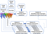
The IST consists of four main software modules (Figure 1). The first module generates virtual populations of AIS patients; the second simulates treatment and brain tissue injury; the third estimates outcome for each individual virtual AIS patient and the final module assembles all results and reports the outcome.

FIGURE 1
www.frontiersin.org

Figure 1. Schematic overview of the INSIST approach to implement an IST for AIS.