Tuesday, April 30, 2024

Quick action on the golf course resulted in rapid recovery from stroke

 Damn lucky to be in the cadre that fully recover.  Don't expect the same when you have a stroke.

Quick action on the golf course resulted in rapid recovery from stroke

By Diane Daniel, American Heart Association News

George Richards (right) and his friend, Mike Davisson, who recognized Richards was having a stroke during a round of golf. (Photo courtesy of Nicole Harnishfeger)

George Richards had just hit his tee shot on the sixth hole. He was playing a nine-hole round with the same golfing buddies he met up with every Thursday afternoon near his home in Leland, North Carolina.

The drive was pretty decent, landing in the fairway about 120 yards from the hole. Richards grabbed a club from his bag with his right hand and walked toward the ball to take his second shot.

But when Richards tried putting his left hand on the grip, he couldn't get it to move. The arm just hung there.

Richards flashed back to a program he'd seen about teaching people with disabilities to swing a club using just one arm. He gave it a try.

Richards swung feebly at the ball. It rolled forward a couple inches.

Then one of his pals, Mike Davisson, saw that Richards' arm was dangling and the left side of his face was drooping.

"Call 911," Davisson shouted. "George is having a stroke."

Another player put Richards in the golf cart and headed toward the clubhouse, keeping one arm around him so he wouldn't topple over.

"I'm fine. I'm 100%," Richards said, repeating the words like a mantra.

Only after they reached the clubhouse did Richards come to understand that he was in fact having a stroke.

Of all people, he would know.

Richards and his wife, Carol, started running educational and support groups for survivors and families of stroke after their son, David Dow, had a stroke at age 10 in 1995. David's stroke, caused by a rare brain disease, resulted in severe aphasia.

Aphasia is a communication impairment caused by brain damage that can impact speaking, understanding, reading, writing and math. In 2013, the family started the nonprofit Aphasia Recovery Connection. Several members of Carol's family also have had strokes.

When Carol and George moved to Leland from Nevada in 2021, one of the first things George did was find out if a certified stroke center was nearby. He was relieved to learn that there was. That's where the first responders took him.

George Richards (right) and his wife, Carol. (Photo courtesy of George Richards)

As soon as George – who was then 77 – arrived at the hospital, he was greeted by a stroke team. A brain scan showed that he had a clot in his right middle cerebral artery. He was given clot-busting medication.

A second scan showed that the clot had not dissolved. Doctors then performed a thrombectomy, a procedure in which a device is used to remove the clot, restoring blood flow. The procedure was performed less than 50 minutes since the stroke symptoms began.

Carol spent that time in the waiting room playing out worst-case scenarios. When she next saw George, he wanted to discuss the results of the scan and whether he might have aphasia or frontal lobe damage.

Carol knew his mind was working fine. A nurse even asked if George worked in neurology.

Within a couple hours, physical therapists tested George's cognitive and motor skills and declared him to have no deficits. Two days later, he was back home.

Doctors think the cause of his stroke was atrial fibrillation, or AFib, which is an irregular heartbeat. The stroke was in September 2022, about a decade after he'd received a pacemaker to help his heart. He'd been due for a routine replacement of the device the week after the stroke.

Since the stroke, George and Carol have ramped up their efforts to educate others. They organized a stroke awareness event in their community that attracted more than 130 people. They plan to sponsor another event later this year. George also spoke at a ceremony last year to open a new neurosciences institute in his region, sharing how specialized care at the ready helped save his life.

George Richards raising stroke awareness at the opening ceremony of a neurosciences institute in North Carolina. (Photo courtesy of George Richards)
George Richards spoke about his experience at the opening ceremony of a neurosciences institute in North Carolina. (Photo courtesy of George Richards)

More than anything, George and Carol said, they were lucky that Davisson knew the signs of stroke and called 911.

"That is the key," George said. "Don't say, 'We need to drive him to the hospital.' Just call 911."

Carol shudders at the notion of how much worse things might have been had Davisson not acted so quickly.

"George could be in an assisted living facility today," she said. "Instead, he's back to golf three times a week. Mike is our hero."

Stories From the Heart chronicles the inspiring journeys of heart disease and stroke survivors, caregivers and advocates.


American Heart Association News Stories

American Heart Association News covers heart disease, stroke and related health issues. Not all views expressed in American Heart Association News stories reflect the official position of the American Heart Association. Statements, conclusions, accuracy and reliability of studies published in American Heart Association scientific journals or presented at American Heart Association scientific meetings are solely those of the study authors and do not necessarily reflect the American Heart Association’s official guidance, policies or positions.

ESOC(European Sroke Organization Conference) 2024

I'd be willing to bet NONE of these will talk about 100% recovery protocols! That is how fucking useless stroke associations are! It's like they have never talked to any stroke survivors without talking down to them using the tyranny of low expectations!

 And since there won't be any stroke survivors presenting they can stay in their insulated cocoons away from stroke reality. Hey, I'm available for any presentation, if you can handle the truth!

 ESOC(European Sroke Organization Conference) 2024

As some of the Major Clinical Trials have been highly anticipated, the ESO is pleased to present a preview of some of the trials covered at this year’s conference:

Ischemic Stroke – Acute Treatment

  • CHARM
  • ACTISAVE

Ischemic Stroke – Acute Treatment – Thrombolysis

  • TASTE
  • TEMPO-2
  • TRACE III
  • ORIGINAL
  • ATTENTION IA
  • TRUTH

Intracerebral Haemorrhage – Acute Treatment

  • INTERACT4
  • SWITCH

Acute Stroke Care

  • TACTICS
  • TWIN2WIN
  • REMOTE-CAT
  • RICH-2
  • RECLAIM II
  • CLEVER

Secondary Analyses

  • INTERACT
  • ANNEXA-I
  • EMBOLISE
  • TENSION
  • SELECT2
  • MR CLEAN No-IV

Learn more about the Large Clinical Trials

First Announcement of Large Clinical Trials

Second Announcement of Large Clinical Trials

Third Announcement of Large Clinical Trials

Cholestane-3β,5α,6β-Triol Inhibits Acid-Sensing Ion Channels and Reduces Acidosis-Mediated Ischemic Brain Injury

Now we just need your competent? doctor and hospital to ensure human testing gets done on this. But I'll bet your doctor and hospital are incompetent  in getting needed research done!

Someday a stroke 'leader' will try to ream me out for making them look bad by being truthful, I look forward to that day.  Here's my email: oc1dean@gmail.com; I await your reply.

Cholestane-3β,5α,6β-Triol Inhibits Acid-Sensing Ion Channels and Reduces Acidosis-Mediated Ischemic Brain Injury

Originally publishedhttps://doi.org/10.1161/STROKEAHA.124.046963Stroke. 2024;0

BACKGROUND:

Activation of the acid-sensing ion channels (ASICs) by tissue acidosis, a common feature of brain ischemia, contributes to ischemic brain injury, while blockade of ASICs results in protection. Cholestane-3β,5α,6β-triol (Triol), a major cholesterol metabolite, has been demonstrated as an endogenous neuroprotectant; however, the mechanism underlying its neuroprotective activity remains elusive. In this study, we tested the hypothesis that inhibition of ASICs is a potential mechanism.

METHODS:

The whole-cell patch-clamp technique was used to examine the effect of Triol on ASICs heterogeneously expressed in Chinese hamster ovary cells and ASICs endogenously expressed in primary cultured mouse cortical neurons. Acid-induced injury of cultured mouse cortical neurons and middle cerebral artery occlusion–induced ischemic brain injury in wild-type and ASIC1 and ASIC2 knockout mice were studied to examine the protective effect of Triol.

RESULTS:

Triol inhibits ASICs in a subunit-dependent manner. In Chinese hamster ovary cells, it inhibits homomeric ASIC1a and ASIC3 without affecting ASIC1β and ASIC2a. In cultured mouse cortical neurons, it inhibits homomeric ASIC1a and heteromeric ASIC1a–containing channels. The inhibition is use-dependent but voltage- and pH-independent. Structure-activity relationship analysis suggests that hydroxyls at the 5 and 6 positions of the A/B ring are critical functional groups. Triol alleviates acidosis-mediated injury of cultured mouse cortical neurons and protects against middle cerebral artery occlusion–induced brain injury in an ASIC1a-dependent manner.

CONCLUSIONS:

Our study identifies Triol as a novel ASIC inhibitor, which may serve as a new pharmacological tool for studying ASICs and may also be developed as a potential drug for treating stroke.

Association Between MR‐Based Thrombus Imaging Characteristics and Endovascular Therapy Outcome in Acute Ischemic Stroke: A Systematic Review and Meta‐Analysis

 If your definition of successful revascularization is not 100% recovery then you'll regret your useless research when you are the 1 in 4 per WHO that has a stroke!

Association Between MR‐Based Thrombus Imaging Characteristics and Endovascular Therapy Outcome in Acute Ischemic Stroke: A Systematic Review and Meta‐Analysis

Originally publishedhttps://doi.org/10.1161/SVIN.123.001142Stroke: Vascular and Interventional Neurology. 2024;4:e001142

Abstract

Background

Prediction of successful revascularization and achieving a favorable functional outcome may help determine the optimal treatment strategy and improve the management of stroke. A growing body of literature has implicated a predictive value for thrombus imaging characteristics for stroke outcomes.(And all this is fucking useless since it does nothing to get to 100% recovery! I'D HAVE YOU ALL FIRED!)

Methods

We conducted an electronic search using PubMed, Ovid MEDLINE, and EMBASE, previously published meta‐analyses, and systematic review studies that intervened by endovascular thrombectomy or intravenous thrombolysis following large vessel occlusion stroke from 2000 to 2023 and involved magnetic resonance‐based thrombus imaging, then screened 2007 studies against our eligibility criteria. We extracted the enrollees’ characteristics and the association between clot features and radiological and functional outcome measures.

Results

Thirty‐three studies were found eligible, with a total number of 6902 enrollees. Susceptibility vessel sign was found in 3531 subjects (51.2%). Nine studies involved only the administration of intravenous thrombolysis, whereas 24 studies intervened by endovascular thrombectomy. Seventeen studies found at least an association between thrombus imaging characteristics and successful revascularization, whereas the others reported no association. only 13 studies found at least one thrombus characteristic associated with functional outcome, while the others showed no association between  the thrombus characteristics and functional outcome after stroke. Pooled meta‐analysis of studies that involved endovascular thrombectomy with or without intravenous thrombolysis showed a statistically significant association between the presence of susceptibility vessel sign and both successful reperfusion (odds ratio [OR]: 1.57 [1.09–2.27]; P = 0.02) and favorable functional outcome (OR: 1.76 [1.17–2.66]; P = 0.007).

Conclusion

The presence of susceptibility vessel sign on magnetic resonance‐based clot imaging was associated with functional outcome and successful reperfusion following thrombectomy.

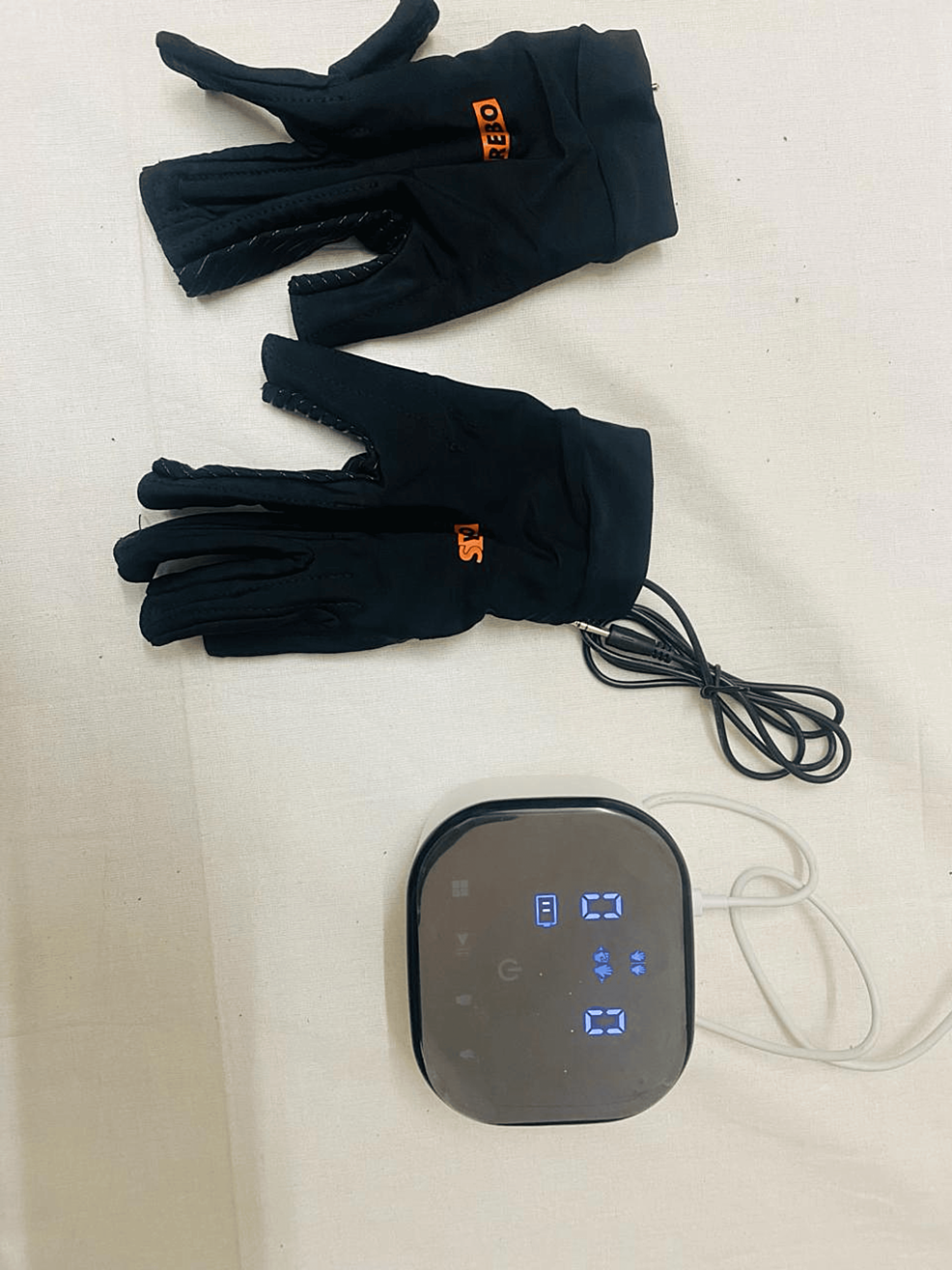
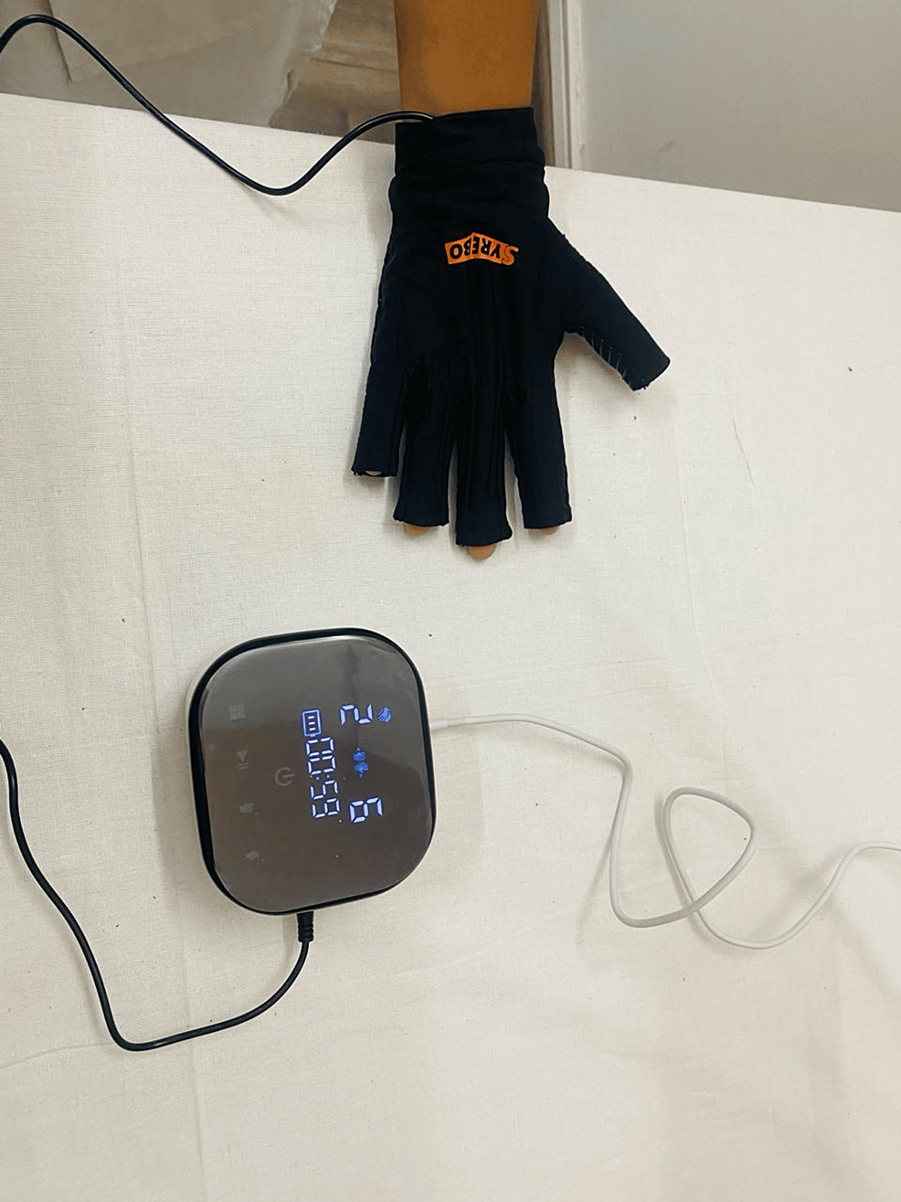
Effectiveness of Syrebo’s Glove Rehabilitation Program in a Patient With Middle Cerebral Artery Infarct: A Case Report

 Which glove is your therapist already using on you? NONE? So you don't have a functioning therapist either along with your non-functioning doctor? With my spastic fingers this glove would require two other persons to get it on, so useless for me.

  • glove (92 posts to October 2011)

    • gloves (5 posts to December 2021)


Effectiveness of Syrebo’s Glove Rehabilitation Program in a Patient With Middle Cerebral Artery Infarct: A Case Report

Vaishnavi S. SharmaH V Sharath Anam R. Sasun

Published: April 29, 2024

DOI: 10.7759/cureus.59314 

  Peer-Reviewed

Cite this article as: Sharma V S, Sharath H, Sasun A R (April 29, 2024) Effectiveness of Syrebo’s Glove Rehabilitation Program in a Patient With Middle Cerebral Artery Infarct: A Case Report. Cureus 16(4): e59314. doi:10.7759/cureus.59314

Abstract

In India, stroke is a significant health concern, with an estimated prevalence of around 1.54% in adults over 20 years old. The incidence of stroke in India varies regionally but is generally high due to factors like hypertension and lifestyle changes. Ischemic strokes comprise the majority, particularly in the middle cerebral artery (MCA) territory. MCA stroke presents with diverse symptoms such as weakness, speech difficulties, and vision problems, emphasizing the need for comprehensive rehabilitation. Physiotherapy plays a vital role in addressing these challenges, focusing on strength, coordination, mobility, and independence through tailored interventions. Additionally, soft robotic gloves, such as Syrebo's rehabilitation, offer promising advancements in neurorehabilitation by enhancing motor recovery and functional abilities, particularly in improving grip strength and hand functionality, thus improving outcomes for stroke patients. This case describes a 66-year-old female presenting with sudden left-sided weakness, slurred speech, and facial deviation indicative of bilateral MCA territory infarct. After admission requiring ventilation and medication, imaging confirmed the diagnosis. Following stabilization, she underwent neurophysiotherapy for rehabilitation. Neurological examination revealed deficits in muscle tone, reflexes, cranial nerve function, language, and swallowing. Outcome measures indicated progress in rehabilitation. The case underscores the significance of timely diagnosis and personalized rehabilitation in optimizing outcomes for MCA territory stroke patients.

Figure 2 shows Syrebo's soft robotics glove equipment. Figure 3 shows the application of Syrebo's soft robotics gloves in patient hand. 

More at link.


A decision tree algorithm to identify predictors of post-stroke complex regional pain syndrome

Change one word in the title and it won't be ABSOLUTELY FUCKING USELESS! Predictors to Preventors! But until we get survivors in charge directing research stroke will NEVER BE SOLVED!

A decision tree algorithm to identify predictors of post-stroke complex regional pain syndrome

Abstract

This prospective cohort study aimed to identify the risk factors for post-stroke complex regional pain syndrome (CRPS) using a decision tree algorithm while comprehensively assessing upper limb and lower limb disuse and physical inactivity. Upper limb disuse (Fugl-Meyer assessment of upper extremity [FMA-UE], Action Research Arm Test, Motor Activity Log), lower limb disuse (Fugl-Meyer Assessment of lower extremity [FMA-LE]), balance performance (Berg balance scale), and physical inactivity time (International Physical Activity Questionnaire-Short Form [IPAQ-SF]) of 195 stroke patients who visited the Kishiwada Rehabilitation Hospital were assessed at admission. The incidence of post-stroke CRPS was 15.4% in all stroke patients 3 months after admission. The IPAQ, FMA-UE, and FMA-LE were extracted as risk factors for post-stroke CRPS. According to the decision tree algorithm, the incidence of post-stroke CRPS was 1.5% in patients with a short physical inactivity time (IPAQ-SF < 635), while it increased to 84.6% in patients with a long inactivity time (IPAQ-SF ≥ 635) and severe disuse of upper and lower limbs (FMA-UE score < 19.5; FMA-LE score < 16.5). The incidence of post-stroke CRPS may increase with lower-limb disuse and physical inactivity, in addition to upper-limb disuse. Increasing physical activity and addressing lower- and upper-limb motor paralysis may reduce post-stroke CRPS.

More at link.


Cognitively healthy centenarians are genetically protected against Alzheimer's disease

 How would your competent? doctor test for these in you? Because if you don't have them your doctor has to give you EXACT DEMENTIA PREVENTION PROTOCOLS!

Cognitively healthy centenarians are genetically protected against Alzheimer's disease

First published: 18 April 2024

Abstract

BACKGROUND

Alzheimer's disease (AD) prevalence increases with age, yet a small fraction of the population reaches ages > 100 years without cognitive decline. We studied the genetic factors associated with such resilience against AD.

METHODS

Genome-wide association studies identified 86 single nucleotide polymorphisms (SNPs) associated with AD risk. We estimated SNP frequency in 2281 AD cases, 3165 age-matched controls, and 346 cognitively healthy centenarians. We calculated a polygenic risk score (PRS) for each individual and investigated the functional properties of SNPs enriched/depleted in centenarians.

RESULTS

Cognitively healthy centenarians were enriched with the protective alleles of the SNPs associated with AD risk. The protective effect concentrated on the alleles in/near ANKH, GRN, TMEM106B, SORT1, PLCG2, RIN3, and APOE genes. This translated to >5-fold lower PRS in centenarians compared to AD cases (= 7.69 × 10−71), and 2-fold lower compared to age-matched controls (= 5.83 × 10−17).

DISCUSSION

Maintaining cognitive health until extreme ages requires complex genetic protection against AD, which concentrates on the genes associated with the endolysosomal and immune systems.

Highlights

  • Cognitively healthy cent enarians are enriched with the protective alleles of genetic variants associated with Alzheimer's disease (AD).
  • The protective effect is concentrated on variants involved in the immune and endolysosomal systems.
  • Combining variants into a polygenic risk score (PRS) translated to > 5-fold lower PRS in centenarians compared to AD cases, and ≈ 2-fold lower compared to middle-aged healthy controls.

1 BACKGROUND

The average human life expectancy continues to grow and by 2050 there will be 3.2 million centenarians in the world.1 At old ages, a major contributor to poor health is cognitive decline and dementia, of which Alzheimer's disease (AD) is the most common type.2, 3 However, AD is not an inevitable consequence of aging, as testified by a small proportion of the population that reaches at least 100 years while maintaining a high level of cognitive and physical functions.4, 5 This raises the question of whether these cognitively healthy centenarians have exceptional features that protect or delay the onset of dementia, and whether such mechanisms may be genetically encoded.

AD is a progressive disorder characterized by loss of cognitive functions, ultimately leading to loss of independence and death, for which an effective treatment is lacking.3, 6 The greatest risk factor for AD is age: the disease is rare at 60 years, and the incidence of AD reaches ≈ 40% per year at 100 years of age.7 Next to aging, heritability plays an important role that changes dramatically with age. While the heritability of AD with age at onset < 65 years is estimated to be 90% to 100%, mostly due to autosomal dominant or strong risk-increasing genetic variants,8 it decreases to 60% to 80% for ages at AD onset of ≈ 75 years (determined by twin studies), based on a unique mix of rare and common risk factors, and further declines with later ages at AD onset.9 Approximately 30% of the genetic risk of AD is attributable to the ε4 allele of the apolipoprotein E (APOE) gene. Large collaborative genome-wide association studies (GWAS) have collectively identified 86 single nucleotide polymorphisms (SNPs) that are associated with a slight modification of the risk of AD.10, 11

Intriguingly, the reverse is also true, as ≈ 60% of the chance to survive to 100 years in good cognitive health depends on inheriting favorable genetic factors,12 comprising a relative depletion of risk-increasing variants and an enrichment of advantageous genetic variants that associate with a prolonged (brain) health.13-15 In fact, in 2018 we reported that the effect size of 29 SNPs that were associated with AD risk was increased on average 2-fold when using cognitively healthy centenarians as controls rather than controls age-matched with the AD cases.16 Consequently, cognitively healthy centenarians had a significantly lower polygenic risk score (PRS), compared to AD cases and age-matched controls.

In the current study, we aimed to further expand on these findings by investigating the prevalence in cognitively healthy centenarians of the 86 SNPs that are currently associated with AD risk, based on the most recent GWAS for AD.10 We studied the effect of individual AD-associated SNPs as well as their combined effect (PRS) on prolonged cognitive health. Furthermore, we identified risk-increasing and protective SNPs that were, respectively, most depleted or enriched in a cohort of cognitively healthy centenarians, which allowed us to highlight the biological mechanisms most strongly involved with resilience against AD.

 
More at link.

Ginseng as a therapeutic target to alleviate gut and brain diseases via microbiome regulation

 What in here will give your doctor information to have a diet protocol created for ginseng? Or doesn't your competent? doctor have the ability to create stroke rehab protocols?

Ginseng as a therapeutic target to alleviate gut and brain diseases via microbiome regulation

, , ,
https://doi.org/10.1016/j.jgr.2024.04.005Get rights and content
Under a Creative Commons license
open access

Abstract

The human gut, which contains a diverse microbiome, plays an important role in maintaining physiological balance and preserving the immune system. The complex interplay between the central nervous system (CNS) and the gut microbiome has gained significant attention due to its profound implications for overall health, particularly for gut and brain disorders. There is emerging evidence that the gut-brain axis (GBA) represents a bidirectional communication system between the CNS and the gastrointestinal tract and plays a pivotal role in regulating many aspects of human health. Ginseng has shown potential to ameliorate conditions associated with dysbiosis, such as gut and CNS disorders by restoring microbial balance and enhancing gut barrier function. This comprehensive review provides valuable insights into the potential of ginseng as a herbal modulator of GBA as a therapeutic intervention for preventing and treating gut and neurological diseases via microbiota regulation to ultimately enhance overall health. Furthermore, we emphasize the therapeutic benefits of ginseng, its ability to enhance beneficial probiotics, such as Firmicutes, Bacteroides, Lactobacillus, Bifidobacterium, and Akkermansia while reducing pathogenic bacteria prevalence, such as Helicobacter, Clostridium, and Proteobacteria. Although the connection between ginseng regulation of microbial communities in response to the gut and neuropsychiatric disorders is lacking, additional investigations are warranted to elucidate the underlying mechanisms, optimize dosages, and explore the clinical relevance of ginseng in promoting GBA balance and ultimately overall health.

Motor Impairment and Recovery in the Upper Limb After Stroke: Behavioral and Neuroanatomical Correlates by Julie Bernhardt

 Even stroke superstars like Julie Bernhardt don't produce research that gets survivors recovered(Creating protocols!). All because there is NO leadership in stroke directing researchers to solve stroke for survivor benefits!

Motor Impairment and Recovery in the Upper Limb After Stroke: Behavioral and Neuroanatomical Correlates by Julie Bernhardt

Leeanne M. Carey, PhD; David F. Abbott, PhD; Gary F. Egan, PhD; Julie Bernhardt, PhD; Geoffrey A. Donnan, MD  
 
Background and Purpose 
 
Motor recovery after stroke is associated with cerebral reorganization. However, few studies have investigated the relationship directly, and findings are equivocal. We therefore aimed to characterize the relationship between motor impairment, motor recovery, and task-related changes in regional cerebral blood flow (rCBF) longitudinally.  
 
Methods 
 
We obtained a profile of motor impairment and recovery in the upper limb and conducted positron emission tomography motor activation studies using a simple finger-tapping task in 9 stroke patients 2 to 7 weeks after stroke and 6 months later. For correlation analysis, mean images of task-related rCBF for each individual were linearly regressed with motor impairment scores. Motor recovery was correlated with longitudinal rCBF images.  
 
Results
 
 Patients (7 males; 72.09.8 years) demonstrated a wide range of impairment severity and variable recovery. Upper-limb motor function was linearly correlated with task-related rCBF. Importantly, sites of correlated rCBF differed over time. Subacutely correlated rCBF was observed in supplementary motor area (SMA), bilateral cingulate, and contralesional insula with a small area in ipsilesional primary sensorimotor cortex (SM1). Conversely, at the 6-month study, correlated rCBF was primarily in ipsilesional SM1, extending to the cingulate gyrus. Better motor recovery was correlated with reduction in contralesional activity and increase in ipsilesional SM1.  
 
Conclusions
 
 Upper-limb motor function and recovery are correlated with rCBF in SMA, cingulate, insula, and SM1, highlighting the role of these areas in the recovery process. The dynamic nature of the relationship suggests ongoing adaptation within motor networks. (Stroke. 2005;36:625-629.)

Brown Fat’s “Off Switch” Discovery Combats Obesity


I was at exactly 25 BMI prior to stroke, then gained 30 pounds because my doctor knew nothing and did nothing to get me recovered enough to continue with all the activities that kept me in shape. And told me nothing about slowing metabolism after age 50.

Brown Fat’s “Off Switch” Discovery Combats Obesity

Summary: Researchers have made significant discoveries about brown adipose tissue (BAT), which is known for its calorie-burning capabilities. Their study identifies a protein, AC3-AT, that acts as an “off switch” for BAT activation, limiting its effectiveness in combating obesity.

By potentially blocking this switch, the researchers believe they can enhance brown fat’s activity, offering a new avenue for treating obesity and related metabolic disorders. The findings, which also showed that mice without AC3-AT were protected from obesity and had increased metabolic rates, suggest promising strategies for human weight management.

Key Facts:

  1. Brown Fat Activation Limitation: The discovery of the AC3-AT protein explains why the calorie-burning effects of brown fat are typically short-lived, providing a target for prolonging its activation.
  2. Protective Effects in Mice: Mice lacking the AC3-AT protein demonstrated resistance to obesity, less fat accumulation, and higher metabolic rates, indicating the protein’s crucial role in energy balance.
  3. Implications for Human Obesity: Given that AC3-AT is also present in humans, these findings open up potential therapeutic applications to enhance brown fat activity and support weight loss in humans.

Source: University of Southern Denmark

Brown fat, also known as brown adipose tissue (BAT), is a type of fat in our bodies that’s different from the white fat around our belly and thighs that we are more familiar with.

Brown fat has a special job—it helps to burn calories from the foods that we eat into heat, which can be helpful, especially when we’re exposed to cold temperatures like during winter swimming or cryotherapy.

This shows an overweight person.
Intriguingly, this study not only identified AC3-AT, which is a shorter, previously unknown form of the AC3protein. Credit: Neuroscience News

For a long time, scientists thought that only small animals like mice and newborns had brown fat. But new research shows that a certain number of adults maintain their brown fat throughout life. Because brown fat is so good at burning calories, scientists are trying to find ways to activate it safely using drugs that boost its heat-producing abilities.

A new study from the research groups of Prof. Jan-Wilhelm Kornfeld from the University of Southern Denmark/the Novo Nordisk Center for Adipocyte Signaling (Adiposign) and Dagmar Wachten from the University Hospital Bonn and the University of Bonn (Germany) has found that brown fat has a previously unknown built-in mechanism that switches it off shortly after being activated.

This limits its effectiveness as treatment against obesity. According to first author of the study, Hande Topel, who is a Senior Postdoc at the University of Southern Denmark and the Novo Nordisk Center for Adipocyte Signaling (Adiposign), the team has now discovered a protein responsible for this switching-off process. It is called ‘AC3-AT’.

Blocking the “off switch” opens up a new strategy

“Looking ahead, we think that finding ways to block AC3-AT could be a promising strategy for safely activating brown fat and tackling obesity and related health problems”, Hande Topel says.

The research team found the switch-off protein using advanced technology predicting unknown proteins.

Hande Topel explains: “When we investigated mice that genetically didn’t have AC3-AT, we found that they were protected from becoming obese, partly because their bodies were simply better at burning off calories and were able to increase their metabolic rates through activating brown fat”.

Two groups of mice were fed a high-fat diet for 15 weeks, which rendered them obese. The group that had their AC3-AT protein removed, gained less weight than the control group and were metabolically healthier.

“The mice that have no AC3-AT protein, also accumulated less fat in their body and increased their lean mass when compared to the control mice”, says co-author, Ronja Kardinal, who is a PhD student at the University of Bonn in the lab of Dagmar Wachten at UKB, continuing: “As AC3-AT is found not only in mice but also in humans and other species, there are direct therapeutic implications for humans”.

Hope for strategies that support weight loss

Although the prevalence of brown fat decreases as humans age, and despite grown-ups not having as much brown fat as newborns, it can still be activated, for instance by cold exposure. When it gets activated, it enhances the rate of metabolism of these individuals, which again may help to stabilize weight loss in conditions where calorie intake is (too) high.

Intriguingly, this study not only identified AC3-AT, which is a shorter, previously unknown form of the AC3protein. The researchers also identified other unknown protein/gene versions, that respond to cold exposure, similar to AC3-AT.

“However, further research is needed to elucidate the therapeutic impact of these alternative gene products and their regulatory mechanisms during BAT activation”, says co-corresponding author Prof. Dagmar Wachten, Co-Director of the Institute of Innate Immunity at the UKB and member of the Cluster of Excellence ImmunoSensation2 and the Transdisciplinary Research Areas (TRA) “Modelling” and “Life & Health” at the University of Bonn.

“Understanding these kinds of molecular mechanisms not only sheds light on the regulation of brown fat but also holds promise for unraveling similar mechanisms in other cellular pathways.

“This knowledge can be instrumental in advancing our understanding of various diseases and in the development of novel treatments”, says co-corresponding author Prof. Jan-Wilhelm Kornfeld, University of Southern Denmark.

This study was conducted in the context of the DFG Collaborative Research Center Transregio-SFB 333 “Brown and Beige Fat – Organ Interactions, Signaling Pathways and Energy Balance (BATenergy)”, which is pursuing a better understanding of the different types of adipose tissue and their role in metabolic diseases and the Novo Nordisk Foundation Center for Adipocyte Signaling (Adiposign) at University of Southern Denmark that aims to understand fat cell dysfunction in model organisms and obese patients.

About this obesity and neuroscience research news

Author: Birgitte Svennevig
Source: University of Southern Denmark
Contact: Birgitte Svennevig – University of Southern Denmark
Image: The image is credited to Neuroscience News

Original Research: Closed access.
Cold-induced expression of a truncated Adenylyl Cyclase 3 acts as rheostat to brown fat function” by Jan-Wilhelm Kornfeld et al. Nature Metabolism

Transient ischaemic attacks: mimics and chameleons

You better hope your doctors can do this because evidence suggests that they're not very good with young adult strokes.

 Transient ischaemic attacks: mimics and chameleons


V Nadarajan1,

  1. R J Perry1,
  2. J Johnson1,
  3. D J Werring1,2
  1. Correspondence to Dr David Werring, Reader in Clinical Neurology and Honorary Consultant Neurologist, Stroke Research Group, UCL Institute of Neurology, Queen Square, London WC1N 3BG, UK; d.werring@ucl.ac.uk

Abstract

Suspected transient ischaemic attack (TIA) is a common diagnostic challenge for physicians in neurology, stroke, general medicine and primary care. It is essential to identify TIAs promptly because of the very high early risk of ischaemic stroke, requiring urgent investigation and preventive treatment. On the other hand, it is also important to identify TIA ‘mimics’, to avoid unnecessary and expensive investigations, incorrect diagnostic labelling and inappropriate long-term prevention treatment. Although the pathophysiology of ischaemic stroke and TIA is identical, and both require rapid and accurate diagnosis, the differential diagnosis differs for TIA owing to the transience of symptoms. For TIA the diagnostic challenge is greater, and the ‘mimic’ rate higher (and more varied), because there is no definitive diagnostic test. TIA heralds a high risk of early ischaemic stroke, and in many cases the stroke can be prevented if the cause is identified, hence the widespread dissemination of guidelines including rapid assessment and risk tools like the ABCD2 score. However, these guidelines do not emphasise the substantial challenges in making the correct diagnosis in patients with transient neurological symptoms. In this article we will mainly consider the common TIA mimics, but also briefly mention the rather less common situations where TIAs can look like something else (‘chameleons’).


This is an Open Access article distributed in accordance with the Creative Commons Attribution Non Commercial (CC BY-NC 3.0) license, which permits others to distribute, remix, adapt, build upon this work non-commercially, and license their derivative works on different terms, provided the original work is properly cited and the use is non-commercial. See: http://creativecommons.org/licenses/by-nc/3.0/

Request Permissions

If you wish to reuse any or all of this article please use the link below which will take you to the Copyright Clearance Center’s RightsLink service. You will be able to get a quick price and instant permission to reuse the content in many different ways.

Mounting Stroke Crisis in India: A Systematic Review

 It's a crisis because you BLITHERING IDIOTS haven't figured out that the solution to this is 100% recovery protocols! Yeah that's a BHAG(Big Hairy Audacious Goal)

but leaders solve those. We have NO leaders in stroke. And the result is 10 million disabled stroke survivors every year!

Explain to me in precise terms where I'm wrong; oc1dean@gmail.com, I'm stroke-addled you know, so simple-minded me needs you to be precise in your explanation.

Looking forward to your excuses!

Mounting Stroke Crisis in India: A Systematic Review

Vedant N. Hedau Tushar Patil

Published: March 27, 2024

DOI: 10.7759/cureus.57058 

  Peer-Reviewed

Cite this article as: Hedau V N, Patil T (March 27, 2024) Mounting Stroke Crisis in India: A Systematic Review. Cureus 16(3): e57058. doi:10.7759/cureus.57058

Abstract

Stroke, a neurological disorder, has emerged as a formidable health challenge in India, with its incidence on the rise. Increased risk factors, which also correlate with economic prosperity, are linked to this rise, including hypertension, diabetes, obesity, sedentary lifestyle, and alcohol intake. Particularly worrisome is the impact on young adults, a pivotal segment of India's workforce. Stroke encompasses various clinical subtypes and cerebrovascular disorders (CVDs), contributing to its multifaceted nature. Globally, stroke's escalating burden is concerning, affecting developing nations. To combat this trend effectively and advance prevention and treatment strategies, comprehensive and robust data on stroke prevalence and impact are urgently required. In India, these encompass individuals with elevated BMIs, and those afflicted by hypertension, diabetes, or a familial history of stroke. Disparities in stroke incidence and prevalence manifest across India, with differences in urban and rural settings, gender-based variations, and regional disparities. Early detection, dietary changes, effective risk factor management, and equitable access to stroke care are required to address this issue. Government initiatives, like the National Programme for Prevention and Control of Cancer, Diabetes, Cardiovascular Diseases, and Stroke (NPCDCS) 2019, provide guidelines, but effective implementation and awareness campaigns are vital. Overcoming barriers to stroke care, especially in rural areas, calls for improved infrastructure, awareness campaigns, and support systems. Data standardization and comprehensive population studies are pivotal for informed public health policies.

Introduction & Background

Cerebrovascular disease (CVD) is a term used to describe all disorders that lead to stroke, which can be either ischemic or hemorrhagic. In low- and middle-income countries (LMICs), including India, the frequency of stroke increased by 100% between 1997 and 2008. This showed a 26% rise in stroke mortality worldwide during the previous 20 years [1]. Stroke continues to be the second most significant cause of death globally due to the increased mortality rate, according to the World Health Organization (WHO) 2020 [2]. Over the past four decades, there has been a statistically significant reduction in stroke incidence rates, with stroke incidence falling by 42% in high-income countries (HICs) and rising by more than 100% in LMICs [1]. Age, sex, low birth weight, ethnicity, and genetic variables are all irreversible risk factors for stroke [3]. A report of India's population-based stroke registries mentions that the majority of first-ever stroke cases reported hypertension (from Tirunelveli, 40.3%, to Cuttack, 75%), diabetes, and current tobacco use (from Varanasi, 19.3%, to Cuttack, 62.4%) [4]. Ischemic stroke (range 41.6% to 77.8%), hemorrhagic stroke (range 42.6% to 74%), and indeterminate stroke (range 2.0% to 35.9%) all had hypertension as their primary risk factor [4]. Young people's risk of stroke is considerably enhanced by smoking, drinking, having a higher BMI, having diabetes, and having high blood pressure [5,6]. A hospital-based multi-center prospective stroke registry in India to identify and recruit 10,000 acute stroke patients from 100 hospitals within the country conducted an interim analysis to determine aetiologies, clinical supervision, and outcomes with 5301 patients. According to the data, stroke patients had a number of highly hazardous factors, including heavy alcohol and cigarette use, diabetes, hypertension, and dyslipidemia [7].

The study found that stroke patients with higher frequencies of risk variables had higher short-term mortality [8]. Adopting coordinated care for stroke in LMICs is limited and insufficient, particularly in a nation like India, where the facilities available for rehabilitation are sparse [9]. The global burden of CVD has been rising, with stroke being the second most significant factor in fatalities worldwide, after ischemic heart disease in 1990 and the WHO 2020 factsheet [2,10]. A sizeable share of stroke deaths occur in LMICs, and these nations also experience more years of life lost to disability-adjusted life than high-income nations [11]. India has a greater cumulative incidence and crude prevalence of stroke than high-income nations [1]. This suggests that stroke is a significant health burden in India. Globally, there were around 25.7 million stroke survivors in 2013, along with 6.5 million stroke fatalities, 113 million years of life lost to disability, and 10.3 million new stroke cases [12]. Worldwide, the incidence of stroke is rising, mostly as a result of an older population and more risk factors such as type 2 diabetes and high blood pressure. Stroke occurrence among young people is increasing in LMICs [13]. There are regional, national, and ethnic differences in cardiovascular disease incidence, prevalence, and mortality. Due to their altered cardio-metabolic profiles and propensity for cardio-metabolic dysfunction, people from South Asian nations, especially India, have a disproportionately increased risk of cardiovascular disease [14]. To address the concerns of stroke prevention and treatment, reliable data on the burden of CVD in the Indian population is needed. To meet this demand, a comprehensive analysis of all community-based studies providing data on the mortality, prevalence, and incidence of stroke in rural, urban, and both population contexts is required. The present article aims to focus on the stroke crisis in India.

A sharper mind: tai chi can improve cognitive function

 Didn't your competent? doctor prescribe tai chi a decade ago? NO? So you don't have a functioning stroke doctor, do you? In my opinion a competent stroke doctor is up-to-date on all stroke research and should know more than me! If your doctor does know all that stuff, why hasn't ANY STROKE PROTOCOLS BEEN WRITTEN? The information is all there in the research! So do something with it, RATHER THAN SITTING ON YOUR ASS!

A sharper mind: tai chi can improve cognitive function

Up until about two decades ago, it was believed that your brain only produced new cells early in life. But research has shown that the brain has the ability to change throughout your entire life span, growing new cells, making new connections, and even increasing in size. These changes can improve cognitive function—and various forms of exercise, including tai chi, can help.
 

In a meta-analysis of 20 studies on tai chi and cognition, tai chi appears to improve executive function—the ability to multitask, manage time, and make decisions—in people without any cognitive decline. In those with mild cognitive impairment, tai chi slowed the progression to dementia more than other types of exercise and improved their cognitive function in a comparable fashion to other types of exercise or cognitive training.

In one study, researchers had nearly 400 Chinese men and women with some cognitive impairment perform either tai chi or a stretching and toning program three times a week. After a year, the tai chi group showed greater improvements, and only 2% of that group progressed to dementia, while 11% from the traditional exercise group did.

In another study, tai chi outperformed walking. Following 40 weeks of either tai chi, walking, social interaction, or no intervention, researchers compared MRI images and discovered that brain volume increased the most in the tai chi group. In addition, that group also performed better on cognitive tests.

To learn more about tai chi, its health benefits, and how to learn its movements, check out Introduction to Tai Chi, an Online Course from Harvard Medical School.