With no explanation of what should be done to prevent a stroke; THIS RESEARCH WAS TOTALLY FUCKING USELESS! You're fired!
Does anyone in the stroke medical world actually think?
Application of hair HPG axis hormone levels before onset in predicting the risk of stroke
 Jiao Yang
 Shuhan Li
 Xuan Li1
 Huihua Deng
 Yuanyuan Jia
 Dianhuai Meng
- 1Rehabilitation Center, The First Affiliated Hospital with Nanjing Medical University, Nanjing, China
- 2School of Biological Science and Medical Engineering, Southeast University, Nanjing, China
- 3Department of Rehabilitation Medicine, The Eighth Affiliated Hospital of Southern Medical University (The First People’s Hospital of Shunde Foshan), Foshan, China
Background: There have been many studies on the relationship between sex hormones and stress, mood, blood pressure, etc., but its impact on the incidence of stroke remains unknown.
Objective: To investigate the expression levels of hypothalamic–pituitary-gonadal (HPG) axis related hormones such as testosterone (T) and progesterone (P) in hair before stroke and their effects on the risk of stroke.
Methods: 48 patients with stroke were recruited from November 2022 to May 2023 as the observation group and 35 healthy subjects were recruited as the control group. There was no obvious difference in age, gender and BMI between the two groups (p > 0.05). T and P levels in hair were tested by LC-MS/MS, and the correlation with the risk of stroke was analyzed.
Results: The T and P levels of hair before the onset of stroke in the observation group were significantly lower than those in the control group (p < 0.01). The T level of men’s hair before the onset of stroke in the observation group was significantly lower than those in the control group (p < 0.05). The T and P levels of women’s hair before the onset of stroke in the observation group were significantly lower than those in the control group (p < 0.01). The results of ROC curve showed that the cut-off value of T level in men’s hair before the onset of stroke was 4.35 pg/mg, the AUC was 0.690 (0.545, 0.835), the sensitivity was 62.50%, and the specificity was 82.61%. The cut-off value of T level in women’s hair before the onset of stroke was 5.00 pg/mg, the AUC was 0.818 (0.658, 0.978), the sensitivity was 75.00%, and the specificity was 83.33%. The cut-off value of P level in women’s hair before the onset of stroke was 8.00 pg/mg, the AUC was 0.891 (0.754, 1.000), the sensitivity was 81.25%, and the specificity was 100.00%.
Conclusion: This preliminary report is the first to suggest that HPG axis hormones such as T and P in hair could have predictive value in screening for stroke risk.
1 Introduction
Cerebrovascular diseases have now ascended to the foremost cause of mortality in China, with stroke constituting the single most prevalent etiology of disability, exerting a substantial burden on both individuals and society (1). Stroke is a preventable and manageable disease, and early screening combined with active intervention can markedly enhance patient prognosis (2).
Emerging evidence suggests associations between hypothalamic–pituitary-gonadal (HPG) axis hormones including testosterone (T), estradiol (E2), and progesterone (P) and stroke incidence (3, 4). Haya et al. (5) conducted a 29-year follow-up study involving 4,615 adult males and 4,724 adult females to investigate endogenous hormone profiles, revealing that extremely low serum T concentrations were significantly associated with elevated ischemic stroke risk in males. However, this study faced methodological limitations, including the absence of precise temporal data on stroke onset in community-based populations, which has led to the need for studies in larger samples. Furthermore, the requirement for continuous serological monitoring and extended follow-up until stroke occurrence resulted in prolonged study duration, substantial costs, and potential confounding from unadjusted variables such as aging.
Hormone levels in human hair serve as endogenous biomarkers for retrospective assessment of hypothalamic–pituitary-adrenocortical (HPA) and HPG axis activity (6). The measurement of hormone levels in hair is retrospective, with 1 cm of hair corresponding to approximately 1 month of hormonal accumulation, and a reliable retrospective window extending up to 6 months (7). Compared to traditional biological matrices such as blood, serum, and urine, hair hormone analysis offers distinct advantages, including cumulative representation of long-term exposure, non-invasive sampling, and the ability to reflect baseline levels over extended periods (similar to how hemoglobin A1c is used to assess serum glucose levels) (7–9). While clinical studies on hair hormones have proliferated (10, 11), current research predominantly focuses on associations between hair hormone levels and psychiatric, emotional, and sleep-related disorders. For instance, Wright et al. (12) demonstrated in a systematic review that cortisol quantification in hair can evaluate chronic stress exposure in elderly populations. Deng et al. (11) investigated functional characteristics of the HPA and HPG axes, as well as their interplay, in patients with schizophrenia by analyzing expression levels of eight biomarkers in hair samples. Similarly, Wang et al. (13) identified a significant correlation between pre-onset HPA axis hyperactivity and post-stroke emotional disorders.
Current research lacks substantial evidence on the correlation between pre-stroke HPG axis hormone levels in hair and stroke risk. Considering the influence of age and sex on HPG axis activity (14), this study investigates the relationship between the expression levels of HPG axis hormones in the hair and the risk of stroke onset among individuals aged over 50, across different genders. The findings aim to provide novel insights and methodologies for early stroke screening and intervention strategies.
2 Materials and methods
This study was reviewed and approved by the Ethics Committee of Jiangsu Provincial People’s Hospital (2022-SR-553), and was prospectively registered with the Chinese Clinical Trial Registry (ChiCTR2200065803). All participants provided written informed consent to participate in the study.
2.1 Participants
Study participants included 48 stroke patients (32 males, 16 females) hospitalized at Jiangsu Provincial People’s Hospital and its Qixia Rehabilitation Branch between November 2022 and May 2023. Subjects in the stroke group had a mean age of 64.67 ± 7.53 years, with a mean disease duration of 31.50 days (IQR 33.00). Stroke group included 35 ischemic and 13 hemorrhagic cases. A control group of 35 healthy individuals (23 males, 12 females; mean age 65.69 ± 8.90 years) was recruited. No statistically significant differences were observed between groups in age, sex, or body mass index (BMI) (p > 0.05) (Table 1).
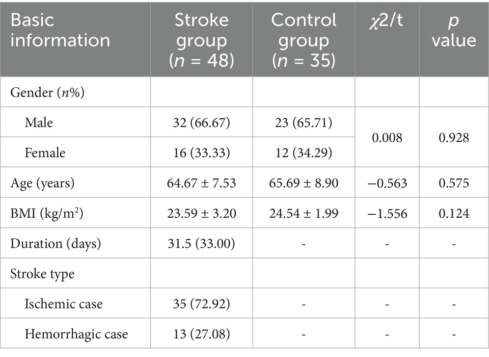
Table 1. Comparison of general data between the two groups [M (IQR)] or M ± SD.
Inclusion Criteria for Stroke Group were as follows: ① Age 50–80 years; ➁ Stroke diagnosis confirmed by neuroimaging and compliant with diagnostic guidelines established by the Chinese Stroke Society; ➂ First-ever unilateral stroke; ➃ Disease duration ≤ 3 months; ➄ Clinically stable with vital signs within normal ranges; ➅ Postmenopausal status (confirmed by amenorrhea ≥ 12 months) for female participants.
Inclusion Criteria for Control Group were as follows: ① Age 50–80 years; ➁ No history of stroke or major neurological disorders; ➂ Postmenopausal status for female participants.
Exclusion Criteria (Both Groups) were as follows: ① Auditory comprehension deficits or inability to follow verbal commands; ➁ Concurrent with other central nervous system lesions (e.g., traumatic brain injury, brain neoplasms); ➂ History of reproductive system disorders or surgeries; ➃ Substance abuse, hormone therapy within the past year, or chronic alcoholism; ➄ Occipital hair length < 3 cm; ➅ Recent hair treatments affecting analysis.
2.2 Sample collection
Hair samples were collected from all subjects on their enrollment day. Hair samples were collected by cutting 1 cm of hair from the occipital region, located at the back of the subject’s head near the scalp. Specimens were wrapped in aluminum foil, labeled, and stored at room temperature protected from light.
2.3 Hormonal analysis
Hormonal concentrations in hair were analyzed via high-performance liquid chromatography–tandem mass spectrometry (LC-MS/MS), where an Agilent 1,200 liquid chromatograph (Agilent Technologies, Inc., United States) was combined with an API 3200 Q-TRAP mass spectrometer (Applied Biosystems, Inc., United States), following a previously developed method (15). Hair samples corresponding to the period before the onset of lesions were selected based on the disease course (If the duration of the patient’s disease is 30 days at the time of admission, the 1 cm of hair closest to the scalp is the total amount of hormones accumulated in the hair in the month when the stroke occurred, and the 2 cm close to the scalp is the total amount of hormones accumulated in the hair in the month before the stroke occurred). Hair samples were rinsed with 5 mL of methanol for 2 min, and then heated to 50°C for drying. In a 2 mL centrifuge tube, hair samples were weighed after measuring 1–2 mm in length and cut into powder. After incubation in methanol for 24 h at 25°C, the mixture was centrifuged at 1.2 × 10⁴ r/min for 5 min. Transfer 800 μL of supernatant into another clean centrifuge tube and evaporate nitrogen at 40°C. The residue was then redissolved in 50 mL of mobile phase for LC-MS/MS analysis.
There was good performance, with limits of detection and quantification of 0.3 and 1.0 pg/mg for T, and 0.2 and 0.5 pg/mg for P, respectively. The recovery, intra-day, and inter-day coefficients of variation met the requirements. Analyses were conducted at the Jiangsu Provincial Key Laboratory of Biomaterials and Devices, Southeast University.
2.4 Statistical analysis
SPSS 25.0 software was used for data analysis. Normality of continuous variables was assessed via the Shapiro–Wilk test. Normally distributed data are presented as mean ± standard deviation, while non-normally distributed data are expressed as median (interquartile range, IQR). Group comparisons for normally distributed continuous variables utilized independent samples t-tests, whereas non-parametric Mann–Whitney U tests were applied to non-normally distributed data. Receiver operating characteristic (ROC) curve analysis was conducted to evaluate the predictive efficacy and the cutoff values of T and P for stroke risk. Categorical variables are reported as frequencies and percentages, with between-group comparisons performed using chi-square tests. A two-tailed p < 0.05 was considered statistically significant.
3 Results
3.1 Comparison of T and P expression levels between the two groups
The T and P levels of hair before the onset of stroke were significantly lower in the stroke group compared to the healthy control group (T: 3.60 vs. 6.90 pg/mg, Z = −3.52, p = 0.001; P: 5.85 vs. 9.80 pg/mg, Z = −3.08, p = 0.002). The median reductions in T and P levels were 48% and 40%, respectively (Table 2).
No comments:
Post a Comment