Changing stroke rehab and research worldwide now.Time is Brain! trillions and trillions of neurons that DIE each day because there are NO effective hyperacute therapies besides tPA(only 12% effective). I have 523 posts on hyperacute therapy, enough for researchers to spend decades proving them out. These are my personal ideas and blog on stroke rehabilitation and stroke research. Do not attempt any of these without checking with your medical provider. Unless you join me in agitating, when you need these therapies they won't be there.

What this blog is for:

My blog is not to help survivors recover, it is to have the 10 million yearly stroke survivors light fires underneath their doctors, stroke hospitals and stroke researchers to get stroke solved. 100% recovery. The stroke medical world is completely failing at that goal, they don't even have it as a goal. Shortly after getting out of the hospital and getting NO information on the process or protocols of stroke rehabilitation and recovery I started searching on the internet and found that no other survivor received useful information. This is an attempt to cover all stroke rehabilitation information that should be readily available to survivors so they can talk with informed knowledge to their medical staff. It lays out what needs to be done to get stroke survivors closer to 100% recovery. It's quite disgusting that this information is not available from every stroke association and doctors group.

Saturday, August 31, 2024

A Reliability Study of the Load Distribution Percentage While Walking Using Curalgia Feet Sx Smart Insoles

 Didn't your competent? doctor start using these measurements of walking over a decade ago? NO? So you don't have a functioning stroke doctor, do you?

How the fuck do you expect to get recovered when your doctor is completely out-of-date?

Send me hate mail on this: oc1dean@gmail.com. I'll print your complete statement with your name and my response in my blog. Or are you afraid to engage with my stroke-addled mind? Your patients need an explanation of why you aren't up-to-date on stroke research.

  Sensoria™ Fitness Socks March 2014

Rapid Rehab Smart Insole Will Train Athletes and Assist Rehab Patients June 2013

 Design of a biofeedback device for gait rehabilitation in post-stroke patients August 2015

Or this:

 A Personalized Self-Management Rehabilitation System for Stroke Survivors: A Quantitative Gait Analysis Using a Smart Insole  2016

The latest here:

A Reliability Study of the Load Distribution Percentage While Walking Using Curalgia Feet Sx Smart Insoles

Deepjyoti DasMaitri Chaturvedi Maneesh AroraSukanya DikshitVishwal Padole

Published: August 30, 2024

DOI: 10.7759/cureus.68232 

  Peer-Reviewed

Cite this article as: Das D, Chaturvedi M, Arora M, et al. (August 30, 2024) A Reliability Study of the Load Distribution Percentage While Walking Using Curalgia Feet Sx Smart Insoles. Cureus 16(8): e68232. doi:10.7759/cureus.68232

Abstract

Background

Gait analysis has evolved through many years of research. Many methods are used to analyze the gait of a subject. Recent times have shown a high demand for wearable sensor-based insoles integrated with smartphone-based devices used for gait analysis due to ease of use. This study utilized Curalgia Feet Sx Smart Insoles and its software toolset, Gait Analysis+, designed and manufactured in India making it an accessible and cost-effective option. The Curalgia Feet Sx Smart Insoles allow for a broad range of biofeedback-based rehabilitation and recovery training for several patients and have many applications, such as sports performance enhancement and neurological disorder rehab (e.g., brain stroke rehab). The system also significantly delays the onset of neurodegenerative illnesses by providing balance and proprioceptive training. The smart insole can help the athlete, the coach, and the sports medicine team get the on-field data in real-time, which will help them understand if any technical or biomechanical alterations are required. This may help in performance enhancement. This study aimed to determine the interrater reliability of the load distribution percentage parameter of the Curalgia Feet Sx Smart Insole for both feet while walking in a controlled setting.

Methodology

A total of 120 subjects were enrolled in the study. In total, 90 subjects were randomly selected using Research Randomizer which included male and female students and staff at Sardar Bhagwan Singh University. The subjects were asked to come to the research lab of the physiotherapy department wearing their sports shoes. Curalgia Feet Sx insoles were inserted into the shoe firmly to fit properly. Two assessors took two readings after the smart insole was connected to the smartphone-based application, GaitAnalysis+, via Bluetooth. The dynamic analysis option was selected, and each subject’s analysis was done one after another with a desirable break in between. Each subject walked for three minutes at their normal speed after pressing “Start Analysis.” At the three-minute mark, the subjects were asked to press “Stop Analysis” and the investigator downloaded the report on the smartphone. The data collected was compiled as the cumulative weight in kg (load distribution) borne and the % weight (load distribution %) borne by each foot for the duration of the walk. Statistical analysis was done using Karl Pearson’s test and interclass correlation calculation.

Results

Assessor 1 and Assessor 2 collected readings for the left foot as “L” and the right foot as “R.” Assessor 1 readings were L1-R1 for load distribution and L1% and R1% for load distribution %. Assessor 2 readings were L2-R2 for load distribution and L2% and R2% for load distribution %. The r value (correlation coefficient) was calculated using the load distribution. The mean value of L1 was 337.46 (SD=94.16). The mean L2 was 313.6 (SD=104.40). The R1 mean was 229.03 (SD=112.88), and the R2 mean was 233.011 (SD=79.84). The r was 0.7171 for the left foot and 0.7502 for the right foot, suggesting an excellent correlation. The ICC was calculated for load distribution %. The means of L1% was 55.94, L2% was 57.59, R1% was 44.06, and R2% was 42.41. The ICC was found to be 0.91 for both feet, suggesting high interrater reliability for the tested parameter.

Conclusions

The findings confirmed that the Curalgia Feet Sx Smart Insoles presented good interrater reliability for the load distribution % parameter.

Introduction

Gait analysis has been extensively employed in numerous contexts, including sports, rehabilitation, and medical diagnostics. It is also utilized in orthopedics and rehabilitation to monitor patients following surgery and is particularly helpful in applications that require orthopedic assistive devices [1]. Gait analysis has evolved through many years of research. Many methods are used to analyze the gait of a subject. Sensor-based insoles are one of the methods used for gait analysis. Moreover, there has been a great demand for wearable sensors integrated with smartphone-based devices used for gait analysis [2]. The Curalgia Feet Sx Smart Insoles and a software toolkit enable a wide variety of biofeedback-based rehabilitation and recovery training for many patients and have several applications, including brain stroke rehab and sports performance improvement. The system also serves as an important tool in delaying the onset of neurodegenerative diseases. For athletes, using this smart insole, the coach and the sports medicine team can obtain the athlete’s on-field data, which will help them on the biomechanical level. This data will be used to implement training methods to reduce injury risk and fatigue and improve athletic performance.

A typical gait analysis is mainly visual, observing a patient as they walk [3]. Although it is a crucial assessment component, this section lacks objective information on the center of force (CoF), step time, swing time, stride length, force, and weight distribution. Information like this is important for the diagnosis and treatment of gait issues, but it cannot be accurately obtained through visual analysis. Here the Curalgia gait analysis tool comes in. These in-shoe sensors add objective, repeatable, and actionable data to the evaluation process. This product has been conceptualized and manufactured in India, making it easily accessible and cost-efficient. The insoles analyze gait using a multisensor system and record various parameters, viz., load distribution percentage, step count, cadence, stride length, ground contact time, gait phase analysis, foot pronation, strike distribution, overstride, and body balance. However, no research has been published to date on the reliability of this product. Evaluation of the product’s reliability plays a very important role, as it helps in determining the consistency, reproducibility, sensitivity, and accuracy of the product. This is crucial to ensure that the data is sound and replicable and that the results are accurate. To ensure the integrity and quality of a measurement tool, evidence of reliability is required.

Determining the interrater reliability indicates how closely and accurately the study’s data reflect the measured variables, making it a crucial study. A few studies [3-6] have been conducted to evaluate the reliability of various smart insoles available on the market; however, the companies producing these products are based outside of India. Given that Curalgia Foot Sx is among the first smart insoles to be developed in India and that no studies have been released on it, it is necessary to investigate the product’s dependability. Additionally, taking assessments requires a lot of work and is cumbersome with the current gold-standard technology. The smart insoles are portable, easy to use, and cost-effective and will help make the assessments easier. This study determined the reliability of the load distribution percentage (%) parameter of the insole system as the nature of foot mechanics and the specific areas of the foot that bear the body weight while ambulation play a vital role in having a normal gait. It is also helpful in preventing injuries related to balance, faulty foot positioning, faulty force distribution, etc.

Soft pneumatic actuators for pushing fingers into extension

 Now your competent? doctor needs to contact stroke leadership for followup research that determines the EXACT NUMBER OF REPETITIONS to break spasticity! Oh, your doctor is not capable of that simple task? You don't have a functioning stroke doctor then! RUN AWAY!

Soft pneumatic actuators for pushing fingers into extension

Abstract

Background

Compliant pneumatic actuators possess many characteristics that are desirable for wearable robotic systems. These actuators can be lightweight, integrated with clothing, and accommodate uncontrolled degrees of freedom. These attributes are especially desirable for hand exoskeletons, where the soft actuator can conform to the highly variable digit shape. In particular, locating the pneumatic actuator on the palmar side of the digit may have benefits for assisting finger extension and resisting unwanted finger flexion, but this configuration requires suppleness to allow digit flexion while retaining sufficient stiffness to assist extension.

Methods

To meet these needs, we designed an actuator consisting of a hollow chamber long enough to span the joints of each digit while sufficiently narrow not to inhibit finger adduction. We explored the geometrical design parameter space for this chamber in terms of shape, dimensions, and wall thickness. After fabricating an elastomer-based prototype for each actuator design, we measured active extension force and passive resistance to bending for each chamber using a mechanical jig. We also created a finite element model for each chamber to enable estimation of the impact of chamber deformation, caused by joint rotation, on airflow through the chamber. Finally, we created a prototype hand exoskeleton with the chamber parameters yielding the best outcomes.

Results

A rectangular cross-sectional area was preferable to a semi-obround shape for the chamber; wall thickness also impacted performance. Extension joint torque reached 0.33 N-m at a low chamber pressure of 48.3 kPa. The finite element model confirmed that airflow for the rectangular chamber remained high despite deformation resulting from joint rotation. The hand exoskeleton created with the rectangular chambers enabled rapid movement, with a cycle time of 1.1 s for voluntary flexion followed by actuated extension.

Conclusions

The developed soft actuators are feasible for use in promoting finger extension from the palmar side of the hand. This placement utilizes pushing rather than pulling for digit extension, which is more comfortable and safer. The small chamber volumes allow rapid filling and evacuation to facilitate relatively high frequency finger movements.

Background

Hand impairment is a common occurrence following injury to the central nervous system. Substantial hand motor deficits are likely to occur after stroke [1], the most common cause of major long-term disability in the U.S. and a primary cause of disability throughout the world [2, 3]. Hand deficits are also associated with cerebral palsy (CP) [4, 5], the most common movement disorder in children [6, 7]. Reduced motor control of the hand has ramifications for self-care, employment, and social interactions.

In these clinical populations, common therapeutic practice for upper extremity rehabilitation involves repetitive practice of movement [8, 9] (e.g., constraint-induced movement therapy [10,11,12] and HABIT [13, 14]). Exoskeletons can facilitate this practice by providing assistance of desired movement [15, 16] and resistance of undesired movement. These devices typically employ rigid actuators, however, that may introduce considerable mass and inertia, potentially disturbing control and movement of the hand. Soft actuators have advantages in terms of weight, comfort, and conformation to different shapes [17,18,19,20]. These actuators may be especially well suited to the hand, where space is limited, additional mass is costly, and there are many degrees of freedom.

Many individuals with hand impairment especially have difficulty independently moving their digits. Therapeutic practice of finger individuation is needed and could be promoted by soft hand exoskeletons. Current soft actuator designs for the hand, however, are typically focused on pushing the digits into flexion from the dorsal side using a bellows-type approach [18, 21,22,23]. For stroke survivors or individuals with CP, finger extension is typically affected to a greater degree than digit flexion [24]. Involuntary coactivation of finger flexor muscles and muscle compartments leads to involuntary flexion of multiple fingers when trying to move only one digit [25]. Thus, active assistance of desired extension and resistance of unwanted flexion may be preferable to assistance of flexion for facilitating task practice. For rehabilitation therapy, the degree of assistance/resistance would ideally be variable and customized to each digit. Additionally, directly driving the finger without the need for external transmission, such as linkages or cables required with some solutions such as McKibben actuators [26], would be beneficial in order to reduce bulk and the number of required components, while increasing comfort. Furthermore, to facilitate therapeutic practice, the provided assistance should allow rapid, independent movement of the digits.

Given these target design criteria, we focused on palmar placement of the actuators, which would directly push (rather than pull) the digits into extension. Pushing reduces compressive joint forces relative to pulling while avoiding rubbing over the joints as the finger flexes. Rigid finger actuators have been positioned on the palm in the past to provide finger extension, but their presence limits finger flexion and precludes grasping of objects [27]. Similarly, stiffer pneumatic actuators such as PneuNets [28] could impose substantial resistance to desired flexion. Formerly, we developed polyurethane-based actuators that could assist digit extension from the palmar surface of the hand [29, 30]. When deflated, the actuators provided little added bulk or flexion resistance. The polyurethane actuators, however, are difficult to fabricate and are susceptible to kinking when bent, reducing airflow and the assistance provided.

The goal of this work was to design and test elastomer-based pneumatic chambers that could directly aid finger extension and resist unwanted flexion for each digit independently from the palmar side of the hand. To explore the design space, we evaluated a set of chambers with varying geometric characteristics: shape, size, and wall thickness. Each chamber was tested over a range of pressures and bending angles. Finite element models (FEMs) were created to estimate airflow through the chamber, as the airflow, and thus assistance provided to the finger, can become compromised as the chamber is distorted during finger flexion. These actuators were then incorporated into a soft glove designed to facilitate therapeutic practice of hand movements, including object grasp-and-release and rapid individuated movements of the digits. We hypothesized that a rectangular cross-sectional shape would yield higher extension force, higher flow rate, and lower passive bending resistance than a semi-obround shape, and that the extension force produced would increase with increased pressure and bending angle. A preliminary analysis of initial experimental results was presented in a conference paper [31].

More at link.

Tiny shards of plastic are increasingly infiltrating our brains, study says

 Go ask your competent? doctor what interventions they have that will still allow you to get 100% recovered!


Your doctor has been working on this from earlier this year? Oh no, you don't have a functioning stroke doctor, do you?

Tiny plastics in carotid plaque tied to elevated risk for heart attack, stroke, death

 March 2024 


The latest here:

Tiny shards of plastic are increasingly infiltrating our brains, study says

CNN  — 

Human brain samples collected at autopsy in early 2024 contained more tiny shards of plastic than samples collected eight years prior, according to a preprint posted online in May. A preprint is a study which has not yet been peer-reviewed and published in a journal.

“The concentrations we saw in the brain tissue of normal individuals, who had an average age of around 45 or 50 years old, were 4,800 micrograms per gram, or 0.5% by weight,” said lead study author Matthew Campen, a regents’ professor of pharmaceutical sciences at the University of New Mexico in Albuquerque.

“Compared to autopsy brain samples from 2016, that’s about 50% higher,” Campen said. “That would mean that our brains today are 99.5% brain and the rest is plastic.”

That increase, however, only shows exposure and does not provide information about brain damage, said Phoebe Stapleton, an associate professor of pharmacology and toxicology at Rutgers University in Piscataway, New Jersey, who was not involved in the preprint.

“It is unclear if, in life, these particles are fluid, entering and leaving the brain, or if they collect in neurological tissues and promote disease,” she said in an email. “Further research is needed to understand how the particles may be interacting with the cells and if this has a toxicological consequence.”

The brain samples contained 7 to 30 times more tiny shards of plastic than samples from the cadavers’ kidneys and liver, according to the preprint.

“Studies have found these plastics in the human heart, the great blood vessels, the lungs, the liver, the testes, the gastrointestinal tract and the placenta,” said pediatrician and biology professor Dr. Philip Landrigan, director of the Program for Global Public Health and the Common Good and the Global Observatory on Planetary Health at Boston College.

What do you think? Join 111 others in the comments

“It’s important not to scare the hell out of people, because the science in this space is still evolving, and nobody in the year 2024 is going to live without plastic,” said Landrigan, who was not involved with the preprint.

“I say to people, ‘Listen, there are some plastics that you can’t escape. You’re not going to get a cell phone or a computer that doesn’t contain plastic.’ But do try to minimize your exposure to the plastic that you can avoid, such as plastic bags and bottles.”

The American Chemistry Council, an industry association, told CNN that while “some studies on microplastics have recently garnered headlines, just last month the FDA noted, ‘Current scientific evidence does not demonstrate that levels of microplastics or nanoplastics detected in foods pose a risk to human health.’

Research underway not only helps address current data gaps in our understanding of exposure to microplastics but it also aims to develop improved tools to measure the toxicity of microplastics to humans, said Kimberly Wise White, the council’s vice president of regulatory and scientific affairs.

“This work is important given the unvalidated methods often applied by researchers which can lead to unreliable or misleading outcomes, the complex nature of microplastics, and the many variables that can affect human health,” she said.

Nanoplastics ‘hijack’ their way into the brain

For the study, researchers examined brain, kidney and liver tissues from 92 people who underwent a forensic autopsy to verify cause of death in both 2016 and 2024. Brain tissue samples were gathered from the frontal cortex, the area of the brain associated with thinking and reasoning, and which is most affected by frontotemporal dementia (FTD) and later stages of Alzheimer’s disease.

“Based on our observations, we think the brain is pulling in the very smallest nanostructures, like 100 to 200 nanometers in length, whereas some of the larger particles that are a micrometer to five micrometers go into the liver and kidneys,” Campen said.

Microplastics are fragments that can range from less than 0.2 inch (5 millimeters) or about the size of a pencil eraser, to 1 nanometer. A strand of human hair is about 80,000 nanometers wide, according to the US Environmental Protection Agency. Anything smaller is a nanoplastic that must be measured in billionths of a meter.

Nanoplastics are the most worrisome plastics for human health, experts say, because the minuscule pieces can take up residence inside individual cells.

“Somehow these nanoplastics hijack their way through the body and get to the brain, crossing the blood-brain barrier,” Campen said. “Plastics love fats, or lipids, so one theory is that plastics are hijacking their way with the fats we eat which are then delivered to the organs that really like lipids — the brain is top among those.”

The human brain is about 60% fat by weight, far more than any other organ.  Essential fatty acids, such as omega 3s, are key to the strength and performance of the brain’s cells. Since the human body can’t produce essential fatty acids on its own, they must come from food or supplements.

Diet is the main route of exposure for micro- and nanoplastics, said Landrigan, who is the lead author of a March 2023 report from the Minderoo – Monaco Commission on Plastics and Human Health, a global consortium of scientists, health-care workers and policy analysts charged with following plastics from creation to final product.

In that report, the consortium determined plastics are associated with harms to human health at every single stage of the plastic lifecycle.

“Some microplastics are also airborne,” Landrigan said. “For example, when people are driving down the highway and their tires are abrading on the surface of the highway, a certain amount of microplastic particles are thrown into the air.

“If you live near the coast, some of the microplastic particles that are in the ocean get kicked into the air through wave action,” he said. “So ingestion is probably the dominant route, but inhalation is also an important route.”

Plastics with ties to cancer

Polyethylene, which is used in plastic bags, films and bottles and is not biodegradable, was the predominant type of plastic found in tissue samples. It was found in greater quantities in the brain than in the liver or kidney, according to the preprint.

Polyethylene was also the predominant type of polymer found in human and dog testicles, according to an August 2024 study by Campen and his team.

The production of various forms of polyethylene, such as polyethylene terephthalate (PET) plastics, are the biggest contributor to the release of the solvent 1,4-dioxane into the environment, according to industry data collected by Defend our Health, an environmental advocacy group.

The US National Toxicity Program and the International Agency for Research on Cancer considers 1,4-dioxane to be possibly carcinogenic to humans. In 2023, the EPA released a draft report saying that the solvent poses an “unreasonable risk of injury to health” for plastics workers and community residents whose drinking water has been polluted by discharges from PET plastics factories.

“The biggest question is, ‘OK, what are these particles doing to us?’ Honestly there’s a lot we still don’t know,” Landrigan said. “What we do know with real certainty is that these microplastic particles are like Trojan horses — they carry with them all the thousands of chemicals that are in plastics and some are very bad actors.”

By invading individual cells and tissues in major organs, nanoplastics can potentially interrupt cellular processes and deposit endocrine-disrupting chemicals such as bisphenolsphthalatesflame retardantsheavy metals and per- and polyfluorinated substances, or PFAS.

Endocrine disruptors interfere with the human reproductive system, leading to genital and reproductive malformations as well as female infertility and a decline in sperm count, according to the Endocrine Society.

“We have some pretty good indications that microplastics and nanoplastics cause harm, even though we are a long way from knowing the full extent of that harm,” Landrigan said. “I would say we have enough information here that we need to start taking protective action.”

Microplastics can sometimes be seen with the naked eye. Nanoplastics cannot.

Learn to use less plastic

There are many steps individuals can take to reduce their exposure to plastics and their plastic footprint, experts say.

“It’s hard to avoid foods wrapped in plastic film but be sure to take the food out of the plastic wrapping before you cook it or put it in the microwave,” Landrigan said. “When you heat plastic, that accelerates the movement of the microplastics out of the wrapping into the food.

Invest in a zippered fabric bag and ask the dry cleaner to return your clothes in that instead of those thin sheets of plastic, suggested the Natural Resources Defense Council, an environmental advocacy group. Bring a travel mug to the local coffee store for takeout and silverware to the office to cut back on plastic cups and utensils.

“Don’t use plastic bags when you go shopping. Use a cloth bag or a paper bag or a recycle bag. Try to avoid plastic water bottles, if you can possibly do so,” Landrigan said.

March 2024 study found 1 liter of bottled water — the equivalent of two standard-size bottled waters typically purchased by consumers — contained an average of 240,000 plastic particles from seven types of plastics. Some 90% of those were nanoplastics.

“Use a metal or glass drinking cup instead of a plastic cup. Store your food in glass containers instead of in plastic ones,” Landrigan said. “Work in your local community to ban plastic bags, as many communities around the United States have now done. There is a lot you can do.”

Ultraprocessed Foods Are Everywhere. How Bad Are They?

 I'm sure your competent? doctor didn't tell you EXACTLY what ultraprocessed foods are, so this can be your guess at them.

Ultraprocessed Foods Are Everywhere. How Bad Are They?

These foods make up about 60% of Americans' diets

A photo of a customer making a purchase at a convenience store in Boston.
(AP Photo/Lisa Poole, file)

Whether they know it or not, most Americans don't go a day -- or often a single meal -- without eating ultraprocessed foods.

From sugary cereals at breakfast to frozen pizzas at dinner, plus in-between snacks of potato chips, sodas, and ice cream, ultraprocessed foods make up about 60% of the U.S. diet. For kids and teens, it's even higher – about two-thirds of what they eat.

That's concerning because ultraprocessed foods have been linked to a host of negative health effects, from obesity and diabetes to heart disease, colon cancer, depression, lupus, dementiaopens in a new tab or window, and more. One recent study suggested that eating these foods may raise the risk of early death.

Nutrition science is tricky, though, and most research so far has found connections, not proof, regarding the health consequences of these foods.

Food manufacturers argue that processing boosts food safety and supplies and offers a cheap, convenient way to provide a diverse and nutritious diet.

Even if the science were clear, it's hard to know what practical advice to give when ultraprocessed foods account for what one study estimates is 73% of the U.S. food supply.

The Associated Press asked several nutrition experts and here's what they said:

What Are Ultraprocessed Foods?

Most foods are processed, whether it's by freezing, grinding, fermentation, pasteurization, or other means. In 2009, Brazilian epidemiologist Carlos Monteiro, PhD, and colleagues first proposed a system that classifies foods according to the amount of processing they undergo, not by nutrient content.

At the top of the four-tier scale are foods created through industrial processes and with ingredients such as additives, colors, and preservatives that you couldn't duplicate in a home kitchen, said Kevin Hall, PhD, a researcher who focuses on metabolism and diet at the National Institutes of Health.

"These are most, but not all, of the packaged foods you see," Hall said.

Such foods are often made to be both cheap and irresistibly delicious, said Neena Prasad, MD, MPH, director of the Bloomberg Philanthropies' Food Policy Program.

"They have just the right combination of sugar, salt, and fat and you just can't stop eating them," Prasad said

However, the level of processing alone doesn't determine whether a food is unhealthy or not, Hall noted. Whole-grain bread, yogurt, tofu, and infant formula are all highly processed, for instance, but they're also nutritious.

Are Ultraprocessed Foods Harmful?

Here's the tricky part. Many studies suggest that diets high in such foods are linked to negative health outcomes. But these kinds of studies can't say whether the foods are the cause of the negative effects -- or whether there's something else about the people who eat these foods that might be responsible.

At the same time, ultraprocessed foods, as a group, tend to have higher amounts of sodium, saturated fat, and sugar, and tend to be lower in fiber and protein. It's not clear whether it's just these nutrients that are driving the effects.

Hall and his colleagues were the first to conduct a small but influential experiment that directly compared the results of eating similar diets made of ultraprocessed versus unprocessed foods.

Published in 2019, the research included 20 adults who went to live at an NIH center for a month. They received diets of ultraprocessed and unprocessed foods matched for calories, sugar, fat, fiber, and macronutrients for 2 weeks each and were told to eat as much as they liked.

When participants ate the diet of ultraprocessed foods, they consumed about 500 calories per day more than when they ate unprocessed foods, researchers found -- and they gained an average of about 2 pounds during the study period. When they ate only unprocessed foods for the same amount of time, they lost about 2 pounds.

Hall is conducting a more detailed study now, but the process is slow and costly and results aren't expected until late next year. He and others argue that such definitive research is needed to determine exactly how ultraprocessed foods affect consumption.

"It's better to understand the mechanisms by which they drive the deleterious health consequences, if they're driving them," he said.

Should Ultraprocessed Foods Be Regulated?

Some advocates, like Prasad, argue that the large body of research linking ultraprocessed foods to poor health should be more than enough to spur government and industry to change policies. She calls for actions such as increased taxes on sugary drinks, stricter sodium restrictions for manufacturers, and cracking down on marketing of such foods to children, the same way tobacco marketing is curtailed.

"Do we want to risk our kids getting sicker while we wait for this perfect evidence to emerge?" Prasad said. Earlier this year, FDA Commissioner Robert Califf, MD, broached the subject, telling a conference of food policy experts that ultraprocessed foods are "one of the most complex things I've ever dealt with."

But, he concluded, "We've got to have the scientific basis, and then we've got to follow through."

How Should Consumers Manage Ultraprocessed Foods at Home?

In countries like the U.S., it's hard to avoid highly processed foods -- and not clear which ones should be targeted, said Aviva Musicus, ScD, science director for the Center for Science in the Public Interest, which advocates for food policies.

"The range of ultraprocessed foods is just so wide," she said.

Instead, it's better to be mindful of the ingredients in foods. Check the labels and make choices that align with the current U.S. Dietary Guidelines, she suggested.

"We have really good evidence that added sugar is not great for us. We have evidence that high-sodium foods are not great for us," she said. "We have great evidence that fruits and vegetables which are minimally processed are really good for us."

It's important not to vilify certain foods, she added. Many consumers don't have the time or money to cook most meals from scratch.

"I think foods should be joyous and delicious and shouldn't involve moral judgment," Musicus said.

Effects of White Matter Hyperintensities on Cognitive Decline and Neurodegeneration


My doctor told me I had a bunch of white matter hyperintensities but never showed me them on any scan, so I don't know the size, location or any intervention needed, because my doctor knew nothing and did nothing.

This told me nothing useful. Like how to reverse white matter hyperintensities.

Effects of White Matter Hyperintensities on Cognitive Decline and Neurodegeneration

  • 1 Nanjing Medical University, Nanjing, China
  • 2 yangzhou friendship hospital, Yang zhou, China

The final, formatted version of the article will be published soon.


Please enter your email address:

Background: 

The relationship between white matter hyperintensities (WMH) and the core features of Alzheimer’s disease (AD) remains controversial. Further, due to the prevalence of co-pathologies, the precise role of WMH in cognition and neurodegeneration also remains uncertain. 


Methods: 

Herein, we analyzed 1803 participants with available WMH volume data, extracted from the ADNI database, including 756 cognitively normal controls, 783 patients with mild cognitive impairment (MCI), and 264 patients with dementia. Participants were grouped according to cerebrospinal fluid (CSF) pathology (A/T profile) severity. Linear regression analysis was applied to evaluate the factors associated with WMH volume. Modeled by linear mixed-effects, the increase rates (Δ) of the WMH volume, cognition, and typical neurodegenerative markers were assessed. The predictive effectiveness of WMH volume was subsequently tested using Cox regression analysis, and the relationship between WMH/ΔWMH and other indicators such as cognition was explored through linear regression analyses. Furthermore, we explored the interrelationship among amyloid-β deposition, cognition, and WMH using mediation analysis. 

Results: 

Higher WMH volume was associated with older age, lower CSF amyloid-β levels, hypertension, and smoking history (all p ≤ 0.001), as well as cognitive status (MCI, p < 0.001; dementia, p = 0.008), but not with CSF tau levels. These results were further verified in any clinical stage, except hypertension and smoking history in the dementia stage. Although WMH could not predict dementia conversion, its increased levels at baseline were associated with a worse cognitive performance and a more rapid memory decline. Longitudinal analyses showed that baseline dementia and positive amyloid-β status were associated with a greater accrual of WMH volume, and a higher ΔWMH was also correlated with a faster cognitive decline. In contrast, except entorhinal cortex thickness, the WMH volume was not found to be associated with any other neurodegenerative markers. To a lesser extent, WMH mediates the relationship between amyloid-β and cognition. 

Conclusion: 

WMH are non-specific lesions that are associated with amyloid-β deposition, cognitive status, and a variety of vascular risk factors. Despite evidence indicating only a weak relationship with neurodegeneration, early intervention to reduce WMH lesions remains a high priority for preserving cognitive function in the elderly.

Friday, August 30, 2024

Tau May Protect Brain Cells from Oxidative Damage

Your competent? doctor has to distinguish between the removal of tau reducing the rate of functional decline in this research vs. the most recent.

AC Immune Announces First Positive Cognitive Results for a Tau-Targeting Monoclonal Antibody in Alzheimer’s Disease September 2021

From above: Semorinemab demonstrated a statistically significant reduction in cognitive decline from baseline by 43.6% compared to placebo (p<0.0025) as measured by the Alzheimer’s Disease Assessment Scale, Cognitive Subscale, 11-item Version (ADAS-Cog11) at week 49 in people with mild-to-moderate AD

 I am assuming your doctor is competent enough to know of this research from 3 years ago! 

The latest here:

Tau May Protect Brain Cells from Oxidative Damage

Summary: Researchers have discovered that the Tau protein, often linked to neurodegenerative diseases like Alzheimer’s, also has a protective role in the brain. Tau helps combat oxidative stress by aiding in the formation of lipid droplets in glial cells, which sequester toxic lipids and protect neurons.

However, when Tau is mutated or absent, this protective mechanism fails, leading to increased brain damage. This finding could open new avenues for treating neurodegenerative conditions by harnessing Tau’s protective abilities.

Key Facts:

  1. Tau helps form lipid droplets in glial cells, reducing oxidative stress in neurons.
  2. Mutations in Tau hinder its protective role, contributing to neurodegenerative diseases.
  3. This discovery suggests new treatment strategies focusing on Tau’s protective functions.

Source: Baylor College of Medicine

A study by researchers at Baylor College of Medicine and the Jan and Dan Duncan Neurological Research Institute (Duncan NRI) at Texas Children’s Hospital, reveals that the protein Tau – a key player implicated in several neurodegenerative conditions including Alzheimer’s disease – also plays a positive role in the brain.

Tau mitigates neuronal damage caused by excessive reactive oxygen species (ROS) or free radicals and promotes healthy aging.

The study was published in Nature Neuroscience.

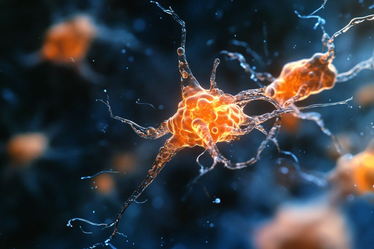
This shows neurons.
The team found that endogenous normal Tau in flies is required for glial lipid droplet formation and for protecting against neuronal ROS. Credit: Neuroscience News

“ROS are natural byproducts of various cellular functions in the body. While low levels of ROS are beneficial, excess ROS is harmful to cells as it triggers the production of toxic forms of other molecules that induce oxidative stress, including peroxidated lipids,” said lead author Dr. Lindsey Goodman, a postdoctoral fellow in the lab of Dr. Hugo Bellen.

“Neurons are particularly susceptible to oxidative stress and are destroyed if peroxidated lipid levels are not tightly controlled.”

Lipid droplets protect the brain from oxidative damage

There is mounting evidence supporting the notion that our brains have developed multiple neuroprotective strategies to combat ROS-induced oxidative damage.

One of the strategies, discovered in 2015 by the Bellen team, consists of neurons exporting these toxic peroxidated lipids to neighboring glial cells, which sequester them into lipid droplets for storage and future energy production.

“This process effectively removes and neutralizes these toxic lipids,” Goodman said. “In the current study we investigated the role of Tau in the formation of glial lipid droplets.”

The team found that endogenous normal Tau in flies is required for glial lipid droplet formation and for protecting against neuronal ROS. Similarly, Tau was required in glial cells obtained from rats and humans to form lipid droplets.

And while expression of normal human Tau was sufficient to restore the process of formation and maturation of glial lipid droplets in flies lacking their own Tau, when this human Tau protein carried disease-causing mutations – which are linked to an increased risk for Alzheimer’s disease – the glia were incapable of forming lipid droplets in response to neuronal ROS.

“This argues that mutations in Tau may reduce the protein’s normal ability to prevent oxidative stress in addition to causing the protein to accumulate into the typical hallmarks of disease, as described by previous work,” said Goodman. “Altogether, the findings support a new neuroprotective role for Tau against the toxicity associated with ROS.”

Further connections with disease were discovered using established fly and rat models of Tau-mediated conditions that overexpress disease-causing human Tau protein in glia. In these scenarios, the investigators again saw defects in glial lipid droplets and glial cell demise in response to neuronal ROS. This demonstrated that Tau is a dosage-sensitive regulator of glial lipid droplets where too much or too little Tau is detrimental.

“By revealing a surprising new neuroprotective role for Tau, the study opens the door to potential new strategies to slow, reverse and treat neurodegenerative conditions,” said Bellen, corresponding author of the work.

He is a distinguished service professor in molecular biology and genetics at Baylor and holds a Chair in Neurogenetics at Duncan NRI. Bellen also is a March of Dimes Professor in Developmental Biology at Baylor.

In summary, contrary to its usual ‘bad guy’ role in neurodegenerative disease, this study demonstrates that Tau also plays a ‘good guy’ role in glia by helping sequester toxic lipids, reducing oxidative damage and, hence protecting our brains. However, when Tau is absent or when defective Tau proteins are present, this protective effect disappears, leading to disease.

Funding: This work was supported by several grants from the National Institutes of Health, the Canadian Institutes of Health and Research Doctoral Award, Sloan Research Fellowship from the Alfred P. Sloan Foundation, Canada Research Chairs program, a CIHR project grant and a Grant-in-Aid for Scientific Research on Challenging Research (Exploratory).

About this neurology research news

Author: Molly Chiu
Source: Baylor College of Medicine
Contact: Molly Chiu – Baylor College of Medicine
Image: The image is credited to Neuroscience News

Original Research: Closed access.
Tau is required for glial lipid droplet formation and resistance to neuronal oxidative stress” by Lindsey Goodman et al. Nature Neuroscience