Changing stroke rehab and research worldwide now.Time is Brain! trillions and trillions of neurons that DIE each day because there are NO effective hyperacute therapies besides tPA(only 12% effective). I have 523 posts on hyperacute therapy, enough for researchers to spend decades proving them out. These are my personal ideas and blog on stroke rehabilitation and stroke research. Do not attempt any of these without checking with your medical provider. Unless you join me in agitating, when you need these therapies they won't be there.

What this blog is for:

My blog is not to help survivors recover, it is to have the 10 million yearly stroke survivors light fires underneath their doctors, stroke hospitals and stroke researchers to get stroke solved. 100% recovery. The stroke medical world is completely failing at that goal, they don't even have it as a goal. Shortly after getting out of the hospital and getting NO information on the process or protocols of stroke rehabilitation and recovery I started searching on the internet and found that no other survivor received useful information. This is an attempt to cover all stroke rehabilitation information that should be readily available to survivors so they can talk with informed knowledge to their medical staff. It lays out what needs to be done to get stroke survivors closer to 100% recovery. It's quite disgusting that this information is not available from every stroke association and doctors group.

Monday, July 31, 2023

Mouth breathing might be ruining your sleep. Here’s how to fix it

I wrote about this over a year ago, does your doctor still incompetently not have a breathing and sleeping protocol for you?  One of my friends puts a little piece of masking tape over his lips to ensure nostril breathing occurs, I probably won't be doing that.

Mouth breathing might be ruining your sleep. Here’s how to fix it

Mouth breathing through the night can lead to diminished sleep quality, snoring and elevated stress.

Editor’s Note: Sign up for CNN’s Sleep, But Better newsletter series. Our seven-part guide has helpful hints to achieve better sleep.

CNN  — 

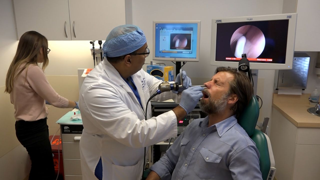
Living with a plugged nose isn’t fun, but James Nestor was ready. Plus, it was for science.

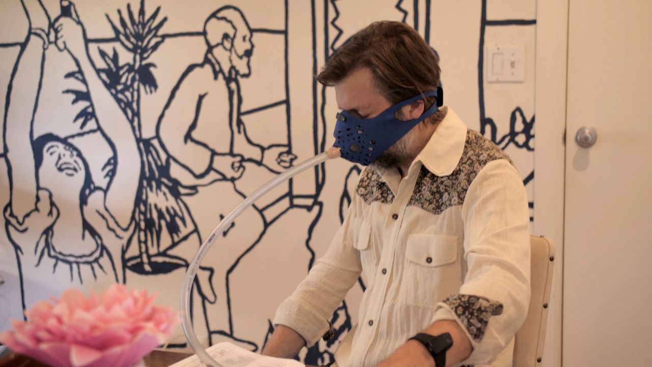
While researching his book, “Breath: The New Science of a Lost Art,” Nestor let Stanford University scientists block his nostrils with silicone and surgical tape to measure the impacts of breathing through his mouth for 10 days.

“We knew it wasn’t going to be good, because there’s a very firm scientific foundation showing all the deleterious effects of mouth breathing, from periodontal disease to metabolic disorders,” Nestor said.

The surprise was just how quickly the experiment affected him.

Nestor’s blood pressure rose 13 points, edging the writer into stage one hypertension. Measurements of heart rate variability showed his body was in a state of stress. His pulse went up, and he stumbled around in a mental fog.

He also snored for hours each night, developing obstructive sleep apnea. His blood oxygen levels dropped.

“We had no idea it was going to be that bad,” Nestor said. “The snoring and sleep apnea was so dramatic, and it came on so quickly, that everyone was pretty floored.”

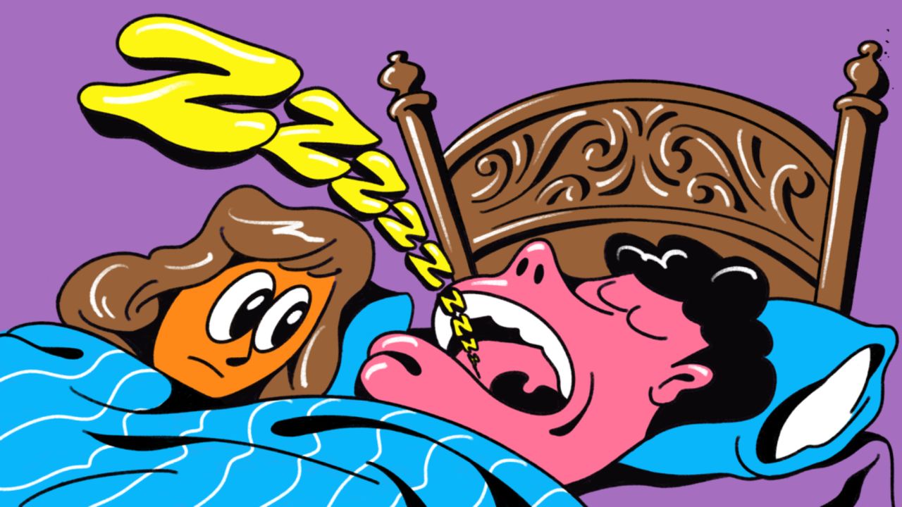
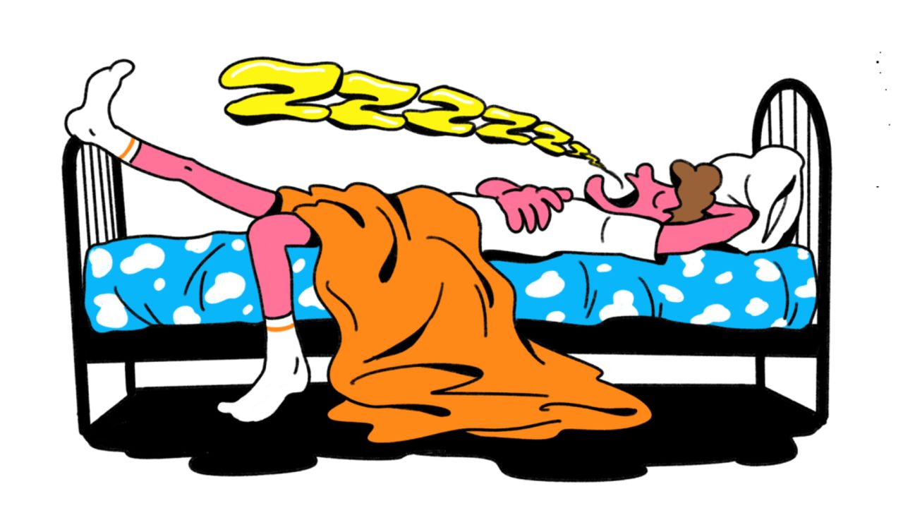
What Nestor learned, aside from the hazards of being a research subject, was that mouth breathing can ruin a good night’s sleep.

James Nestor undergoes an endoscopy at Stanford University in the lead-up to the mouth-breathing experiment.

Breathing through your mouth at night puts you at higher risk for sleep disorders including snoring, sleep apnea and hypopnea, the partial blockage of air, scientists have found. Each of those, in turn, can lead to daytime fatigue.

That doesn’t mean you’re doomed to wake up in a daze because you’re prone to mouth breathing when you sleep.

Experts have a long list of strategies designed to turn you into a nasal breather — including a low-cost breathing hack you can pick up at the corner store.

What causes nighttime mouth breathing?

There is a long list of reasons why people breathe through their mouths at night, said Dr. Steven Park, a surgeon with a specialty in sleep medicine.

20200819-mouth-breathing-in-story-1

“The most common reason is if your nose is stuffy,” Park said. “From allergies, or if you have a deviated septum. Lots of medications can also cause nasal congestion.”

Those problems are aggravated by lying down, he explained.

To calculate how mouth breathing affected the body and mind, Nestor measured physiological data three times a day.

“Generally when you lie down the blood vessels inside your nose fill up with blood,” he said, explaining that the rush of blood causes swelling and constriction. If you can’t breathe easily through your nose, you’re likely to open your mouth for air, Park said. That triggers a positive feedback loop.

“You would think that if you open your mouth you would breathe better, but actually the reverse happens,” he said. Opening your jaws causes the tongue to slump backward, obstructing your airway. “Even if you don’t have sleep apnea, or you have mild sleep apnea, opening your mouth makes it much, much worse.”

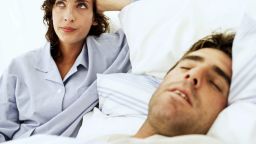
How do you know if you’re mouth breathing at night? Many people, Park noted, are tipped off by a spouse or partner who notices that they’re breathing through the mouth.

If you wake up with a dry mouth or lingering tiredness, they may be warning signs. Another indication of sleep apnea or other disturbed sleep is if you have to visit the bathroom multiple times in the night, said Park. Interrupted breathing stresses the heart, Park explained, triggering the release of hormones that cause you to produce more urine.

How to stop mouth breathing

When Nestor blocked his nose for science, he experienced an extreme version of mouth breathing. But in retrospect, he realized he’d been waking up with a dry mouth for some time, a sign he’d been ditching nasal breathing for at least part of the night.

20200820-mouth-breathing-in-story-2

If you’re tackling your nighttime mouth breathing, Park suggested you start by taking care of your nose to minimize congestion.

“Number one, avoid eating close to bedtime,” he said. That’s because stomach juices can come up into your nose, sinuses, ears and mouth, causing congestion and inflammation.

Park also recommended nasal saline irrigation, flushing the nose with salt water in a squeeze bottle. “That’s a mild decongestant, because the salt water draws out clear water from the membrane,” he explained. (Over-the-counter decongestant sprays can cause habituation and rebound symptoms, Park said, and should be reserved for short-term use.)

Breathing problems are so widespread that they’ve spurred a whole industry dedicated to opening your nose. Park said some people find relief from nasal strips, which open up the nose from the outside, or nasal dilators that expand air passages from within.

But even if you get your nose to clear, nighttime mouth breathing can be a hard habit to break. That’s led some to seek out products that secure their lips closed at night.

Many experts warn against mouth taping while you sleep, because it can be dangerous.

“If you have obstructive sleep apnea, yes, this can be very dangerous,” said sleep specialist Dr. Raj Dasgupta, an associate professor of clinical medicine at the Keck School of Medicine at the University of Southern California, in a previous CNN article.

“There is limited evidence on the benefits of mouth taping and I would be very careful — and even talk to your health care provider before attempting it,” Dasgupta added.

Dasgupta recommended seeing an ear, nose and throat doctor or a sleep specialist to get a diagnosis and treatment plan.

Sunday, July 30, 2023

HandCARE: A Cable-Actuated Rehabilitation System to Train Hand Function After Stroke

In the 14 years since this came out was it ever written into a protocol and distributed? Why doesn't your doctor and hospital know that answer? Aren't they in the business of getting stroke survivors recovered? 

Do you prefer your doctor and hospital incompetence NOT KNOWING? OR NOT DOING?

HandCARE: A Cable-Actuated Rehabilitation System to Train Hand Function After Stroke


Abstract:
We have developed a robotic interface to train hand and finger function. HandCARE is a Cable-actuated rehabilitation system, in which each finger is attached to an instrumented cable loop allowing force control and a predominantly linear displacement. The device, whose designed is based on biomechanical measurements, can assist the subject in opening and closing movements and can be adapted to accommodate various hand shapes and finger sizes. Main features of the interface include a differential sensing system, and a clutch system which allows independent movement of the five fingers with only one actuator. The device is safe, easily transportable, and offers multiple training possibilities. This paper presents the biomechanical measurements carried out to determine the requirements for a finger rehabilitation device, and the design and characterization of the complete system.
Page(s): 582 - 591

Long-term MIND Diet Commitment Key to Boosting Brain Health

But there is nothing EXACT  about this diet so nothing is scientifically repeatable so you can't be sure that your dementia risk declines.  A diet protocol is needed instead of hope that something works.

Long-term MIND Diet Commitment Key to Boosting Brain Health

Summary: A recent study unveils the significance of long-term commitment to the MIND diet in enhancing brain health.

The first randomized clinical trial of its kind, the study observed that short-term cognitive improvements were seen within the first two years, however, no significant difference was noted between the MIND diet and a calorie-restricted control diet over a three-year period. Despite this, the researchers underline that the benefits of the MIND diet likely emerge over a longer period, consistent with earlier observational data.

The MIND diet has been celebrated for its positive effects on brain health, slowing cognitive decline and reducing Alzheimer’s risk.

Key Facts:

  1. The three-year clinical trial showed cognitive improvements in the initial two years for the MIND diet group, but no significant difference when compared to the control diet group by the end of the study.
  2. The study, the first randomized clinical trial designed to test the effects of a diet on cognitive abilities, enlisted individuals 65 years or older without cognitive impairment.
  3. The MIND diet, a combination of the Mediterranean and DASH diets, has been ranked among the top five diets by U.S. News & World Report for six consecutive years.

Source: Rush University

New research shows the importance of long-term commitment to the MIND diet for reaping the greatest benefit to brain health.

“The benefits within the new study’s three-year clinical trial weren’t as impressive as we’ve seen with the MIND diet observational studies in the past, but there were improvements in cognition in the short-term, consistent with the longer-term observational data,” said lead study author Lisa Barnes, PhD, associate director of the Alzheimer’s Disease Research Center at RUSH.

This shows fruits, veggies and fish.
The trial compared two different diet interventions, both of which included dietary counseling with mild calorie restriction of 250 calories per day for weight loss. Credit: Neuroscience News

Results from the study, published in The New England Journal of Medicine, showed that within a three-year period, there was no significant statistical difference in change in cognition for participants in the MIND diet group compared to the usual diet control group; both groups were coached to reduce calories by 250 kilocalories per day. But there was a significant improvement during the first two years of the study.

“What we saw was improvement in cognition in both groups, but the MIND diet intervention group had a slightly better improvement in cognition, although not significantly better,” Barnes said.

“Both groups lost approximately 5 kilograms over three years, suggesting that it could have been weight loss that benefited cognition in this trial.”

‘Exciting’ improvement

This is the first randomized clinical trial designed to test the effects of a diet thought to be protective for brain health, on the decline of cognitive abilities among a large group of individuals 65 years or older who did not have cognitive impairment. The MIND diet has been ranked among the top five diets by U.S. News & World Report annually for the last six years.

“There is established research that shows that a person’s diet affects health,” Barnes said. “The participants in this study had to have sub-optimal diets as determined by a score of 8 or less on a diet screening instrument before the study even began. It is reasonable to think that either they were going to maintain their cognition or decrease the rate of cognitive decline in the future.”

“It was exciting to see that there was improvement in cognition over the first year or so, but it could have been due to practice effects on the cognitive tests, and we saw it for the control diet as well, which focused on just caloric restriction.”

Previous research by the late Martha Clare Morris, ScD, showed that there was a slower rate of decline among those who ate specific foods. Morris was a nutritional epidemiologist at RUSH and the original principal investigator of the MIND diet study that was funded by a $14.5 million National Institutes of Health grant and involved two clinical sites, RUSH in Chicago and Harvard School of Public Health in Boston.

In 2015, Morris and her colleagues at RUSH and Harvard University developed the MIND diet — which is short for Mediterranean-DASH Intervention for Neurodegenerative Delay — in preparation for the trial.

The diet is based on the most compelling research on the foods and nutrients that affect brain health. As the name suggests, the MIND diet is a hybrid of the Mediterranean and DASH (Dietary Approaches to Stop Hypertension) diets.

Both diets have been found to reduce the risk of cardiovascular conditions, such as hypertension, diabetes, heart attack and stroke. In two studies published in 2015, Morris and colleagues found that the MIND diet could slow cognitive decline and lower a person’s risk of developing Alzheimer’s disease significantly, even if the diet was not followed meticulously.

Study tracked 604 participants over three years

The latest trial of the MIND Diet for Prevention of Cognitive Decline in Older Persons, was a randomized, Phase III trial that enrolled 604 people who were overweight and had a suboptimal diet and a family history of Alzheimer’s disease. 

The trial compared two different diet interventions, both of which included dietary counseling with mild calorie restriction of 250 calories per day for weight loss.

Participants of both groups had individualized diet guidelines developed by dietitians, and they received regular phone and in-person consultations, as well as occasional group sessions over the three-year life of the study.

Participants were seen five times during the three years to evaluate their mental abilities, blood pressure, diet, physical activity, health conditions and medication use.

“Both groups of participants got a lot of support and accountability by trained registered dietitians,” said Jennifer Ventrelle, assistant professor in the Departments of Preventive Medicine and Clinical Nutrition and lead dietitian on the MIND diet trial at RUSH.

“The good news is that this helped all participants improve on average, but unfortunately hindered the ability to detect significant differences between the two groups in this relatively short period of time.

“Current and future research plans to look at people coached to follow the diet in this format compared to individuals following a usual diet in a format closer to usual care such as brief clinical encounters or a self-guided program with less support.”

“By the end of the study, the average weight loss was approximately 5.5% of initial body weight for all participants, exceeding the study target of 3%, the amount recognized as clinically significant to prevent or improve adverse health outcomes,” Ventrelle said.

“The average MIND score at the end of three years for the MIND group was 11.0 and 8.3 for the control group, placing both groups in a therapeutic range to slow cognitive decline and lower a risk for Alzheimer’s disease, according to previous studies.

“The significant weight loss and improved MIND scores suggest that the control group also improved their diet and may suggest that following the MIND diet at a score of at least 8.3, coupled with at least a 250 calorie reduction to produce weight loss, may improve cognition. More research is needed to confirm this.”

Fish, chicken, berries, nuts and leafy greens

The MIND diet has 14 dietary components, including nine “brain-healthy food groups” – such as chicken and fish, green leafy vegetables and berries, and nuts – and five unhealthy groups: red meat, butter and stick margarine, full fat cheese, pastries and sweets, and fried foods.

“Randomized trials are gold standards for establishing a cause-and-effect relationship between diet and incidence of Alzheimer’s disease,” Barnes said.

“These individuals were healthy at the start of the trial and had no cognitive impairment, and their cognition got slightly better over time,” Barnes said.

“Why there was no difference between the two diet groups at the end of the trial could be a result of many factors including that the control group had a relatively healthy diet.

“Moving forward, we will look at specific food groups and their associations with biomarkers that were measured in the blood to see if certain nutrients and food groups are more important than others since the two groups were pretty healthy from a dietary perspective at the start.”

About this diet and cognition research news

Author: Nancy DiFiore
Source: Rush University
Contact: Nancy DiFiore – Rush University
Image: The image is credited to Neuroscience News

Original Research: Closed access.
Trial of the MIND Diet for Prevention of Cognitive Decline in Older Persons” by Lisa Barnes et al. NJEM


Spasticity after stroke: Part 1

Oh God, more blathering about how spasticity isn't that bad! So you believe your leader, Dr. William M. Landau?

But there is no need to treat spasticity, Dr. William M. Landau says so in his uninformed 'expert' opinion.  Survivors would immediately disabuse him of that notion. When schadenfreude hits him with his stroke he'll regret his ideas on the matter. 

His statement from here:

Spasticity After Stroke: Why Bother? Aug. 2004 

My comment to them:

You won't have this blase opinion on spasticity when you have a stroke and spasticity is preventing your 100% recovery.  I totally disagree with Dr. William M. Landau's opinion on spasticity when he wrote a letter to the editor in the AHA journal Stroke;  

Spasticity After Stroke: Why Bother?  Aug. 2004
No point in listening to me, I'm just a 17 year stroke survivor. Amy Farber  has this to say. For the past five years Farber has been battling not only her own disease but also the wall of resistance erected by those who believe that a patient can make about as much of a meaningful contribution to the process of scientific discovery as a laboratory rat.
I write Deans' Stroke Musings; 26,217 posts, 5,408,632 views. 

The latest here:

Spasticity after stroke: Part 1

Spasticity is a word that is often heard when someone’s movement problems after a stroke are being described.  Yet strangely there is little or no agreement as to what spasticity actually is!

Here are a few quotes gleaned from medical sites on the internet:

“Spasticity is a condition in which certain muscles are continuously contracted. This contraction causes stiffness or tightness of the muscles and can interfere with normal movement, speech and gait”

“Spasticity is a feature of altered skeletal muscle performance with a combination of paralysis, increased tendon reflex activity, and hypertonia.”

Spasticity is a muscle control disorder that is characterized by tight or stiff muscles and an inability to control those muscles. In addition, reflexes may persist for too long and may be too strong (hyperactive reflexes)”.

From these quotes spasticity might be: continuously contracting muscles, or paralysed muscles, might be stiff muscles, or there may be changes in reflex activity, or a combination of all of these features!  Not very helpful if you don’t agree on what you are describing!  This failure to clearly describe/define spasticity is problematic for clinicians and stroke survivors.  If spasticity isn’t clearly defined or described:

  1. it can’t be Identified or distinguished from other muscle responses which may occur after stroke (such as stiffness, hyperreflexia etc),
  2. it can’t be Measured objectively, and importantly
  3. interventions for spasticity cannot be shown to be effective (or ineffective) because of the uncertainty as to what is being measured.

There are many theories and rationales about just what spasticity is and why it occurs (eg Lance 1980, Pandyan 2005).  The lack of consensus about the definition of spasticity only adds to the confusion for clinicians and more importantly, for stroke survivors.

What is the incidence of spasticity?

The ongoing preoccupation with spasticity is surprising when the incidence of spasticity after stroke is actually quite low.(That's not low to those who have spasticity! Don't try to justify not solving spasticity because of your incorrect feeling it has a low occurrence rate.)  The incidence of spasticity (depending on how it is defined and measured) seems to be somewhere between 17% (Lundstrom 2008) to 38% (Watkins 2002) (see Sommerfield 2012 for a review).  However, it must be acknowledged that for some individual stroke survivors, the consequences of spasticity can have profoundly adverse consequences for their activities of daily living such as being unable to open their hand to be able to clean it.

Is there a relationship between spasticity and function?

Even more surprising, the research that has measured the relationship between spasticity and function has found that spasticity is not correlated with loss of functional ability(Well, then you did bad research because spasticity is definitely preventing correct movements. DON'T YOU EVER TALK TO SURVIVORS? Or do you stay in your echo chamber, not joining the real world?) (Dietz et al 1981, Ada et al Upper Limb 2006, Ada et al Lower Limb 1998, Williams et al Lower Limb – in traumatic brain injury – 2015, Fayazi 2014, Shaw et al 2011).  So even if spasticity is present, it does not appear to be the main problem limiting stroke survivors’ ability to move.  Weakness, on the other hand, is very strongly correlated with loss of function.  (Look for future blogs to discuss the relationship between weakness and poor function in blogs on the StrokeEd website over the coming months.)

This is the first in a series of blogs about spasticity in an attempt to try to make sense of what continues to be a very fraught issue – despite the strong evidence that spasticity is not the main problem preventing stroke survivors from moving.  These blogs will present some of the problems with the prevailing beliefs about spasticity and importantly, provide some evidence based strategies clinicians may use to assist stroke survivors learn to move even in the presence of spasticity.

Your comments, both from clinicians, researchers and stroke survivors, are very welcome.  This blog is intended to help, not hinder, clinical decision making.  So if you want to comment please keep the contributions about spasticity open and informative to progress the discussion for the benefit of all.  Comments will be moderated to ensure a civil conversation!

Evolution of Brain Activation with Good and Poor Motor Recovery after Stroke

 My conclusion is that NOTHING HERE HELPS SURVIVORS RECOVER! Useless. You're fired.

Evolution of Brain Activation with Good and Poor Motor Recovery after Stroke

  • PDF / ePub
  • Abstract

    Objective
     
    To characterize the evolution of brain activation in stroke patients with variable motor recovery and quantify changes relative to healthy controls.  
     
    Methods
     
    Serial PET activation studies, using a simple finger-tapping task, and quantitative measures of motor performance were obtained in 9 patients (2-7 weeks poststroke and 6 months later) and compared with serial healthy volunteer data. 
     
    Results. 
    Patients with moderate impairment and good recovery (n = 5) activated the primary sensorimotor cortex (SM1) contralateral to the paretic hand moved, bilateral supplementary motor area (SMA), contralateral cingulate gyrus, and ipsilateral lateral premotor cortex. Activation in the bilateral SMA was greater at the initial study but reduced over time compared to healthy controls and poor recoverers. Patients with severe impairment and poor recovery (n =4) showed limited activation of contralateral SM1 and SMA at both studies and no significant change over time. A posterior shift in SM1 activation was evident in good and poor recoverers. 
     
    Conclusions. 
     
    Activation of typical motor regions and recruitment of additional sites occur subacutely poststroke, with evolution to normal patterns in moderately impaired patients who recover well. In comparison, severely impaired, poor-recovery patients show persistent, reduced activation. Dynamic changes in SMA, differentially observed in good recoverers over 6 months, highlight its importance in recovery.

    Saturday, July 29, 2023

    Research Casts Doubt on Value of Daily Aspirin for Healthy Adults

    You're not solving the correct problem. How do you identify those persons that are at risk of bleeding from aspirin?

    Research Casts Doubt on Value of Daily Aspirin for Healthy Adults

    Daily use of low-dose aspirin offers no significant protection against stroke and was linked to a higher rate of bleeding in the brain, according to new research published in JAMA.

    The research matches other evidence advising that healthy older adults without a history of heart conditions or warning signs of stroke should not take low-dose aspirin. 

    The findings also support the recommendation from the U.S. Preventive Services Task Force that low-dose aspirin should not be prescribed for preventing a first heart attack or stroke in healthy older adults, The New York Times reported.

    "We can be very emphatic that healthy people who are not on aspirin and do not have multiple risk factors should not be starting it now," said Randall Stafford, MD, of Stanford University, who was not involved in the study, in The Times.

    It's not as clear for others, he said.

    "The longer you've been on aspirin and the more risk factors you have for heart attacks and strokes, the murkier it gets," he said.

    Some cardiac and stroke experts say daily aspirin should remain part of the regimen for people who have had a heart attack or stroke.

    The JAMA report was based on data from a randomized control trial of 19,000 people from Australia and America. Participants were over the age of 70 and did not have heart disease. 

    The data covered an average of almost 4.7 years and revealed that aspirin lowered the rate of ischemic stroke but not significantly(To me any lowering is great, SO SOLVE THE BLEEDING RISK PROBLEM YOU BLITHERING IDIOTS!). An ischemic stroke happens when a clot forms in a blood vessel that sends blood to the brain. 

    There was also a 38% higher rate of brain bleeds for people who took aspirin daily compared to those who took a placebo.

    The Times wrote, "In the past, some doctors regarded aspirin as something of a wonder drug, capable of protecting healthy patients against a future heart attack or stroke. But recent studies have shown that the powerful drug has limited protective power among people who have not yet had such an event, and it comes with dangerous side effects."

    Sources

    JAMA: "Low-Dose Aspirin and the Risk of Stroke and Intracerebral Bleeding in Healthy Older People Secondary Analysis of a Randomized Clinical Trial."

    The New York Times: "For Adults With No Heart Attack or Stroke History, Evidence Says Not to Start Baby Aspirin."


    Social Connections Combat Dementia and Prolong Lifespan

    Well, the solution to this is 100% recovery before you lose the first two groups of friends as described by Aristotle.  Your doctor's responsibility.

    Aristotle believes that there are three different kinds of friendship; that of utility, friendship of pleasure, and virtuous friendship. 

    I'm in two different wine groups, one weekly Zoom meeting and one monthly pot luck one, Weekly trivia at a local bar. 2-3 nights live jazz music, numerous friends there, I'm responsible for getting there early enough to get a table.

    The latest here:


    Social Connections Combat Dementia and Prolong Lifespan

    Summary: Spending time with family and friends not only gives us a positive psychological boost, it can also reduce dementia risks and improve longevity, a new study reports. Researchers found those with strong social connections have a lower risk of mild cognitive impairment, dementia, and death. The study reports improving social connections and frequently taking time to spend with others has positive implications for health and cognition.

    Source: University of New South Wales

    Spending time with loved ones can have significant health benefits as we age, according to a new meta-analysis study from the Centre for Healthy Brain Ageing (CHeBA) at UNSW Sydney.

    The researchers studied the link between social connections in older people and the risk of mild cognitive impairment (MCI), dementia and mortality. They pooled together the results of 13 international studies, which followed people aged 65 years and above over long periods of time.

    The research is published today in Alzheimer’s & Dementia.

    “We know from previous research that social connections are important for our health and being isolated puts us at higher risk of dementia and death,” said first author Dr. Suraj Samtani, who is a clinical psychologist and researcher at CHeBA.

    “Our goal was to find which social connections protect us from dementia and death.”

    Studying aging populations

    The researchers obtained results from studies in low, middle and high-income countries across the world. These included Australia, North America, and several nations in Europe, South America, Asia, and Africa. The study population is more diverse than previous meta-analyses, which have mainly focused on North America and Europe.

    Then the researchers analyzed information about the social connections of the study participants. They were interested in the social connection type (e.g., being in a relationship or married, engaging with a community group), function (e.g., social support, having a confidante), and quality (e.g., level of relationship satisfaction).

    Finally, the researchers looked at whether the participants developed MCI or dementia, or passed away, during the studies. They controlled for other variables which could influence these outcomes including age, sex, education level, lifestyle factors and other chronic diseases.

    “We looked at social variables across these studies, such as living with others, interacting with friends and family, engaging in community activities, and social support,” Dr. Samtani said. “We wanted to know which of these are associated with risk of getting dementia over time or dying.”

    Protective effects of social connections

    Among the study participants, good social connections were associated with a lower risk of MCI, dementia and death.

    “We found that frequent interactions—monthly or weekly—with family and friends and having someone to talk to reduced the risk of getting dementia. We also found that living with others and doing community activities reduced the risk of dying,” Dr. Samtani said.

    Why would social interaction be linked to MCI, dementia and death? Many other studies show that poor social connections are associated with poorer lifestyle and poorer health.

    For example, close relationships can have a stress buffering effect, as we confide in and receive support from these individuals. Controlling stress is important for brain and overall health.

    Another example is that our family, friends and community members may influence us to take on healthy behaviors. This phenomenon, exemplified by your friends dragging you to park run at 7am on a Saturday, is known as ‘social contagion’.

    This shows a happy older couple
    Among the study participants, good social connections were associated with a lower risk of MCI, dementia and death. Credit: Neuroscience News

    One limitation of the findings is the ‘chicken or the egg’ problem. Participants who had already MCI or dementia at the beginning of the studies were excluded from the analysis. However, it is possible that they had some undetected cognitive and physical health issues. These could impact their social interactions, rather than the other way around.

    Tips to stay healthy

    The researchers recommend that we prioritize social connection to reduce risk of cognitive decline and live longer.

    “Try to meet with friends and family at least once a month, take part in community activities like volunteering or a rotary club, and open your heart to someone when you feel stressed. Living with others, for example in an intergenerational household, is also helpful,” Dr. Samtani said.

    “Connecting with others helps us to keep our bodies and minds healthy.”

    The researchers at CHeBA are now looking at interventions to improve the social connections of older adults, to protect their brain and overall health. Dr. Samtani has recently been awarded a Dementia Australia Research Foundation post-doctoral fellowship to trial a social cognition intervention for older adults with memory concerns.

    “We hope that helping people to stay engaged in conversations and maintain healthy friendships and relationships will help them to stay healthy and happy,” Dr. Samtani said.

    About this social neuroscience and cognition research news

    Author: Press Office
    Source: University of New South Wales
    Contact: Press Office – University of New South Wales
    Image: The image is credited to Neuroscience News

    Original Research: Open access.
    Social connections and risk of incident mild cognitive impairment, dementia, and mortality in 13 longitudinal cohort studies of ageing” by Gowsaly Mahalingam et al. Alzheimer’s & Dementia

    Stimulation of the Cerebellum Improves Episodic Memory in Older People

    Doesn't your doctor already having you use some tDCS rehab?

    Transcranial Brain Stimulation: No Benefit for Stroke Rehab


    Stimulation of the Cerebellum Improves Episodic Memory in Older People

    Summary: Transcranial direct current stimulation (tDCS) of the right cerebellum improved episodic memory in elderly people, a new study reports. The findings open the door to developing new, non-invasive therapies to treat age-related memory and cognitive problems.

    Source: BIAL Foundation

    A recent study demonstrated that non-invasive stimulation of the right cerebellum led to improvements in episodic memory performance in healthy elderly individuals, at the end of a 12-day neurostimulation program, and also at the point of a 4-month follow-up.

    The steady increase in average life expectancy poses significant challenges to individuals, families, and societies across multiple dimensions.

    Estimating that by 2050 one in every six individuals will be over the age of 65, the study of aging and its association with cognitive decline, neurodegenerative diseases and overall frailty is becoming increasingly important.

    Therefore, it has been an important goals of neurosciences research to understand the relationship between the aging brain and episodic memory deficits and to develop interventions to mitigate the age-related decline in our ability to remember personal past events (episodic memory).

    The research team led by Jorge Almeida (Faculty of Psychology and Educational Sciences, University of Coimbra) published the article “The cerebellum is causally involved in episodic memory under aging,” in GeroScience, which demonstrated that the cerebellum is one of the neuronal regions causally involved in episodic memory during aging.

    If in the past the cerebellum was considered exclusively as the basis of motor coordination, controlling, for example, our balance and posture, in recent decades studies have proven that this brain region, located at the back of the brain, also decisively influences cognitive and emotional processes.

    This shows the cerebellum
    Simulations were run on SimNIBS 3.2. Credit: The Researchers / GeroScience

    In this study, the team of researchers from universities of Portugal, Brazil, the U.S., and Iran delivered a 12-day neurostimulation program to the right cerebellum of 56 healthy elderly individuals aged 60 years old or over, and registered improvements in their episodic memory performance that lasted at least four months beyond the stimulation period.

    The results demonstrate the causal relevance of the cerebellum in processes associated with long-term episodic memory, highlighting its role in regulating and maintaining cognitive processing.

    According to Jorge Almeida, this work “opens up the possibility of developing non-pharmacological interventions to ameliorate typical age-related cognitive frailty that induce long-lasting improvements that, at least, outlast the four months tested herein.”

    About this aging, neurotech, and memory research news

    Author: Press Office
    Source; BIAL Foundation
    Contact: Press Office – BIAL Foundation
    Image: The image is credited to the researchers/GeroScience

    Original Research: Open access.
    The cerebellum is causally involved in episodic memory under aging” by Jorge Almeida et al. GeroScience

    Gait training with a wearable powered robot during stroke rehabilitation: a randomized parallel-group trial

     So no real difference.

    Gait training with a wearable powered robot during stroke rehabilitation: a randomized parallel-group trial


    Abstract

    Background

    We have developed a wearable rehabilitation robot, “curara®,” and examined its immediate effect in patients with spinocerebellar degeneration and stroke, but its rehabilitative effect has not been clarified. The purpose of this study was to examine the effect of this device on gait training in stroke patients.

    Methods

    Forty stroke patients were enrolled in this study. The participants were divided randomly into two groups (groups A and B). The participants assigned to group A received RAGT with curara® type 4, whereas those in group B received conventional therapist-assisted gait training. The clinical trial period was 15 days. The participants performed 10 sessions of gait training (5 times per week) each lasting 30 ± 5 min per day. The 10-m walking time (10mWT), and 6-minute walking distance (6MWD) were evaluated as the main outcomes. Timed up and go and Berg Balance Scale (BBS) were also examined. Gait parameters (stride duration and length, standard deviation of stride duration and length, cadence, ratio of the stance/swing phases, minimum/maximum knee joint angle, and minimum/maximum hip joint angle) were measured using a RehaGait®. The items other than BBS were measured on days 0, 7, and 14, whereas BBS was measured on days 0 and 14. The improvement rate was calculated as the difference of values between days 14 and 0 divided by the value on day 0. The improvement rates of the 10mWT and 6MWD were set as the main outcomes.

    Results

    The data of 35 participants were analyzed. There was no significant difference in the main outcomes between both groups at the end of gait training. As for intragroup changes, gait speed, stride length, stride duration, and cadence were improved significantly between days 0 and 14 in each group. When examining the interaction effect between the day of measurement and group, stride duration (p = 0.006) and cadence (p = 0.012) were more significantly improved in group A than in group B.

    Conclusions

    This novel wearable powered robot may have the potential to improve gait speed of individuals in stroke rehabilitation.

    Trial registration

    Japan Registry of Clinical Trials (jRCTs032180163). Registered on February 22, 2019; https://jrct.niph.go.jp/en-latest-detail/jRCTs032180163.

    UMIN Clinical Trials Registry (UMIN000034237)

    Registered on September 22, 2018; https://center6.umin.ac.jp/cgi-open-bin/icdr/ctr_view.cgi?recptno=R000038939.

    Background

    Recently, robot-assisted gait training (RAGT) has been applied widely to individuals with stroke to regain and improve walking ability [1]. In the 1990s, body weight-supported treadmill training was introduced in the clinical setting [2]. Currently, rehabilitation robots with different assistive forms for lower limb movements have become popular, e.g., Gait Trainer® and Haptic Walker®, which assist with foot movements, and Lokomat®, which assists with lower limb orthosis on a treadmill. These devices enable the joints of the lower limbs to move in a state close to normal during walking without voluntary efforts from the patient [3,4,5].

    In addition to stationary rehabilitation robots, wearable rehabilitation robots have been developed recently and used widely in gait training, e.g., Hybrid Assistive Limb (HAL®) and ReWALK® [6]. Generally, wearable rehabilitation robots are smaller and lighter than stationary rehabilitation robots; therefore, a great advantage of these devices is that individuals wearing them can move around freely. This makes it possible for people with a wearable rehabilitation robot to perform overground training in daily life. Systematic reviews have reported that rehabilitation robots improve balance and ankle spasticity in patients with brain diseases including stroke [7, 8]. Concerning the rehabilitative effect of HAL®, the degree of walking independence evaluated by the Functional Ambulation Category was significantly improved in the HAL®-wearing group compared to the non-wearing group [9, 10]. In addition, there are reports that RAGT showed more significant improvement in the time up and go (TUG) test, 10mWT and 6MWD than conventional walking training [11, 12].

    We started clinical research in 2017 for the practical application of a wearable rehabilitation robot, “curara®”. We have examined the effect of the device in patients with spinocerebellar degeneration [13, 14] and stroke [15]. We have shown that the use of the device improves walking speed, stride length, walking rate, and asymmetry in stroke patients temporarily [15], but we have not yet clarified its rehabilitative effect on gait training. Therefore, the purpose of this study was to examine the effect of the device on gait training in stroke patients.

    More at link.