Use the labels in the right column to find what you want. Or you can go thru them one by one, there are only 32,650 posts. Searching is done in the search box in upper left corner. I blog on anything to do with stroke. DO NOT DO ANYTHING SUGGESTED HERE AS I AM NOT MEDICALLY TRAINED, YOUR DOCTOR IS, LISTEN TO THEM. BUT I BET THEY DON'T KNOW HOW TO GET YOU 100% RECOVERED. I DON'T EITHER BUT HAVE PLENTY OF QUESTIONS FOR YOUR DOCTOR TO ANSWER.
Changing stroke rehab and research worldwide now.Time is Brain!trillions and trillions of neuronsthatDIEeach day because there areNOeffective hyperacute therapies besides tPA(only 12% effective). I have 523 posts on hyperacute therapy, enough for researchers to spend decades proving them out. These are my personal ideas and blog on stroke rehabilitation and stroke research. Do not attempt any of these without checking with your medical provider. Unless you join me in agitating, when you need these therapies they won't be there.
What this blog is for:
My blog is not to help survivors recover, it is to have the 10 million yearly stroke survivors light fires underneath their doctors, stroke hospitals and stroke researchers to get stroke solved. 100% recovery. The stroke medical world is completely failing at that goal, they don't even have it as a goal. Shortly after getting out of the hospital and getting NO information on the process or protocols of stroke rehabilitation and recovery I started searching on the internet and found that no other survivor received useful information. This is an attempt to cover all stroke rehabilitation information that should be readily available to survivors so they can talk with informed knowledge to their medical staff. It lays out what needs to be done to get stroke survivors closer to 100% recovery. It's quite disgusting that this information is not available from every stroke association and doctors group.
Following
TIA and minor stroke, the risk of recurrent stroke can be significantly
reduced with short duration dual antiplatelet therapy (DAPT). We wish
to investigate if 10 days of DAPT is as effective as 21 days treatment.
Study design:
This
is an open-label, randomized, parallel-group study comparing whether 10
days of DAPT treatment (ASA+Clopidogrel) is non-inferior to 21-day of
DAPT. in patients with minor ischemic stroke (AIS) or high-risk
transient ischemic attack (TIA). In both groups DAPT is started within
24 hours of symptom onset.
This study is being conducted in approximately 15 study sites in the Kingdom of Saudi Arabia. The planned sample size if 1932.
Outcomes:
Noninferiority
of 10 days compared to 21 days of DAPT in the prevention of the
composite endpoint of stroke and death at 90 days in AIS/TIA patients.
The primary safety outcome is major intracranial and systemic
hemorrhage.
Study period:
Enrolment started in the second quarter of 2023, and the completion of the study is expected in the fourth quarter of 2025.
Discussion:
The
trial is expected to show that 10 days of DAPT is non-inferior for the
prevention of early recurrence of vascular events in patients with
high-risk TIAs and minor strokes.
Get full access to this article
View all access and purchase options for this article.
You currently have no access to this content. Visit the access options
page to authenticate.
Endovascular
reperfusion therapy is the primary strategy for acute ischemic stroke.
No-reflow is a common phenomenon, which is defined as the failure of
microcirculatory reperfusion despite clot removal by thrombolysis or
mechanical embolization. It has been reported that up to 25% of ischemic
strokes suffer from no-reflow, which strongly contributes to an
increased risk of poor clinical outcomes. No-reflow is associated with
functional and structural alterations of cerebrovascular
microcirculation, and the injury to the microcirculation seriously
hinders the neural functional recovery following macrovascular
reperfusion. Accumulated evidence indicates that pathology of no-reflow
is linked to adhesion, aggregation, and rolling of blood components
along the endothelium, capillary stagnation with neutrophils, astrocytes
end-feet, and endothelial cell edema, pericyte contraction, and
vasoconstriction. Prevention or treatment strategies aim to alleviate or
reverse these pathological changes, including targeted therapies such
as cilostazol, adhesion molecule blocking antibodies, peroxisome
proliferator-activated receptors (PPARs) activator, adenosine, pericyte
regulators, as well as adjunctive therapies, such as extracorporeal
counterpulsation, ischemic preconditioning, and alternative or
complementary therapies. Herein, we provide an overview of
pathomechanisms, predictive factors, diagnosis, and intervention
strategies for no-reflow, and attempt to convey a new perspective on the
clinical management of no-reflow post-ischemic stroke.
For patients with
suspected acute stroke, researchers noted higher image quality for deep
learning-accelerated MRI, which can be completed over 11 minutes sooner
than similar sequences for conventional MRI.
Emerging
research suggests that deep learning (DL) accelerated magnetic
resonance imaging (MRI) offers similar signal properties for acute
ischemic stroke, provides higher image quality, and reduces imaging time
by more than 75 percent in comparison to conventional MRI.
For the prospective study, recently reported in Radiology,
researchers reviewed data from 211 study participants (mean age of 65)
with suspected acute stroke who had conventional MRI and DL accelerated
MRI, which incorporated multi-shot echo-plantar imaging. The MRI
sequences included T1-weighted, T2-weighted, diffusion-weighted imaging
(DWI) and T2 fluid-attenuated inversion recovery (FLAIR) sequences,
according to the study.
The study authors found that
DL accelerated MRI and conventional MRI were interchangeable for the
detection of acute ischemic lesions. While conventional MRI had slightly
higher interrater agreement for acute ischemic lesion diagnosis in the
left anterior circulation (94 percent vs. 93 percent) and right anterior
circulation (94 percent vs. 91 percent), DL accelerated MRI had
slightly higher interrater agreement in the posterior circulation (94
percent vs. 93 percent) and for relevant secondary findings (80 percent
vs. 77 percent).
Here
one can see a comparison of conventional MRI and deep learning
accelerated MRI that reveal acute infarction in a 75-year-old man.
Researchers noted excellent image quality with deep learning accelerated
MRI in 77.1 percent of readings in comparison to 61.9 percent of
readings with conventional MRI. (Images courtesy of Radiology.)
Three
reviewing radiologists also noted higher image quality with DL
accelerated MRI. Out of 633 readings with DL accelerated MRI, the
reviewing radiologists noted excellent image quality for 488 readings
(77.1 percent) in comparison to 61.9 percent (392 of 633 readings) for
conventional MRI.
The researchers also noted
significant reductions in scanning time for DL accelerated MRI. The
T2-weighted transverse plane sequence had an average acquisition time of
2 minutes, 39 seconds with conventional MRI vs. 24 seconds with DL
accelerated MRI. T2 FLAIR sequences were completed in three minutes, 20
seconds on average with conventional MRI in comparison to one minute 22
seconds with DL accelerated MRI. Overall, the study authors noted a
significant reduction in image acquisition time with DL accelerated MRI
(14 minutes 18 seconds) in contrast to conventional MRI (3 minutes 4
seconds) for the detection of acute ischemic infarction.
Three Key Takeaways
Similar signal properties for acute ischemic stroke detection. The
study suggests that deep learning (DL) accelerated magnetic resonance
imaging (MRI) provides similar signal properties for detecting acute
ischemic stroke when compared to conventional MRI. This indicates that
DL accelerated MRI can be a reliable alternative for acute ischemic
lesion diagnosis.
Higher image quality with DL accelerated MRI. Three
reviewing radiologists noted higher image quality with DL accelerated
MRI. The study found that DL accelerated MRI had excellent image quality
for 77.1 percent of readings, compared to 61.9 percent for conventional
MRI. This suggests that DL acceleration not only maintains but may even
enhance image quality in certain cases.
Significant reduction in imaging time. DL
accelerated MRI demonstrated a substantial reduction in imaging time
with over 75 percent reduction compared to conventional MRI. The study
highlights specific sequences, such as T2-weighted transverse plane and
T2 FLAIR, showing significantly shorter acquisition times with DL
accelerated MRI. The potential to achieve faster imaging is crucial for
improving efficiency in clinical settings, making it a valuable
consideration for patients with suspected stroke.
“The
deployment of DL-accelerated MRI reconstructions is becoming
successively prevalent in MRI applications due to its potential to
substantially reduce examination times while maintaining diagnostic
image quality. Given the increasing demand for medical examinations and
increasing financial constraints placed on health care systems,
implementing this technique may be of great value,” wrote lead study
author Sebastian Altmann, M.D., who is affiliated with the Department of
Neuroradiology at the University Medical Center Mainz in Mainz,
Germany, and colleagues.
While acknowledging that
computed tomography (CT) is more often utilized in cases of suspected
acute ischemic stroke due to availability and cost-effectiveness, the
researchers emphasized stronger sensitivity of MRI for diagnosing early
and small ischemic lesions.
“This may be of
increasing relevance as randomized clinical trials have shown that
patients with acute ischemic stroke of unknown onset and without a clear
window of time could benefit from intravenous thrombolysis selected at
DWI and (FLAIR) imaging,” pointed out Altmann and colleagues.
Beyond
the inherent limitations of a single-center study, the authors noted
possible bias with manual assessment of DWI and ADC values for acute
ischemic infarctions. The researchers did not assess other pathologic
abnormalities aside from acute ischemic stroke and acknowledged that
they did not acquire contrast-enhanced MRI views.
My left hand will not open due to
spasticity which my doctor never did one damn thing about, except to
tell me spasticity wouldn't go away. What a fucking useless doctor. When the little cup of sour cream is full I can dip into in successfully, but getting the last dregs out is impossible. I can't get my left hand open and at the same time get the little cup in it without crushing it.
New image-guided therapy system is a complete
interventional solution for confident diagnosis, image guidance, and
therapy assessment of patients with stroke or other neurovascular
diseases
Amsterdam, the Netherlands – Royal Philips
(NYSE: PHG, AEX: PHIA), a global leader in health technology, today
announced major enhancements to its Image Guided Therapy System –
Azurion – with the launch of its new Azurion neuro biplane system.
Designed to streamline neurovascular procedures and help care teams
make the right decisions faster, treat more patients, and achieve better
outcomes, the new interventional system features enhanced 2D and 3D
imaging and X-ray detector positioning flexibility. As healthcare
providers strive to deliver high-quality care to patients, at #ECR2024 Philips is partnering with its customers to improve productivity and access to more sustainable healthcare.
Minimally invasive procedures enabled by interventional systems like
Azurion are a key part of the diagnosis and treatment pathway for
stroke, where every minute counts in conserving the patient’s
quality-of-life. Interventional systems are also used to precisely plan
and carry out complex neurovascular procedures such as the repair of
brain aneurysms and birth defects. By allowing neuro interventionists to
treat more patients, more efficiently, with potentially better
outcomes, the new Philips Azurion neuro biplane system enhances both the
staff and patient experience and contributes to lower cost of care.
“Working closely with leading interventionists, we designed the
latest Azurion neuro biplane to meet their requirements of superior
patient care, optimized angio suite performance, and efficient return on
investment,” said Mark Stoffels, Business Leader of Image Guided
Therapy Systems at Philips. “Together, I am confident we can continue to
reduce the impact of stroke, helping more patients to recover faster
and reducing long-term impact on their health.”
Philips’ Azurion neuro biplane image-guided therapy system is designed
to smooth and optimize procedure workflows where a combination of 2D and
3D imaging is needed for confident diagnosis and precision treatment.
Used with the company’s latest Neuro Suite software and services, it
provides neuro interventionists with a fully integrated solution that
combines Philips’ world-class ClarityIQ low dose imaging with a range of
neuro dedicated tools* and value-added services that offer
unprecedented levels of efficiency, flexibility, and control.
New features in the Azurion neuro biplane system include enhanced
C-arm rotation, angulation (imaging angles), and parking facilities that
allow rapid transitioning between 2D to 3D imaging, comprehensive
table-side control that eliminates the need to leave the sterile field,
automatic beam rotation to obtain correctly oriented images for every
angulation and rotation, and a new head immobilizer to support enhanced
stroke care.
Accelerating treatment for stroke
One in four adults over the age of 25 will have a stroke at some point
in their lives [1]. Globally, the direct and indirect cost has been
estimated at around USD 891 billion per year [2]. The key to reducing
the personal, societal, and financial impact of stroke is to follow the
axiom ‘time-is-brain’. The faster stroke is treated, the better the
potential outcome.
For ischemic stroke, which accounts for 87% of all strokes [3], the
benefits of delivering intra-arterial treatment (IAT) – catheter-based
procedures to mechanically disrupt or remove blood clots (mechanical
thrombectomy) and/or inject clot-busting agents – within 6 hours of
symptom onset, are widely accepted [4]. With mechanical thrombectomy
becoming the standard for treating large vessel occlusion (LVO) ischemic
stroke, the demand for high utilization angio suites that allow
interventionists to operate with speed and efficiency has rapidly
increased.
In addition to the efficiency enhancing features of its Azurion
platform, Philips is maximizing the up-time of its angio suite solutions
by using AI and machine learning to monitor system performance through
the new remote connection services of the Philips ServiceHub. These
services communicate, monitor, and proactively respond to potential
service issues – for example, predicting likely component failures at
least 7 days in advance so that pre-emptive action can be taken.
With its comprehensive stroke care portfolio, Philips is connecting
the dots between caregivers – wherever they are – at every vital step in
the stroke care pathway. The result is smart stroke solutions designed to support connected care. Visit Philips at #ECR2024 for more information.
Studies
suggest the BCG jab discovered a century ago could provide a cheap and
effective way of boosting the immune system to protect people from
developing the condition
Sun 25 Feb 2024 08.00 ESTLast modified on Mon 26 Feb 2024 04.10 EST
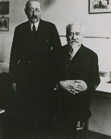
Scientific
discoveries can emerge from the strangest places. In early 1900s
France, the doctor Albert Calmette and the veterinarian Camille Guérin
aimed to discover how bovine tuberculosis was transmitted. To do so,
they first had to find a way of cultivating the bacteria. Sliced
potatoes – cooked with ox bile and glycerine – proved to be the perfect
medium.
As the bacteria grew, however, Calmette and Guérin were surprised to find that each generation lost some of its virulence.
Animals infected with the microbe (grown through many generations of
their culture) no longer became sick but were protected from wild TB. In
1921, the pair tested this potential vaccine on their first human
patient – a baby whose mother had just died of the disease. It worked,
and the result was the Bacille Calmette-Guérin (BCG) vaccine that has
saved millions of lives.
French
veterinarian Camille Guérin and physician Albert Calmette developed the
BCG jab in 1921 using sliced potatoes cooked with ox bile and
glycerine. Photograph: Musée Pasteur
Calmette
and Guérin could have never imagined that their research would inspire
scientists investigating an entirely different kind of disease more than
a century later. Yet that is exactly what is happening, with a string
of intriguing studies suggesting that BCG can protect people from
developing Alzheimer’s disease.
If these
preliminary results bear out in clinical trials, it could be one of the
cheapest and most effective weapons in our fight against dementia.
According to the World Health Organization, 55 million people
now have dementia, with about 10 million new cases each year.
Alzheimer’s disease is by far the most common form, accounting for about
60%-70% of cases. It is characterised by clumps of a protein called
amyloid beta that accumulate within the brain, killing neurons and
destroying the synaptic connections between the cells.
Exactly what causes the plaques to develop has been a mystery, but multiple lines of evidence
implicate problems with the immune system. When we are young, our
body’s defences can prevent bacteria, viruses or fungi from reaching the
brain. As we get older, however, they become less efficient, which may
allow microbes to work their way into our neural tissue. According to
this theory, the amyloid beta is produced to kill those invaders as a
short-term defence against infection. If the brain’s own immune cells –
known as microglia – were working optimally, they could clear away the
protein once the threat has passed. But in many cases of Alzheimer’s
disease, they seem to malfunction, triggering widespread inflammation
that leads to further neural carnage.
If
this theory is correct, attempts to boost the immune system’s overall
functioning could prevent the development of the disease.
New approaches are certainly needed. After decades of research on ways to clear the plaques, only two new drugs have been approved by
the US Food and Drug Administration. Both are based on antibodies that
bind to the amyloid beta proteins, triggering an immune response that
clears them out of the brain. This appears to slow disease progression
in some patients, but the improvement in overall quality of life is
often limited.
Anti-amyloid antibodies also
come with a hefty price tag. “The cost of treatment is likely to lead to
an enormous health equity gap in lower-income countries,” says Marc
Weinberg, who researches Alzheimer’s at Massachusetts general hospital
in Boston. (He emphasises his opinions are personal and do not reflect
those of his institution.)
Could existing vaccines such as BCG offer an alternative solution?
The
idea may sound far-fetched, but decades of research show that BCG can
have surprising and wide-ranging benefits that go way beyond its
original purpose. Besides protecting people from TB, it seems to reduce the risk of many other infections, for instance. In a recent clinical trial, BCG halved the odds of developing a respiratory infection over the following 12 months, compared with the people receiving a placebo.
BCG
is also used as a standard treatment for forms of bladder cancer. Once
the attenuated bacteria have been delivered to the organ, they trigger
the immune system to remove the tumours, where previously they had
passed below the radar. “It can result in remarkable disease-free
recoveries,” says Prof Richard Lathe, a molecular biologist at Edinburgh
University.
These remarkable effects are
thought to emerge from a process called “trained immunity”. After an
individual has received BCG, you can see changes in the expression of
genes associated with the production of cytokines – small molecules that
can kick our other defences, including white blood cells, into action.
As a result, the body can respond more efficiently to a threat – be it a
virus or bacteria entering the body, or a mutant cell that threatens to
grow uncontrollably. “It can be likened to upgrading the security
system of a building to be more responsive and efficient, not just
against known threats but against any potential intruders,” says
Weinberg.
There are good reasons to believe
that trained immunity could reduce the risk of Alzheimer’s. By
bolstering the body’s defences, it could help keep pathogens at bay
before they reach the brain. It could also prompt the brain’s own immune
cells to clear away the amyloid beta proteins more effectively, without
causing friendly fire to healthy neural tissue.
Animal studies provide some tentative evidence. Laboratory mice immunised with BCG have reduced brain inflammation,
for example. This results in notably better cognition, when other mice
of the same age begin to show a steady decline in their memory and
learning. But would the same be true of humans?
To find out, Ofer Gofrit of the Hadassah-Hebrew University Medical Centre in Jerusalem and his colleagues collected the data of 1,371 people
who had or had not received BCG as part of their treatment for bladder
cancer. They found that just 2.4% of the patients treated with BCG
developed Alzheimer’s over the following eight years, compared with 8.9%
of those not given the vaccine.
Since the results were published in 2019, other researchers have replicated the findings. Weinberg’s team, for instance, examined the records of about 6,500 bladder cancer patients in
Massachusetts. Crucially, they ensured that the sample of those who had
received BCG and those who hadn’t were carefully matched for age,
gender, ethnicity and medical history. The people who had received the
injection, it transpired, were considerably less likely to develop
dementia.
The precise level of protection varies between studies, with a recent meta-analysis
showing an average risk reduction of 45%. If this can be proven with
further studies, the implications would be huge. “Simply delaying the
development of Alzheimer’s by a couple of years would lead to tremendous
savings – both in suffering and our money,” says Prof Charles
Greenblatt of the Hebrew University of Jerusalem, who was a co-author of
Gofrit’s original paper.
Plenty
of caution is necessary. The existing papers have all examined patients
with bladder cancer, but as yet there is little data on the general
population. One obvious strategy may be to compare people who have
received the BCG vaccine during childhood with those who hadn’t, but the
effects of BCG may dwindle over the decades – long before most people would be in danger of developing Alzheimer’s.
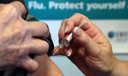
While
BCG is thought to provide the most potent immune training, other
vaccines such as the flu jab may also stimulate the body’s defences. Photograph: David Cheskin/PA
We
can, however, examine the effects of other vaccines delivered in old
age. With its live (but attenuated) bacteria, BCG is thought to provide
the most potent immune training, but other vaccines may also stimulate the body’s defences. Consider the flu jab.
Nicola Veronese of the University of Palermo in Italy and her
colleagues recently analysed the results of nine studies, many of which
controlled for lifestyle factors, including income, education, smoking,
alcohol consumption and hypertension. The team found that the influenza
vaccine was associated with a 29% reduced risk of dementia. “Two studies
also showed an association between the number of doses, over previous
years, and the incidence of dementia,” says Veronese.
Such
studies still cannot prove causality. “In this kind of epidemiological
research, it may be that there’s a confounding factor that’s lurking
that isn’t properly accounted for,” says Jeffrey Lapides of Drexel
University College of Medicine in Pennsylvania, though he agrees that
the vaccine effects on dementia are plausible and deserve more research.
The
clinching evidence would come from a randomised controlled trial in
which patients are either assigned the active treatment or the placebo.
Since dementia is very slow to develop, it will take years to collect
enough data to prove that BCG – or any other vaccine – offers the
expected protection from full-blown Alzheimer’s compared with a placebo.
In
the meantime, scientists have started to examine certain biomarkers
that show the early stages of disease. Until recently, this was
extraordinarily difficult to do without expensive brain scans, but new
experimental methods allow scientists to isolate and measure levels of amyloid beta proteins in blood plasma, which can predict a subsequent diagnosis with reasonable accuracy.
A pilot study
by Coad Thomas Dow of the University of Wisconsin-Madison and his
colleagues suggests that BCG injections can effectively reduce plasma
amyloid levels, particularly among those carrying the gene variants
associated with a higher risk of Alzheimer’s. Although the sample size
was small – just 49 participants in total – it has bolstered hopes that
immune training will be an effective strategy for fighting the disease.
“These results were encouraging,” says Weinberg, who was not involved in
the study.
Weinberg
has his own grounds for optimism. Working with Dr Steven Arnold and Dr
Denise Faustman, he has collected samples of the cerebrospinal fluid
that washes around the central nervous system of people who have or have
not received the vaccine. Their aim was to see whether the effects of
trained immunity could reach the brain – and that is exactly what they
found. “The response to pathogens is more robust in specific populations
of these immune cells after BCG vaccination,” says Weinberg.
We
can only hope that these early results will inspire further trials. For
Weinberg, it’s simple. “The BCG vaccine is safe and globally
accessible,” he says. It is also incredibly cheap compared with the
other options, costing just a few pence a dose. Even if it confers just a
tiny bit of protection, he says: “It wins the cost-effectiveness
contest hands down.”
As Calmette and Guérin discovered with their potato slices more than a century ago, progress may come when you least expect it.
This article was amended on 26 February 2024 to correct an instance of a misspelling of Marc Weinberg’s surname.
Neuropsychiatric symptoms are a common accompaniment of dementia. These include agitation, depression, apathy, delusions, hallucinations, and sleep impairment.
Neuropsychiatric
symptoms (NPS) may inform dementia risk assessment in conjunction with
cognitive testing and imaging and laboratory Alzheimer’s disease (AD)
biomarkers, and was independently associated with the risk of mild
cognitive impairment (MCI)-dementia progression, over and beyond the
contributions of CSF biomarkers, according to a study published in the Journal of Alzheimer’s Disease.
“It's
hard to predict which patients will have a more rapid progression and
receive a diagnosis of dementia,” said Maria Vittoria Spampinato, MD,
Medical University of South Carolina, Charleston, South Carolina. “It’s
important to know who is likely to progress to dementia, as they will
need a lot of support and assistance from their family and other
caregivers.”
“Although it’s important to do lab testing to measure
the number of amyloid plaques and tau disease, NPS testing is important
in identifying which patients are at greater risk,” she said.
To
test whether NPS could help to predict MCI to AD progression, the
researchers identified 300 patients aged 65 years and older with MCI
from the Alzheimer’s Disease Neuroimaging Initiative database. Patients
were given the Neuropsychiatric Inventory (NPI) to document symptoms,
such as anxiety, depression, delusions, hallucinations, abnormal
movement behaviour, and sleep disorders as potential early signs of
preclinical AD to establish a prediction model for AD.
The study
findings showed that more than a quarter of the patients with MCI went
on to develop AD. For each 1-point increase in NPI score, there was a 3%
increase in the risk of mental decline leading to the diagnosis of AD.
Surprisingly, the study showed that NPS predicted the risk of mental decline better than certain established risk factors of AD.
The
prediction model developed by Dr. Spampinato and colleagues shows
promise for identifying which patients with MCI will progress to AD;
however, it will need to be validated in a larger group of patients
recruited from memory care institutions before being used in the clinic.
Anytime I see the word 'care' in
stroke I know that we don't have the right goals anywhere in stroke.
100% recovery is the only goal in stroke. NOT 'care'.
This is why the AHA/ASA are totally incompetent in solving stroke to 100% recovery, they don't even have it as a goal
Three measurements will tell me if the stroke hospital is possibly not
completely incompetent; DO YOU MEASURE ANYTHING? I would start cleaning
the hospital by firing the board of directors, you can't let
incompetency continue for years at a time.
There is no quality here if you don't measure the right things.
Elaine L. Miller, PhD, RN, CRRN, FAHA, Chair; Laura Murray, PhD, CCC-SLP;Lorie Richards, PhD, OTR/L, OT, FAHA; Richard D. Zorowitz, MD, FAHA; Tamilyn Bakas, PhD, RN, FAHA;Patricia Clark, PhD, RN, FAHA; Sandra A. Billinger, PhD, PT, FAHA; on behalf of the American HeartAssociation Council on Cardiovascular Nursing and the Stroke Council I. Introduction In the United States, the incidence rate of new or recurrent stroke is approximately 795 000 per year, and stroke prevalence for individuals over the age of 20 years is estimated at6.5 million. 1 Mortality rates in the first 30 days after stroke have decreased because of advances in emergency medicine and acute stroke care. In addition, there is strong evidence that organized postacute, inpatient stroke care delivered within the first 4 weeks by an interdisciplinary health care team results in an absolute reduction in the number of deaths. 2,3 Despite these positive achievements, stroke continues to represent the leading cause of long-term disability inAmericans: An estimated 50 million stroke survivors world-wide currently cope with significant physical, cognitive, andemotional deficits, and 25% to 74% of these survivors requiresome assistance or are fully dependent on caregivers foractivities of daily living (ADLs). 4,5 Notwithstanding the substantial progress in acute stroke care over the past 15 years(I don't see ANY PROGRESS TOWARDS 100% RECOVERY!), the focus of stroke medical advances and healthcare resources has been on acute and subacute recovery phases, which has resulted in substantial health disparities in later phases of stroke care. Additionally,healthcare providers (HCPs) are often unaware of not only patients’ potential for improvement during more chronic recovery phases but also common issues that stroke survivors and their caregivers experience. Furthermore, even with evidence that documents neuroplasticity potential regardless of age and time after stroke, 6 the mean lifetime cost of ischemic stroke (which accounts for 87% of all strokes) in the United States is an estimated $140 000 (for inpatient,rehabilitation, and follow-up costs), with 70% of first-year stroke costs attributed to acute inpatient hospital care 1 ;therefore, fewer financial resources appear to be dedicated to providing optimal care during the later phases of stroke recovery.Because there remains a need to educate nursing and other members of the interdisciplinary team about the potential for recovery in the later or more chronic phases of stroke care,the present scientific statement summarizes the best available evidence and recommendations for interdisciplinary management of the needs of stroke survivors and their families during inpatient and outpatient rehabilitation and in chronic care and end-of-life settings. The guidelines for making decisions regarding classes and levels of evidence are listed in Table 1 and are the same as those used by previous American Heart Association (AHA) writing groups. 7 Before reviewing the evidence pertaining to stroke rehabilitation, we first briefly review the World Health Organization’s (WHO)international classification of functioning, disability, and health (ICF), 8 which serves as an organizational scaffold for the present statement; provide an overview of the interdisciplinary team approach to rehabilitation; and define the different care settings in which stroke survivors may receive services during the more chronic phases of their recovery. Asa reference, a list of abbreviations used within this statement can be found in Table 2.
You fucking blithering idiots; survivors don't want 'care', they want recovery and results. This is why the WSO is worthless, they do nothing for survivors. Survivors want 100% recovery! GET THERE!
If their results were good they would publish them but since they obviously are not good they focus on 'care'!
Explore the
remarkable achievements of Asian Hospital and Medical Center, including
the Diamond Status Award from the World Stroke Organization.
Asian Hospital Clinches Prestigious Awards for Stroke Care, Communication Campaigns and HR Innovations
Asian Hospital and
Medical Center, a leading healthcare institution, has garnered
significant recognition for its outstanding healthcare services and
programs throughout 2023 and the early months of 2024. These accolades
underscore the hospital's dedication to healthcare excellence and
innovation, setting a benchmark in providing world-class care and
services.
Diamond Status Award
In
the last quarter of 2023, the World Stroke Organization (WSO) awarded
its prestigious Diamond Status to Asian Hospital's Brain Attack Team,
distinguishing it as the only private hospital in the Philippines to
achieve this recognition for exceptional stroke care(NOT RESULTS!) within the critical
'golden hour'. This accolade is part of the WSO Angels Award, aimed at
promoting and recognizing excellence in stroke care(NOT RESULTS!) worldwide.
"Thanks
to the relentless dedication of our Brain Attack Team in providing
exemplary stroke care(NOT RESULTS!), Asian Hospital has been honored with the Diamond
Status. This highest accolade underscores our commitment to meeting the
rigorous standards set by the World Stroke Organization for stroke care(NOT RESULTS!)
excellence," remarked Dr. Jennifer Manzano, Programme Director of Asian
Brain Institute.
I'm sure my first thousand blog posts were barely read at all. But a few stroke survivors positively commented so I kept going, I've never had ANY communications from ANY stroke medical 'professional' so I guess they don't give a shit about what survivors think. Someday the stroke medical world will listen to me, but only after they become the 1 in 4 per WHO that has a stroke.
My journey on this blog will continue, I'm having too much fun tweaking supposedly smarter people than me.
Oops, I'm not playing by the polite rules of Dale Carnegie, 'How to Win Friends and Influence People'.
Telling supposedly smart stroke medical persons they know nothing about stroke is a no-no even if it is true.
Politeness
will never solve anything in stroke. Yes, I'm a bomb thrower and proud
of it. Someday a stroke 'leader' will try to ream me out for making them look bad by being truthful, I
look forward to that day.
Seth's Blog : The leap
In action movies, there’s a lot of leaping. Brave shifts in which the hero gets from here to there, all at once.
It’s easy to imagine that sudden leaps are how we make our impact.
This is blog post #9000 (give or take).
When did the leap happen?
It wasn’t an external leap. The first hundred blog posts were read by fewer than a dozen people.
It was an internal one. The decision to be a blogger. And then redeciding, each day, not to stop.
Every four years, we have a worldwide holiday to celebrate this sort
of leap. The leap of choice. Not to suddenly get from here to there, but
to choose to go on the journey.
It’s only once every 1,460 days, you can do it.
Leap today.
Perhaps we begin by visualizing it. In the most concrete terms you
can find, write it down. If you took a leap today, what would it look
like? Who would benefit?
And then, share it with just one other person.
Often, the act of physically writing it down is the most difficult part.
Are these flushing neurons still working post stroke? Then, if not, is this the reason for heightened dementia risk post stroke? Ask your doctor EXACTLY how they will be testing to see if these brainwaves are still functioning properly! Your doctor has had years of knowledge on this to competently come up with a solution, but I bet you don't have a functioning stroke doctor!
Summary: A new study unveiled a crucial role of
sleep: brainwaves facilitating the cleansing of the brain by flushing
out waste. This discovery not only underscores the brain’s non-dormant
state during sleep but also highlights a sophisticated system where
neurons’ synchronized activity powers the flow of cerebrospinal fluid,
effectively removing metabolic waste and potentially neurodegenerative
disease-causing toxins.
This insight opens up possibilities for
enhancing brain cleaning processes to combat neurological diseases and
improve sleep efficiency, hinting at a future where optimized sleep
could lead to better health outcomes.
Key Facts:
Brainwaves Propel Cleansing Fluids:
During sleep, neurons coordinate to produce rhythmic waves that drive
the movement of fluid through the brain, washing away waste.
Potential for Disease Prevention:
Understanding and enhancing this cleansing process could delay or
prevent diseases like Alzheimer’s and Parkinson’s by ensuring the
effective removal of brain waste.
Implications for Sleep Quality:
This research suggests that improving the brain’s waste removal
efficiency could allow for healthier brains even with less sleep,
offering new avenues for treating sleep disorders and enhancing overall
well-being.
Source: Washington University
There
lies a paradox in sleep. Its apparent tranquility juxtaposes with the
brain’s bustling activity. The night is still, but the brain is far from
dormant. During sleep, brain cells produce bursts of electrical pulses
that cumulate into rhythmic waves – a sign of heightened brain cell
function.
But why is the brain active when we are resting?
Slow
brain waves are associated with restful, refreshing sleep. And now,
scientists at Washington University School of Medicine in St. Louis have
found that brain waves help flush waste out of the brain during sleep.
Individual nerve cells coordinate to produce rhythmic waves that propel
fluid through dense brain tissue, washing the tissue in the process.
Cerebrospinal
fluid surrounding the brain enters and weaves through intricate
cellular webs, collecting toxic waste as it travels. Credit:
Neuroscience News
“These neurons are miniature
pumps. Synchronized neural activity powers fluid flow and removal of
debris from the brain,” explained first author Li-Feng Jiang-Xie, PhD,
a postdoctoral research associate in the Department of Pathology &
Immunology.
“If we can build on this process, there is the
possibility of delaying or even preventing neurological diseases,
including Alzheimer’s and Parkinson’s disease, in which excess waste –
such as metabolic waste and junk proteins – accumulate in the brain and
lead to neurodegeneration.”
The findings are published Feb. 28 in Nature.
Brain
cells orchestrate thoughts, feelings and body movements, and form
dynamic networks essential for memory formation and problem-solving. But
to perform such energy-demanding tasks, brain cells require fuel. Their
consumption of nutrients from the diet creates metabolic waste in the
process.
“It is critical that the brain disposes of metabolic waste that can
build up and contribute to neurodegenerative diseases,” said Jonathan
Kipnis, PhD, the Alan A. and Edith L. Wolff Distinguished Professor of
Pathology & Immunology and a BJC Investigator. Kipnis is the senior
author on the paper.
“We knew that sleep is a time when the brain
initiates a cleaning process to flush out waste and toxins it
accumulates during wakefulness. But we didn’t know how that happens.
These findings might be able to point us toward strategies and potential
therapies to speed up the removal of damaging waste and to remove it
before it can lead to dire consequences.”
But cleaning the dense
brain is no simple task. Cerebrospinal fluid surrounding the brain
enters and weaves through intricate cellular webs, collecting toxic
waste as it travels. Upon exiting the brain, contaminated fluid must
pass through a barrier before spilling into the lymphatic vessels in the
dura mater – the outer tissue layer enveloping the brain underneath the
skull. But what powers the movement of fluid into, through and out of
the brain?
Studying the brains of sleeping mice, the researchers
found that neurons drive cleaning efforts by firing electrical signals
in a coordinated fashion to generate rhythmic waves in the brain,
Jiang-Xie explained. They determined that such waves propel the fluid
movement.
The research team silenced specific brain regions so
that neurons in those regions didn’t create rhythmic waves. Without
these waves, fresh cerebrospinal fluid could not flow through the
silenced brain regions and trapped waste couldn’t leave the brain
tissue.
“One of the reasons that we sleep is to cleanse the brain,” Kipnis said.
“And
if we can enhance this cleansing process, perhaps it’s possible to
sleep less and remain healthy. Not everyone has the benefit of eight
hours of sleep each night, and loss of sleep has an impact on health.
“Other
studies have shown that mice that are genetically wired to sleep less
have healthy brains. Could it be because they clean waste from their
brains more efficiently? Could we help people living with insomnia by
enhancing their brain’s cleaning abilities so they can get by on less
sleep?”
Brain wave patterns change throughout sleep cycles. Of note, taller
brain waves with larger amplitude move fluid with more force. The
researchers are now interested in understanding why neurons fire waves
with varying rhythmicity during sleep and which regions of the brain are
most vulnerable to waste accumulation.
“We think the brain-cleaning process is similar to washing dishes,” neurobiologist Jiang-Xie explained.
“You
start, for example, with a large, slow, rhythmic wiping motion to clean
soluble wastes splattered across the plate. Then you decrease the range
of the motion and increase the speed of these movements to remove
particularly sticky food waste on the plate.
“Despite the varying
amplitude and rhythm of your hand movements, the overarching objective
remains consistent: to remove different types of waste from dishes.
Maybe the brain adjusts its cleaning method depending on the type and
amount of waste.”
About this sleep and neuroscience research news
Author: Marta Wegorzewska Source: Washington University Contact: Marta Wegorzewska – Washington University Image: The image is credited to Neuroscience News
And how the fuck are you supposed to do that when YOU THE AHA/ASA have completely failed at creating anything that gets survivors even partially close to 100% recovery? You're supposed to solve stroke, not just dump everything on the survivors, you're hopeless as a stroke association. Contact me at oc1dean@gmail.com and you can explain in detail how you are helping survivors get to 100% recovery.
NATIONWIDE — Stroke survivors
were more likely to remain physically active or even exercise more after
their stroke if they lived in neighborhoods with easy access to
recreational centers and gyms, according to a preliminary study
presented at the American Stroke Association’s International Stroke
Conference in February.
“We know that stroke survivors need
to be physically active as part of their recovery. Our findings suggest
that it’s important to have a conversation with stroke patients about
physical activity resources available in their area so they are able to
continue their recovery after hospital discharge,” said lead study
author Jeffrey Wing, Ph.D., M.P.H., an assistant professor of
epidemiology at Ohio State University in Columbus. “If their
neighborhood does not offer fitness resources, neurologists should
consider discharging the patient to a rehabilitation facility where they
can participate in physical activities.”
In this study, researchers examined
the potential link between available fitness/exercise centers, pools and
gyms and physical activity among 333 people living in New York City who
had a mild stroke. The data was geocoded, assigned to the U.S. census
tracts, and merged with data from the National Neighborhood Data Archive
(which collects information about the number of physical activity
resources at the census tract level).
Geocoding is the process of
transforming a description of a location—such as an address or the name
of a place—to a location on the earth's surface. Researchers then
examined the association between the number of fitness and recreational
centers, such as pools, gyms and skating rinks per square mile, and the
self-reported change in physical activity levels—more active, about the
same or less active—one year after stroke.
The analysis found:
About 17 percent of participants
reported being more physically active one year after stroke, and 48
percent reported having about the same level of physical activity as
before the stroke.
The odds of being more active were 57
percent higher among participants who lived in areas with more
recreational and fitness resources (about 58 fitness resources) compared
to people living in neighborhoods with fewer or no fitness resources,
after controlling for age, gender, race, ethnicity, education, health
insurance and body mass index.
Similarly, the odds of reporting the
same level of physical activity one year after stroke were 47 percent
higher in participants who lived in areas with more recreational centers
and fitness resources compared to those who lived in areas with fewer
or no resources available.
Previous research has shown that even
moderate physical activity is beneficial for stroke recovery and can
include walking, Wing said. “However, it’s important to recognize the
availability or limited availability of exercise resources in a person’s
immediate neighborhood and to be able to feel safe while participating
in exercise activities.”
Previous research has found that the
characteristics of the built environment of a neighborhood, such as
access to healthy food or recreational spaces promoting physical
activity, were also linked to lower incidence of stroke, Wing noted.
“The takeaway from this analysis is
that it’s not that people should move to a location where there are more
resources to engage in physical activity, but to urge people to find
ways to be active in their own neighborhood,” said study co-author Julie
Strominger, a Ph.D. student of epidemiology at Ohio State. “It’s the
action that will lead to better outcomes, so just the action of being
physically active is what really matters.”
According to the authors of the study, the findings might not be generalizable to non-urban neighborhoods in the U.S.
Study details and background:
The analysis included 333 adults
hospitalized for mild stroke and enrolled in the Discharge Educational
Strategies for Reduction of Vascular Events (DESERVE) study.
The DESERVE study was a randomized clinical trial of 546 stroke survivors and conducted in New York City from 2012-2016.
Participants were 52 percent women,
with an average age of 65 years; they self-identified as 35 percent
Hispanic adults, 31 percent Black adults, 28 percent white adults and 6
percent as “other” race.
The main limitations of the study,
according to the authors, are that the findings might not be
generalizable to non-urban neighborhoods in the U.S. In addition, the
data was extracted from a clinical trial that included only stroke
survivors who had a mild stroke, therefore, this association may not
hold true for survivors of severe stroke.
Also, while people in certain
neighborhoods reported more physical activity, that does not necessarily
mean that they used the fitness and recreational resources in their
neighborhood.
Co-authors, disclosures and funding sources are listed in the abstract.
Statements and conclusions of studies
that are presented at the American Heart Association’s scientific
meetings are solely those of the study authors and do not necessarily
reflect the Association’s policy or position. The Association makes no
representation or guarantee as to their accuracy or reliability.
Abstracts presented at the Association’s scientific meetings are not
peer-reviewed, rather, they are curated by independent review panels and
are considered based on the potential to add to the diversity of
scientific issues and views discussed at the meeting. The findings are
considered preliminary until published as a full manuscript in a
peer-reviewed scientific journal.
Contributed by the American Heart Association.
Keywords
American Heart Association,
American Stroke Association,
stroke,
stroke recover,
exercise


/bnn/media/media_files/bef2d5e3ef0ec2c7524b83184e3476df79d1d3979b8fa06bf6fbf8884b347252.jpg)