Use the labels in the right column to find what you want. Or you can go thru them one by one, there are only 33,175 posts. Searching is done in the search box in upper left corner. I blog on anything to do with stroke. DO NOT DO ANYTHING SUGGESTED HERE AS I AM NOT MEDICALLY TRAINED, YOUR DOCTOR IS, LISTEN TO THEM. BUT I BET THEY DON'T KNOW HOW TO GET YOU 100% RECOVERED. I DON'T EITHER BUT HAVE PLENTY OF QUESTIONS FOR YOUR DOCTOR TO ANSWER.
Changing stroke rehab and research worldwide now.Time is Brain!trillions and trillions of neuronsthatDIEeach day because there areNOeffective hyperacute therapies besides tPA(only 12% effective). I have 523 posts on hyperacute therapy, enough for researchers to spend decades proving them out. These are my personal ideas and blog on stroke rehabilitation and stroke research. Do not attempt any of these without checking with your medical provider. Unless you join me in agitating, when you need these therapies they won't be there.
What this blog is for:
My blog is not to help survivors recover, it is to have the 10 million yearly stroke survivors light fires underneath their doctors, stroke hospitals and stroke researchers to get stroke solved. 100% recovery. The stroke medical world is completely failing at that goal, they don't even have it as a goal. Shortly after getting out of the hospital and getting NO information on the process or protocols of stroke rehabilitation and recovery I started searching on the internet and found that no other survivor received useful information. This is an attempt to cover all stroke rehabilitation information that should be readily available to survivors so they can talk with informed knowledge to their medical staff. It lays out what needs to be done to get stroke survivors closer to 100% recovery. It's quite disgusting that this information is not available from every stroke association and doctors group.
Tuesday, January 19, 2016
Dissolvable wireless sensors monitor brain injury
Which brain monitoring device is your stroke medical professionals using to map your damage and listen in on neuronal communications? ANYTHING AT ALL?
An international team of researchers has developed a miniaturized
wireless electronic device that can monitor temperature and pressure
when implanted into the brains of mice, and then dissolve to be
naturally resorbed into the soft tissue once they are no longer needed.
Electronic implants are used widely in the treatment of numerous
medical conditions, ranging from pacemakers and defibrillators given to
cardiac patients, electrode arrays used for deep brain stimulation
in patients with Parkinson’s Disease, and devices used to monitor
intracranial temperature and pressure inside the skulls of people with severe traumatic brain injuries.
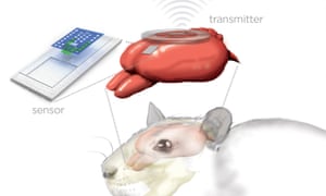
Artist’s rendition of the sensor and wireless transmitter monitoring a rat’s brain. Image: Julie McMahon
Such devices are sometimes used for short periods of time, and often
are implanted permanently. But implantation always carries some risk –
the devices can be somewhat cumbersome and their wires and metallic
components are breeding grounds for bacteria, so the implantation site
can become infected. And removing the device, or replacing it if it
malfunctions, involves another surgical procedure – and more distress –
for the patient.
The new device, developed by a research team that includes engineers,
materials scientists, and neurosurgeons in America and South Korea, and
described in the journal Nature, could potentially overcome
these limitations. It consists of a pressure and temperature sensor,
each one smaller than a grain of rice, integrated on a biodegradable
silicon chip that sits on the surface of the brain, and connected to a
wireless transmitter attached to the outside of the skull.
The researchers tested the device in rats, and showed that it can
monitor intracranial pressure, and the temperature changes that occur as
the rats drift in and out of consciousness following administration of
an anaesthetic, at least as accurately as existing devices. But this
device is unique because its components are made from so-called “green electronics”
– natural materials that are fully biodegradable and biocompatible,
which are designed to work for a few weeks, and then completely
dissolve, over the course of about a day, when immersed in watery fluids
such as cerebrospinal fluid.
When they examined the brain tissue afterwards, the researchers found no indication of an inflammatory response,
or of scarring around the implantation site, confirming that the device
is fully biocompatible. They then modified the device to show that it
can also be used to take the same measurements from sites about 5mm
below the surface of the rat brain.
The researchers say the device can easily be modified in other ways
to monitor other important physiological parameters of brain function,
such as acidity and the motion of fluids. It could also be used to
deliver drugs to the brain, and, with the incorporation of
microelectrodes, to stimulate or record neuronal activity.
As well as being fully biocompatible – and, therefore, safer – the
fabrication process is also cheaper and more environmentally-friendly
than that used for existing technologies, and the researchers are now
aiming to test it in human clinical trials.
Reference
Kang, S. -K., et al. (2016). Bioresorbable silicon electronic sensors for the brain. Nature, DOI: 10.1038/nature16492 [Abstract]
No comments:
Post a Comment