Changing stroke rehab and research worldwide now.Time is Brain! trillions and trillions of neurons that DIE each day because there are NO effective hyperacute therapies besides tPA(only 12% effective). I have 523 posts on hyperacute therapy, enough for researchers to spend decades proving them out. These are my personal ideas and blog on stroke rehabilitation and stroke research. Do not attempt any of these without checking with your medical provider. Unless you join me in agitating, when you need these therapies they won't be there.

What this blog is for:

My blog is not to help survivors recover, it is to have the 10 million yearly stroke survivors light fires underneath their doctors, stroke hospitals and stroke researchers to get stroke solved. 100% recovery. The stroke medical world is completely failing at that goal, they don't even have it as a goal. Shortly after getting out of the hospital and getting NO information on the process or protocols of stroke rehabilitation and recovery I started searching on the internet and found that no other survivor received useful information. This is an attempt to cover all stroke rehabilitation information that should be readily available to survivors so they can talk with informed knowledge to their medical staff. It lays out what needs to be done to get stroke survivors closer to 100% recovery. It's quite disgusting that this information is not available from every stroke association and doctors group.

Showing posts with label artificial brains. Show all posts

Sunday, February 4, 2024

Revolutionary 3D-Printed Brain Tissue Mimics Human Function

Which built brain should our researchers be using?. I expect our researchers to be using the best one.

Nearly complete human brain grown in US lab: scientist August 2015

Multiregional brain on a chip  Jan 2017
Draper Laboratory developing “Brain-on-a-Chip”  October 2012

"Alzheimer's-in-a-Dish" Docs Win Top Smithsonian Ingenuity Award Nov. 2015 

A patient’s budding cortex — in a dish?  June 2015 


,Cell cultures in petri dishes open new doors to brain research  April 2017

Scientists create 3D-printed brain-like tissue from stem cells July 2017

3D Mini-Brains Accelerate Research for Repairing Brain Function December 2017

 The latest here:

Revolutionary 3D-Printed Brain Tissue Mimics Human Function

Summary: Researchers developed the world’s first 3D-printed brain tissue that grows and behaves similarly to natural brain tissue, marking a significant leap forward for neurological and neurodevelopmental disorder research.

This novel 3D-printing technique uses a horizontal layering approach and a softer bio-ink, allowing neurons to interconnect and form networks akin to human brain structures.

The ability to precisely control cell types and arrangements provides unparalleled opportunities to study brain functions and disorders in a controlled environment, offering new avenues for drug testing and understanding brain development and diseases like Alzheimer’s and Parkinson’s.

Key Facts:

  1. The 3D-printed brain tissue can form networks and communicate through neurotransmitters, similar to human brain interactions.
  2. This new printing method allows for precise control over cell types and arrangements, surpassing the capabilities of traditional brain organoids.
  3. The technique is accessible to many labs, not requiring special equipment or culture methods, and can significantly impact the study of various neurological conditions and treatments.

Source: University of Wisconsin

A team of University of Wisconsin–Madison scientists has developed the first 3D-printed brain tissue that can grow and function like typical brain tissue.

It’s an achievement with important implications for scientists studying the brain and working on treatments for a broad range of neurological and neurodevelopmental disorders, such as Alzheimer’s and Parkinson’s disease.

“This could be a hugely powerful model to help us understand how brain cells and parts of the brain communicate in humans,” says Su-Chun Zhang, professor of neuroscience and neurology at UW–Madison’s Waisman Center.
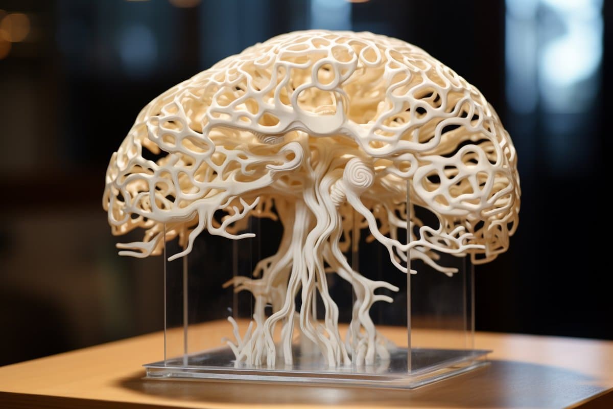
This shows a brain.
“Our tissue stays relatively thin and this makes it easy for the neurons to get enough oxygen and enough nutrients from the growth media,” Yan says. Credit: Neuroscience News

“It could change the way we look at stem cell biology, neuroscience, and the pathogenesis of many neurological and psychiatric disorders.”

Printing methods have limited the success of previous attempts to print brain tissue, according to Zhang and Yuanwei Yan, a scientist in Zhang’s lab. The group behind the new 3D-printing process described their method today in the journal Cell Stem Cell.

Instead of using the traditional 3D-printing approach, stacking layers vertically, the researchers went horizontally. They situated brain cells, neurons grown from induced pluripotent stem cells, in a softer “bio-ink” gel than previous attempts had employed.

“The tissue still has enough structure to hold together but it is soft enough to allow the neurons to grow into each other and start talking to each other,” Zhang says.

The cells are laid next to each other like pencils laid next to each other on a tabletop.

“Our tissue stays relatively thin and this makes it easy for the neurons to get enough oxygen and enough nutrients from the growth media,” Yan says.

The results speak for themselves — which is to say, the cells can speak to each other. The printed cells reach through the medium to form connections inside each printed layer as well as across layers, forming networks comparable to human brains.

The neurons communicate, send signals, interact with each other through neurotransmitters, and even form proper networks with support cells that were added to the printed tissue.

“We printed the cerebral cortex and the striatum and what we found was quite striking,” Zhang says. “Even when we printed different cells belonging to different parts of the brain, they were still able to talk to each other in a very special and specific way.”

The printing technique offers precision — control over the types and arrangement of cells — not found in brain organoids, miniature organs used to study brains. The organoids grow with less organization and control.

“Our lab is very special in that we are able to produce pretty much any type of neurons at any time. Then we can piece them together at almost any time and in whatever way we like,” Zhang says.

“Because we can print the tissue by design, we can have a defined system to look at how our human brain network operates. We can look very specifically at how the nerve cells talk to each other under certain conditions because we can print exactly what we want.”

That specificity provides flexibility. The printed brain tissue could be used to study signaling between cells in Down syndrome, interactions between healthy tissue and neighboring tissue affected by Alzheimer’s, testing new drug candidates, or even watching the brain grow.

“In the past, we have often looked at one thing at a time, which means we often miss some critical components. Our brain operates in networks. We want to print brain tissue this way because cells do not operate by themselves. They talk to each other. This is how our brain works and it has to be studied all together like this to truly understand it,” Zhang says.

“Our brain tissue could be used to study almost every major aspect of what many people at the Waisman Center are working on. It can be used to look at the molecular mechanisms underlying brain development, human development, developmental disabilities, neurodegenerative disorders, and more.”

The new printing technique should also be accessible to many labs. It does not require special bio-printing equipment or culturing methods to keep the tissue healthy, and can be studied in depth with microscopes, standard imaging techniques and electrodes already common in the field.

The researchers would like to explore the potential of specialization, though, further improving their bio-ink and refining their equipment to allow for specific orientations of cells within their printed tissue..

“Right now, our printer is a benchtop commercialized one,” Yan says. “We can make some specialized improvements to help us print specific types of brain tissue on-demand.”

Funding: This study was supported in part by NIH-NINDS (NS096282, NS076352, NS086604), NICHD (HD106197, HD090256), the National Medical Research Council of Singapore (MOH-000212, MOH-000207), Ministry of Education of Singapore (MOE2018-T2-2-103), Aligning Science Across Parkinson’s (ASAP-000301), the Bleser Family Foundation, and the Busta Foundation.

About this neurotech research news

Author: Emily Leclerc
Source: University of Wisconsin
Contact: Emily Leclerc – University of Wisconsin
Image: The image is credited to Neuroscience News

Original Research: Open access.
3D bioprinting of human neural tissues with functional connectivity” by Su-Chun Zhang et al. Cell Stem Cell


Abstract

3D bioprinting of human neural tissues with functional connectivity

Highlights

  • Functional human neural tissues assembled by 3D bioprinting
  • Neural circuits formed between defined neural subtypes
  • Functional connections established between cortical-striatal tissues
  • Printed tissues for modeling neural network impairment

Summary

Probing how human neural networks operate is hindered by the lack of reliable human neural tissues amenable to the dynamic functional assessment of neural circuits. We developed a 3D bioprinting platform to assemble tissues with defined human neural cell types in a desired dimension using a commercial bioprinter.

The printed neuronal progenitors differentiate into neurons and form functional neural circuits within and between tissue layers with specificity within weeks, evidenced by the cortical-to-striatal projection, spontaneous synaptic currents, and synaptic response to neuronal excitation.

Printed astrocyte progenitors develop into mature astrocytes with elaborated processes and form functional neuron-astrocyte networks, indicated by calcium flux and glutamate uptake in response to neuronal excitation under physiological and pathological conditions.

These designed human neural tissues will likely be useful for understanding the wiring of human neural networks, modeling pathological processes, and serving as platforms for drug testing.

Thursday, December 7, 2017

3D Mini-Brains Accelerate Research for Repairing Brain Function

Which built brain should our researchers be using?. I expect our researchers to be using the best one.

Nearly complete human brain grown in US lab: scientist.

Multiregional brain on a chip  Jan 2017
Draper Laboratory developing “Brain-on-a-Chip”  October 2012

"Alzheimer's-in-a-Dish" Docs Win Top Smithsonian Ingenuity Award Nov. 2015 

A patient’s budding cortex — in a dish?  June 2015 


,Cell cultures in petri dishes open new doors to brain research  April 2017

Scientists create 3D-printed brain-like tissue from stem cells

 The latest here:


3D Mini-Brains Accelerate Research for Repairing Brain Function

 The Houston Methodist Research Institute is making mini brains from human stem cells that put researchers on a fast track to repair the nervous system after injury or disease of the brain and spinal cord.
Houston Methodist neuroscientist Robert Krencik, Ph.D., and his team have developed a new system to reduce the time it takes to grow these brain models, which will give them the ability to screen drugs and study what's behind disease-causing mutations more quickly. Their findings are described in an article titled "Systematic three-dimensional coculture rapidly recapitulates interactions between human neurons and astrocytes," in the Dec. 12 issue of Stem Cell Reports.
"We always felt like what we were doing in the lab was not precisely modeling how the cells act within the human brain," Krencik said. "So, for the first time, when we put these cells together systematically, they dramatically changed their morphological complexity, size and shape. They look like cells as you would see them within the human brain, so now we can study cells in the lab in a more natural environment."
And why is this important? Krencik says cells grown in traditional lab cultures are put on a flat petri dish, broken up and otherwise manipulated, disturbing their interactions. This results in not being able to reproduce the form, structure and developmental growth of the brain's cells in the lab, leading to very simplistic-looking and immature cells. In the human brain, however, these cells are very complex-looking and interact in intricate ways with each other and the environment. New technologies are now focused on 3-D culture systems, but the exhaustive time for these studies is not feasible for accelerating discoveries. "Normally, growing these 3-D mini brains takes months and years to develop," Krencik said. "We have new techniques to pre-mature the cells separately and then combine them, and we found that within a few weeks they're able to form mature interactions with each other. So, the length of time to get to that endpoint for studies is dramatically reduced with our system." Krencik's lab focused on a star-shaped cell type called astrocytes, because they are a key factor in getting the brain's neurons to connect and talk to each other by helping to increase the number and strength of neuronal connections in the brain and spinal cord. They are involved in most neural diseases and also are responsible for maintaining a healthy nervous system. With the model Krencik's team bioengineered, the incorporation of astrocytes accelerated the connections of the surrounding neurons. Krencik's group is the first to specifically engineer astrocytes into these 3-D mini brains. By doing so, this led to the accelerated maturation of both the astrocytes and the surrounding neurons. Introducing them for the first time in this paper, he coined these bioengineered mini brains "asteroids" to distinguish them from other types of 3-D sphere cultures, known as organoids. Krencik's "asteroids" contain specific populations of astrocytes, whereas organoids have undefined numbers and types of cells. "Using our system, we can generate mature astrocytes and have them interact intimately with neurons to a greater extent than has been done before," Krencik said. "Unlike other cells in the brain and in the rest of the body, astrocytes have unique properties in humans. It's thought they are partly responsible for the unique cognitive functions of humans and also may underlie aspects of human diseases, such as Alzheimer's and autism spectrum disorders." Ultimately, Krencik is using these "asteroids" to form functional neural circuits that researchers can experimentally manipulate for developing treatments and deciphering what makes diseases tick. Krencik says they can make induced pluripotent stem cells, commonly termed iPS cells, from any disease or patient and then form these mini brains to study the disease process, as well as screen therapeutic compounds on them to aid in the development of drugs. Within about five years, his goal is to use this system to develop clinical trials to improve or regenerate a person's impaired nervous system.

Thursday, December 15, 2016

A European university just landed $3.3M to 3D print artificial brains for research

By matching this up with objective 3d scans of dead and damaged areas we will finally be able to show survivors exactly the extent of their damage. With that knowledge we can start mapping stroke protocols to  recovery from such damage. I'm sure this will take years but we have to start somewhere other than the crap of awareness, prevention and F.A.S.T.
http://www.digitaltrends.com/cool-tech/meso-brain-project/?utm_source=Sailthru&utm_medium=email&


Meso-Brain sounds a bit like a soup you really don’t want to order. In fact, it’s an intriguing new project taking place at Aston University in the U.K., which just received 3.3 million euros in funding from the European Commission’s Future and Emerging Technology (FET) scheme.
Bringing together top researchers in the fields of photonics, imaging, biology, and neuroscience, the goal of the innovative project is to attempt to replicate the brain’s neural structures using 3D nanoprinting technology.
“The Meso-Brain project will use recently-developed 3D laser nano-printing technology to support one of the major aims of neuroscience: to form neural networks with specific biological architectures,” Professor Edik Rafailov told Digital Trends. “If we would be able to use 3D nano-printing to improve the connection of neurons in an area of the brain which has been damaged, we will be in a position to develop much more effective ways to treat those with dementia or brain injuries.”
More: Brain scanner can tell what emotions you are feeling when your mind wanders
The core technology involved with the project involves pluripotent stem cells, which are cells taken from tissues and then genetically modified to behave like an embryonic stem cell. These cells can then be 3D-printed to create precise neural networks according to brain architectures. One day, the results could help repair damaged parts of the brain in those individuals suffering from different types of brain trauma or neurological disorder.
In addition to Aston University, Meso-Brain will also boast contributions from Axol Bioscience Ltd., Laser Zentrum Hannover, The Institute of Photonic Sciences, the University of Barcelona, and Kite Innovations.
Meso-Brain isn’t the only high-profile, publicly funded project designed to reverse-engineer the human brain in recent times. There has also been the United States’ MICrONS project (dedicated to building more biofidelic algorithms) and the European Commission’s Human Brain Project (intended to build a complete computer simulation of the human brain over a ten-year period), among others.
Taken together, we should be looking for some invaluable brain-related insights over the coming years.

Tuesday, February 17, 2015

Artificial Brain Edges Closer to Reality

I'm sure we'll all be dead by the time this is far enough along to help us. But tweak your doctors and ask when this will become available. 

Artificial Brain Edges Closer to Reality