Changing stroke rehab and research worldwide now.Time is Brain! trillions and trillions of neurons that DIE each day because there are NO effective hyperacute therapies besides tPA(only 12% effective). I have 523 posts on hyperacute therapy, enough for researchers to spend decades proving them out. These are my personal ideas and blog on stroke rehabilitation and stroke research. Do not attempt any of these without checking with your medical provider. Unless you join me in agitating, when you need these therapies they won't be there.

What this blog is for:

My blog is not to help survivors recover, it is to have the 10 million yearly stroke survivors light fires underneath their doctors, stroke hospitals and stroke researchers to get stroke solved. 100% recovery. The stroke medical world is completely failing at that goal, they don't even have it as a goal. Shortly after getting out of the hospital and getting NO information on the process or protocols of stroke rehabilitation and recovery I started searching on the internet and found that no other survivor received useful information. This is an attempt to cover all stroke rehabilitation information that should be readily available to survivors so they can talk with informed knowledge to their medical staff. It lays out what needs to be done to get stroke survivors closer to 100% recovery. It's quite disgusting that this information is not available from every stroke association and doctors group.

Showing posts with label wireless. Show all posts

Thursday, January 21, 2021

Nonsurgical treatment for cerebral infarction using wearable wireless ultrasound devices

 Now we just need our stroke doctors and hospitals to contact researchers and get this tested in humans. Only 7 days of intervention to get to mostly a normal level. Does your stroke hospital even follow stroke research?

Nonsurgical treatment for cerebral infarction using wearable wireless ultrasound devices
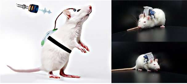
Photographs of animal model using the developed wireless wearable brain stimulation system. Credit: Korea Institute of Science and Technology (KIST)

Cerebral infarction, commonly known as ischemic stroke, has a high mortality rate and causes severe damage to nervous cells in the brain owing to the loss of oxygen, which results in limiting body movements. Several technologies, including physiotherapy and brain stimulation techniques, are being developed and tested for the rehabilitation of brain nervous cells damaged by a stroke. In particular, low-intensity focused ultrasound is expected to be effective for rehabilitating neurological diseases such as stroke, as it can excite or inhibit nerve cells by delivering mechanical energy with high precision at the desired position, while ultrasound is penetrating the cranium without requiring a surgical operation.

Korea Institute of Science and Technology (KIST) announced that the research team at the Center for Bionics, led by Dr. Kim Hyungmin, developed a wireless rehabilitation treatment technology for brain nervous system damaged by a stroke by fabricating a wearable, wireless low-intensity focused ultrasound brain stimulator. Additionally, to verify its effectiveness, this brain stimulator was applied to animal models of stroke.

In 2018, the research team developed a treatment method in which a low-intensity focused ultrasound was applied to the cerebellar core to recover that deteriorated owing to hemiplegia, and in 2020, they proved that the low-intensity focused ultrasound applied to the cerebellar core after a stroke is closely related to the changes in a delta wave in the brain.

To harness this technology efficiently in rehabilitating stroke patients in various environments, the ultrasound device should be easy to be used in everyday life. However, the existing brain stimulator is heavy and fixed, and is therefore only applicable to experiments involving anesthetized or restrained animals, thereby making it difficult to be commercialized.
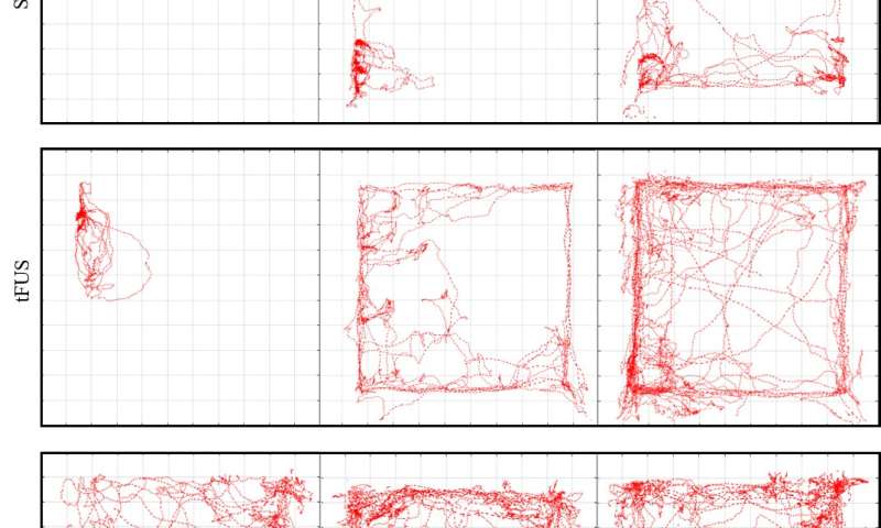
Nonsurgical treatment for cerebral infarction using wearable wireless ultrasound devices

Representative trajectories for stroke model rats treated with sham and tFUS, compared to a healthy rat (bottom row) Credit: Korea Institute of Science and Technology (KIST)

Based on the findings of previous studies, the research team at KIST developed a wearable light-weight brain stimulator, which weighs approximately 20 g, that can be wirelessly controlled. In addition, they verified the rehabilitation effectiveness after assessing the ability of a rat stroke model with the device. When ultrasound was applied with a pressure of approximately 426 kPa to the part of the brain that controls motor functions in a rat stroke model, the motor functions were significantly improved after three days compared to the rat stroke model to which ultrasound was not applied; the motor functions were improved to a level similar to that of normal rats after seven days of rehabilitation.

The brain stimulator developed by Dr. Kim's research team is light in weight and operates wirelessly, which does not limit the physical movements of a body as it continues stimulating the part of the brain that controls motor functions via ultrasound, thereby restoring the nervous system damaged by a . The results obtained from the study are expected to soon contribute to the development of a wearable ultrasound brain stimulation technology at a clinical level.

According to Dr. Kim Hyungmin at KIST, "Recently, the wearable stimulation technology is advancing rapidly, and the significance of this study is in verifying the possibility of non-invasive neurorehabilitation therapy using a wearable device," and he added that "In order to ensure the safety and effectiveness of the treatement, further research is required to optimize stimulation protocol by determining the molecular and cellular mechanisms of ultrasonic neuromodulation."


Explore further

Wearable brain stimulation could safely improve motor function after stroke

More information: Evgenii Kim et al, Wearable transcranial ultrasound system for remote stimulation of freely moving animal, IEEE Transactions on Biomedical Engineering (2020). DOI: 10.1109/TBME.2020.3038018
Provided by National Research Council of Science & Technology
 

Tuesday, January 19, 2016

Dissolvable wireless sensors monitor brain injury

Which brain monitoring device is your stroke medical professionals using to map your damage and listen in on  neuronal communications? ANYTHING AT ALL? 

New invasive imaging technique to monitor brain function


A Brain-Recording Device that Melts into Place 

Ultra-flexible, thin-film electrode arrays for chronic neural recording and stimulation of brain cavity wall

New technology facilitates studies of brain cells in stroke

Nanowire Tetrodes

First signals from brain nerve cells with ultrathin nanowires


Three-dimensional macroporous nanoelectronic networks as minimally invasive brain probes


 The latest here:


Dissolvable wireless sensors monitor brain injury


An international team of researchers has developed a miniaturized wireless electronic device that can monitor temperature and pressure when implanted into the brains of mice, and then dissolve to be naturally resorbed into the soft tissue once they are no longer needed.
Electronic implants are used widely in the treatment of numerous medical conditions, ranging from pacemakers and defibrillators given to cardiac patients, electrode arrays used for deep brain stimulation in patients with Parkinson’s Disease, and devices used to monitor intracranial temperature and pressure inside the skulls of people with severe traumatic brain injuries.
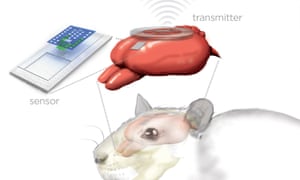
Artist’s rendition of the sensor and wireless transmitter monitoring a rat’s brain.
Pinterest
Artist’s rendition of the sensor and wireless transmitter monitoring a rat’s brain. Image: Julie McMahon
Such devices are sometimes used for short periods of time, and often are implanted permanently. But implantation always carries some risk – the devices can be somewhat cumbersome and their wires and metallic components are breeding grounds for bacteria, so the implantation site can become infected. And removing the device, or replacing it if it malfunctions, involves another surgical procedure – and more distress – for the patient.
The new device, developed by a research team that includes engineers, materials scientists, and neurosurgeons in America and South Korea, and described in the journal Nature, could potentially overcome these limitations. It consists of a pressure and temperature sensor, each one smaller than a grain of rice, integrated on a biodegradable silicon chip that sits on the surface of the brain, and connected to a wireless transmitter attached to the outside of the skull.
The researchers tested the device in rats, and showed that it can monitor intracranial pressure, and the temperature changes that occur as the rats drift in and out of consciousness following administration of an anaesthetic, at least as accurately as existing devices. But this device is unique because its components are made from so-called “green electronics” – natural materials that are fully biodegradable and biocompatible, which are designed to work for a few weeks, and then completely dissolve, over the course of about a day, when immersed in watery fluids such as cerebrospinal fluid.
When they examined the brain tissue afterwards, the researchers found no indication of an inflammatory response, or of scarring around the implantation site, confirming that the device is fully biocompatible. They then modified the device to show that it can also be used to take the same measurements from sites about 5mm below the surface of the rat brain.
The researchers say the device can easily be modified in other ways to monitor other important physiological parameters of brain function, such as acidity and the motion of fluids. It could also be used to deliver drugs to the brain, and, with the incorporation of microelectrodes, to stimulate or record neuronal activity.
As well as being fully biocompatible – and, therefore, safer – the fabrication process is also cheaper and more environmentally-friendly than that used for existing technologies, and the researchers are now aiming to test it in human clinical trials.

Reference

Kang, S. -K., et al. (2016). Bioresorbable silicon electronic sensors for the brain. Nature, DOI: 10.1038/nature16492 [Abstract]

Saturday, April 18, 2015

Walking Quality During Inpatient Stroke Rehabilitation Assessed by Wireless Sensing

With this your PT could objectively determine your specific walking problems and use specific muscle stroke protocols to correct those problems. Instead of the crappy 'Walk this way' demonstration that I got. That was totally useless. And it has the fairly famous Bruce Dobkin.
http://www.neurology.org/content/84/14_Supplement/P5.176.short
  1. Bruce Dobkin3
  1. Neurology vol. 84 no. 14 Supplement P5.176

Abstract

OBJECTIVE: Demonstrate that the quality of gait, in addition to walking speed and distance, can be derived from inertial sensors worn at both ankles.  (better yet would be at the knees, toes and hips)
BACKGROUND: The international Stroke Inpatient Rehabilitation Reinforcement of ACTivity (SIRRACT) trial deployed wireless sensors and activity-recognition algorithms to monitor and provide feedback about the quantity of stroke patients’ daily walking activity (epub, Neurorehabil Neural Repair, 2014). This follow-up study aimed to characterize the quality of walking by calculating spatiotemporal gait metrics. 
DESIGN/METHODS: In SIRRACT, 135 trial participants wore sensors throughout the average 3-week inpatient rehabilitation admission. Stopwatch-timed 10-meter walks (performed weekly for system calibration) were selected for the current analysis. Individual steps were delimited by identifying heel strike and toe-off times. Metrics including gait cycle duration, symmetry of stance and swing times, and double-limb support time were calculated for both the paretic and non-paretic limbs. The magnitude of peak lower leg acceleration during the swing phase of gait was also calculated. 
RESULTS: Data from five trial participants are presented as exemplars. Walking speeds averaged 0.24 ± 0.14m/s on study entry and 0.81 ± 0.35m/s at the time of discharge. Gait cycle duration decreased during rehabilitation (median:334ms [211,853]), as did double-limb support time (median:188ms [72,681]). Stance and swing time asymmetries were present throughout rehabilitation. Peak swing-time acceleration of the paretic leg was greater at discharge (median:0.20g-units [0.15,0.31]). 
CONCLUSIONS: The laboratory-quality gait metrics calculated by our wireless sensor system were sensitive to functional improvements during a period of known clinical recovery. Commercial sensor systems, for which steps counts are inaccurate at the slow speeds typical of hemi-paretic walking, have difficulty producing outcomes related to motor control in persons disabled by neurologic disease. Measurement of the quantity and quality of movements performed during daily activities enables clinicians and researchers to supervise gait training and skills practice during rehabilitation.


Saturday, September 27, 2014

An International Randomized Clinical Trial of Activity Feedback During Inpatient Stroke Rehabilitation Enabled by Wireless Sensing

What a novel idea. Objective measurements of muscle activity. What took so fucking long to think of this?
Would this
3-D Body Suit Put to Use in Healthcare Research
have been even better?
Or this?
Rapid Rehab Smart Insole Will Train Athletes and Assist Rehab Patients
And Bruce Dobkin is known for this book:
'The Clinical Science of Neurologic Rehabilitation'.
 From this lack of using the most up-to-date technology for research our stroke researchers are failing us.

An International Randomized Clinical Trial of Activity Feedback During Inpatient Stroke Rehabilitation Enabled by Wireless Sensing


  1. Andrew K. Dorsch, MD1
  2. Seth Thomas1
  3. Xiaoyu Xu, PhD1
  4. William Kaiser, PhD1
  5. Bruce H. Dobkin, MD1
  6. on behalf of the SIRRACT investigators
  1. 1University of California, Los Angeles, CA, USA
  1. Bruce H. Dobkin, Department of Neurology, Geffen School of Medicine, University of California Los Angeles, RNRC, Room 1-129, 710 Westwood Plaza, Los Angeles, CA 90095, USA. Email: bdobkin@mednet.ucla.edu

Abstract

Background. Walking-related disability is the most frequent reason for inpatient stroke rehabilitation. Task-related practice is a critical component for improving patient outcomes.  

Objective. To test the feasibility of providing quantitative feedback about daily walking performance and motivating greater skills practice via remote sensing. 

Methods. In this phase III randomized, single blind clinical trial, patients participated in conventional therapies while wearing wireless sensors (triaxial accelerometers) at both ankles. Activity-recognition algorithms calculated the speed, distance, and duration of walking bouts. Three times a week, therapists provided either feedback about performance on a 10-meter walk (speed only) or walking speed feedback plus a review of walking activity recorded by the sensors (augmented). Primary outcomes at discharge included total daily walking time, derived from the sensors, and a timed 15-meter walk.  

Results. Sixteen rehabilitation centers in 11 countries enrolled 135 participants over 15 months. Sensors recorded more than 1800 days of therapy, 37 000 individual walking bouts, and 2.5 million steps. No significant differences were found between the 2 feedback groups in daily walking time (15.1 ± 13.1 vs 16.6 ± 14.3 minutes, P = .54) or 15-meter walking speed (0.93 ± 0.47 vs 0.91 ± 0.53 m/s, P = .96). Remarkably, 30% of participants decreased their total daily walking time over their rehabilitation stay.  

Conclusions. In this first trial of remote monitoring of inpatient stroke rehabilitation, augmented feedback beyond speed alone did not increase the time spent practicing or improve walking outcomes. Remarkably modest time was spent walking. Wireless sensing, however, allowed clinicians to audit skills practice and provided ground truth regarding changes in clinically important, mobility-related activities.

Monday, August 12, 2013

Mobile and wireless inertial sensor platform for motion capturing in stroke rehabilitation sessions

If your therapist isn't following this they should be reminded of keeping up with current research. It would be wonderful if we had objective analysis of our movement deficits. Then we could  have decent writeups of what works.
http://ieeexplore.ieee.org/xpl/login.jsp?tp=&arnumber=6576085&url=http%3A%2F%2Fieeexplore.ieee.org%2Fxpls%2Fabs_all.jsp%3Farnumber%3D6576085
Fully mobile and wireless motion capturing is a mandatory requirement for undisturbed and non-reactive analysis of human movements. Therefore, inertial sensor platforms are used in applications like analysis of training sessions in sports or rehabilitation, and allow non-restricted motion capturing. The computation of the required reliable orientation estimation based on the inertial sensor RAW data is a demanding computational task. Highly customized and thus low-power wearable computation platforms require low-level, platform independent communication protocols and connectivity. State-of-the-art small sized commercial inertial sensors either lack the availability of an open, platform independent protocol, wireless connectivity or extension interfaces for additional sensors. Therefore, a extensible, wireless inertial sensor called (IM)2SU∗, featuring onboard inertial sensor fusion, for use in home based stroke rehabilitation is proposed. To evaluate orientation estimation accuracy an optical system is used as golden reference. The proposed IMU provides high orientation estimation accuracy, low costs, a platform independent, wireless connection and extensibility.