Changing stroke rehab and research worldwide now.Time is Brain! trillions and trillions of neurons that DIE each day because there are NO effective hyperacute therapies besides tPA(only 12% effective). I have 523 posts on hyperacute therapy, enough for researchers to spend decades proving them out. These are my personal ideas and blog on stroke rehabilitation and stroke research. Do not attempt any of these without checking with your medical provider. Unless you join me in agitating, when you need these therapies they won't be there. Did your doctor somehow miss the recovery protocol that Pedro Bach-y-Rita used to recover fully back in 1958 with only a partial brain?

What this blog is for:

My blog is not to help survivors recover, it is to have the 10 million yearly stroke survivors light fires underneath their doctors, stroke hospitals and stroke researchers to get stroke solved. 100% recovery. The stroke medical world is completely failing at that goal, they don't even have it as a goal. Shortly after getting out of the hospital and getting NO information on the process or protocols of stroke rehabilitation and recovery I started searching on the internet and found that no other survivor received useful information. This is an attempt to cover all stroke rehabilitation information that should be readily available to survivors so they can talk with informed knowledge to their medical staff. It lays out what needs to be done to get stroke survivors closer to 100% recovery. It's quite disgusting that this information is not available from every stroke association and doctors group.

Showing posts with label nanoRobots. Show all posts

Wednesday, March 30, 2022

Miniature medical robots step out from sci-fi

Our stroke researchers should have been heavily involved in this for a decade.

 With NO LEADERSHIP IN STROKE, it takes forever to make research usable. That is a disgusting timeline. Every stroke 'leader' should be fired.

If we had any leadership at all in stroke, when these nanorobots were introduced we would have had drug delivery and roto-rooter abilities already accomplished.

Remote-Controlled Nanospears Will Attack Cancer Cells June 2018 

Or maybe this solution from March, 2015

Magnetic nanoparticles could stop blood clot-caused strokes

Or this from May, 2012

Future of med devices: Nanorobots in your blood stream


 The latest here:

Miniature medical robots step out from sci-fi


Cartoon of a blood vessel in which tiny ships head towards a malignant invader

Illustration: Jan Kallwejt

Cancer drugs usually take a scattergun approach. Chemotherapies inevitably hit healthy bystander cells while blasting tumours, sparking a slew of side effects. It is also a big ask for an anticancer drug to find and destroy an entire tumour — some are difficult to reach, or hard to penetrate once located.

A long-dreamed-of alternative is to inject a battalion of tiny robots into a person with cancer. These miniature machines could navigate directly to a tumour and smartly deploy a therapeutic payload right where it is needed. “It is very difficult for drugs to penetrate through biological barriers, such as the blood–brain barrier or mucus of the gut, but a microrobot can do that,” says Wei Gao, a medical engineer at the California Institute of Technology in Pasadena.
Among his inspirations is the 1966 film Fantastic Voyage, in which a miniaturized submarine goes on a mission to remove a blood clot in a scientist’s brain, piloted through the bloodstream by a similarly shrunken crew. Although most of the film remains firmly in the realm of science fiction, progress on miniature medical machines in the past ten years has seen experiments move into animals for the first time.

There are now numerous micrometre- and nanometre-scale robots that can propel themselves through biological media, such as the matrix between cells and the contents of the gastrointestinal tract. Some are moved and steered by outside forces, such as magnetic fields and ultrasound. Others are driven by onboard chemical engines, and some are even built on top of bacteria and human cells to take advantage of those cells’ inbuilt ability to get around. Whatever the source of propulsion, it is hoped that these tiny robots will be able to deliver therapies to places that a drug alone might not be able to reach, such as into the centre of solid tumours. However, even as those working on medical nano- and microrobots begin to collaborate more closely with clinicians, it is clear that the technology still has a long way to go on its fantastic journey towards the clinic.
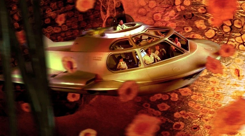
Film still of a tiny spaceship flying through the inside of a human body

In the 1966 film Fantastic Voyage, a miniaturized medical team goes on a mission to remove a blood clot in a scientist’s brain.Contributor: Collection Christophel/Alamy Stock Photo

Poetry in motion

One of the key challenges for a robot operating inside the human body is getting around. In Fantastic Voyage, the crew uses blood vessels to move through the body. However, it is here that reality must immediately diverge from fiction. “I love the movie,” says roboticist Bradley Nelson, gesturing to a copy of it in his office at the Swiss Federal Institute of Technology (ETH) Zurich in Switzerland. “But the physics are terrible.” Tiny robots would have severe difficulty swimming against the flow of blood, he says. Instead, they will initially be administered locally, then move towards their targets over short distances.

When it comes to design, size matters. “Propulsion through biological media becomes a lot easier as you get smaller, as below a micron bots slip between the network of macromolecules,” says Peer Fischer, a robotics researcher at the Max Planck Institute for Intelligent Systems in Stuttgart, Germany. Bots are therefore typically no more than 1–2 micrometres across. However, most do not fall below 300 nanometres. Beyond that size, it becomes more challenging to detect and track them in biological media, as well as more difficult to generate sufficient force to move them.

Scientists have several choices for how to get their bots moving. Some opt to provide power externally. For instance, in 2009, Fischer — who was working at Harvard University in Cambridge, Massachusetts, at the time, alongside fellow nanoroboticist Ambarish Ghosh — devised a glass propeller, just 1–2 micrometres in length, that could be rotated by a magnetic field1. This allowed the structure to move through water, and by adjusting the magnetic field, it could be steered with micrometre precision. In a 2018 study2, Fischer launched a swarm of micropropellers into a pig’s eye in vitro, and had them travel over centimetre distances through the gel-like vitreous humour into the retina — a rare demonstration of propulsion through real tissue. The swarm was able to slip through the network of biopolymers within the vitreous humour thanks in part to a silicone oil and fluorocarbon coating applied to each propeller. Inspired by the slippery surface that the carnivorous pitcher plant Nepenthes uses to catch insects, this minimized interactions between the micropropellers and biopolymers.
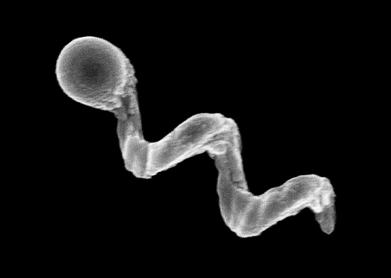
Extreme close-up of a nanopropeller

An electron microscope image of a glass nanopropeller.Credit: Conny Miksch, MPI-IS

Another way to provide propulsion from outside the body is to use ultrasound. One group placed magnetic cores inside the membranes of red blood cells3, which also carried photoreactive compounds and oxygen. The cells’ distinctive biconcave shape and greater density than other blood components allowed them to be propelled using ultrasonic energy, with an external magnetic field acting on the metallic core to provide steering. Once the bots are in position, light can excite the photosensitive compound, which transfers energy to the oxygen and generates reactive oxygen species to damage cancer cells.

This hijacking of cells is proving to have therapeutic merits in other research projects. Some of the most promising strategies aimed at treating solid tumours involve human cells and other single-celled organisms jazzed up with synthetic parts. In Germany, a group led by Oliver Schmidt, a nanoscientist at Chemnitz University of Technology, has designed a biohybrid robot based on sperm cells4. These are some of the fastest motile cells, capable of hitting speeds of 5 millimetres per minute, Schmidt says. The hope is that these powerful swimmers can be harnessed to deliver drugs to tumours in the female reproductive tract, guided by magnetic fields. Already, it has been shown that they can be magnetically guided to a model tumour in a dish.

Credit: Leibniz IFW, Dresden

“We could load anticancer drugs efficiently into the head of the sperm, into the DNA,” says Schmidt. “Then the sperm can fuse with other cells when it pushes against them.” At the Chinese University of Hong Kong, meanwhile, nanoroboticist Li Zhang led the creation of microswimmers from Spirulina microalgae cloaked in the mineral magnetite. The team then tracked a swarm of them inside rodent stomachs using magnetic resonance imaging5. The biohybrids were shown to selectively target cancer cells. They also gradually degrade, reducing unwanted toxicity.

Another way to get micro- and nanobots moving is to fit them with a chemical engine: a catalyst drives a chemical reaction, creating a gradient on one side of the machine to generate propulsion. Samuel Sánchez, a chemist at the Institute for Bioengineering of Catalonia in Barcelona, Spain, is developing nanomotors driven by chemical reactions for use in treating bladder cancer. Some early devices relied on hydrogen peroxide as a fuel. Its breakdown, promoted by platinum, generated water and oxygen gas bubbles for propulsion. But hydrogen peroxide is toxic to cells even in minuscule amounts, so Sánchez has transitioned towards safer materials. His latest nanomotors are made up of honeycombed silica nanoparticles, tiny gold particles and the enzyme urease6. These 300–400-nm bots are driven forwards by the chemical breakdown of urea in the bladder into carbon dioxide and ammonia, and have been tested in the bladders of mice. “We can now move them and see them inside a living system,” says Sánchez.

Breaking through

A standard treatment for bladder cancer is surgery, followed by immunotherapy in the form of an infusion of a weakened strain of Mycobacterium bovis bacteria into the bladder, to prevent recurrence. The bacterium activates the person’s immune system, and is also the basis of the BCG vaccine for tuberculosis. “The clinicians tell us that this is one of the few things that has not changed over the past 60 years,” says Sánchez. There is a need to improve on BCG in oncology, according to his collaborator, urologic oncologist Antoni Vilaseca at the Hospital Clinic of Barcelona. Current treatments reduce recurrences and progression, “but we have not improved survival”, Vilaseca says. “Our patients are still dying.”

The nanobot approach that Sánchez is trying promises precision delivery. He plans to insert his bots into the bladder (or intravenously), to motor towards the cancer with their cargo of therapeutic agents to target cancer cells, using abundant urea as a fuel. He might use a magnetic field for guidance, if needed, but a more straightforward replacement of BCG with bots that do not require external control, perhaps using an antibody to bind a tumour marker, would please clinicians most. “If we can deliver our treatment to the tumour cells only, then we can reduce side effects and increase activity,” says Vilaseca.
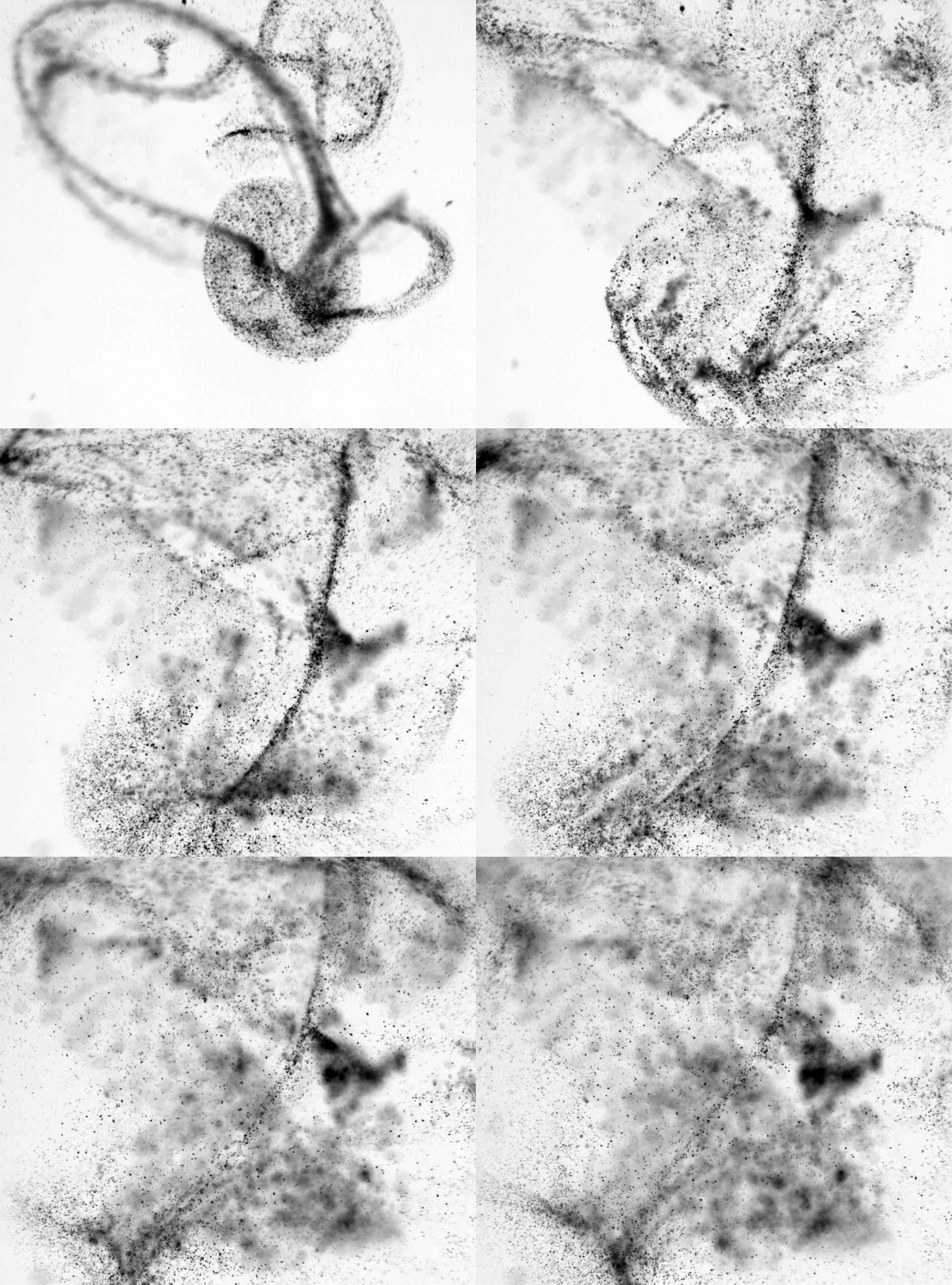
Close-up of urease-powered nanomotors

An optical microscopy video showing a swarm of urease-powered nanomotors swimming in urea solution.Credit: Samuel Sánchez Ordóñez

Not all cancers can be reached by swimming through liquid, however. Natural physiological barriers can block efficient drug delivery. The gut wall, for example, allows absorption of nutrients into the bloodstream, and offers an avenue for getting therapies into bodies. “The gastrointestinal tract is the gateway to our body,” says Joseph Wang, a nanoengineer at the University of California, San Diego. However, a combination of cells, microbes and mucus stops many particles from accessing the rest of the body. To deliver some therapies, simply being in the intestine isn’t enough — they also need to be able to burrow through its defences to reach the bloodstream, and a nanomachine could help with this.

In 2015, Wang and his colleagues, including Gao, reported the first self-propelled robot in vivo, inside a mouse stomach7. Their zinc-based nanomotor dissolved in the harsh stomach acids, producing hydrogen bubbles that rocketed the robot forwards. In the lower gastrointestinal tract, they instead use magnesium. “Magnesium reacts with water to give a hydrogen bubble,” says Wang. In either case, the metal micromotors are encapsulated in a coating that dissolves at the right location, freeing the micromotor to propel the bot into the mucous wall.

Some bacteria have already worked out their own ways to sneak through the gut wall. Helicobacter pylori, which causes inflammation in the stomach, excretes urease enzymes to generate ammonia and liquefy the thick mucous that lines the stomach wall. Fischer envisages future micro- and nanorobots borrowing this approach to deliver drugs through the gut.

Solid tumours are another difficult place to deliver a drug. As these malignancies develop, a ravenous hunger for oxygen promotes an outside surface covered with blood vessels, while an oxygen-deprived core builds up within. Low oxygen levels force cells deep inside to switch to anaerobic metabolism and churn out lactic acid, creating acidic conditions. As the oxygen gradient builds, the tumour becomes increasingly difficult to penetrate. Nanoparticle drugs lack a force with which to muscle through a tumour’s fortifications, and typically less than 2% of them will make it inside8. Proponents of nanomachines think that they can do better.

Sylvain Martel, a nanoroboticist at Montreal Polytechnic in Canada, is trying to break into solid tumours using bacteria that naturally contain a chain of magnetic iron-oxide nanocrystals. In nature, these Magnetococcus species seek regions that have low oxygen. Martel has engineered such a bacterium to target active cancer cells deep inside tumours8. “We guide them with a magnetic field towards the tumour,” explains Martel, taking advantage of the magnetic crystals that the bacteria typically use like a compass for orientation. The precise locations of low-oxygen regions are uncertain even with imaging, but once these bacteria reach the right location, their autonomous capability kicks in and they motor towards low-oxygen regions. In a mouse, more than half the bacteria injected close to tumour grafts broke into this tumour region, each laden with dozens of drug-loaded liposomes. Martel cautions, however, that there is still some way to go before the technology is proven safe and effective for treating people with cancer.

In the Netherlands, chemist Daniela Wilson at Radboud University in Nijmegen and colleagues have developed enzyme-driven nanomotors powered by DNA that might similarly be able to autonomously home in on tumour cells9. The motors navigate towards areas that are richer in DNA, such as tumour cells that undergoing apoptosis. “We want to create systems that are able to sense gradients by different endogenous fuels in the body,” Wilson says, suggesting that the higher levels of lactic acid or glucose typically found in tumours could also be used for targeting. Once in place, the autonomous bots seem to be picked up by cells more easily than passive particles are — perhaps because the bots push against cells.
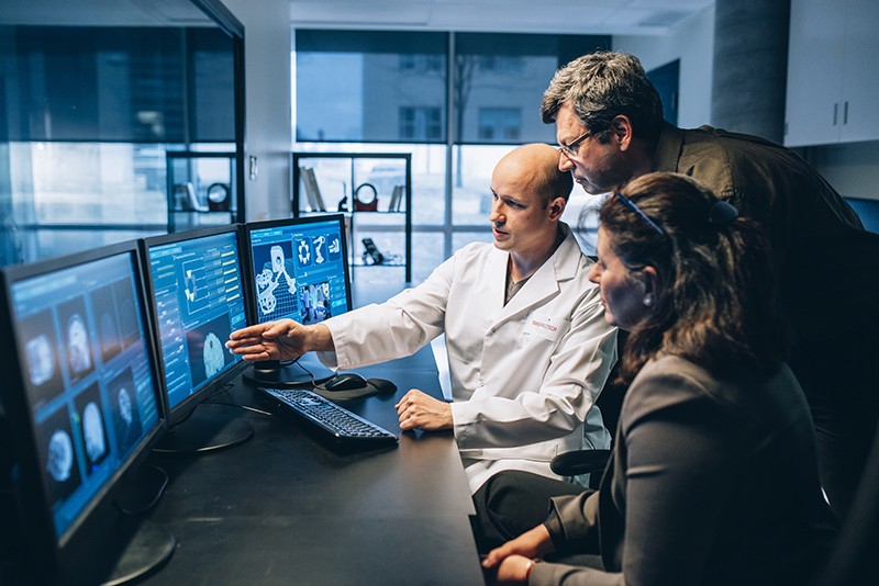
Sylvain Martel and his colleagues review information on a bank of computer screens

Nanoroboticist Sylvain Martel (middle) discusses a new computer interface with two members of his team.Credit: Caroline Perron

Fiction versus reality

Inspirational though Fantastic Voyage might have been for many working in the field of medical nanorobotics, there are some who think the film has become a burden. “People think of this as science fiction, which excites people, but on the other hand they don’t take it so seriously,” says Martel. Fischer is similarly jaded by movie-inspired hype. “People sometimes write very liberally as if nanobots for cancer treatment are almost here,” he says. “But this is not even in clinical trials right now.”

Nonetheless, advances in the past ten years have raised expectations of what is possible with current technology. “There’s nothing more fun than building a machine and watching it move. It’s a blast,” says Nelson. But having something wiggling under a microscope no longer has the same draw, without medical context. “You start thinking, ‘how could this benefit society?’” he says.

With this in mind, many researchers creating nanorobots for medical purposes are working more closely with clinicians than ever before. “You find a lot of young doctors who are really interested in what the new technologies can do,” Nelson says. Neurologist Philipp Gruber, who works with stroke patients at Aarau Cantonal Hospital in Switzerland, began a collaboration with Nelson two years ago after contacting ETH Zurich. The pair share an ambition to use steerable microbots to dissolve clots in people’s brains after ischaemic stroke — either mechanically, or by delivering a drug. “Brad knows everything about engineering,” says Gruber, “but we can advise about the problems we face in the clinic and the limitations of current treatment options.”

Sánchez tells a similar story: while he began talking to physicians around a decade ago, their interest has warmed considerably since his experiments in animals began three to four years ago. “We are still in the lab, but at least we are working with human cells and human organoids, which is a step forward,” says his collaborator Vilaseca.

As these seedlings of clinical collaborations take root, it is likely that oncology applications will be the earliest movers — particularly those that resemble current treatments, such as infusing microbots instead of BCG into cancerous bladders. But even these therapeutic uses are probably at least 7–10 years away. In the nearer term, there might be simpler tasks that nanobots can be used to accomplish, according to those who follow the field closely.

For example, Martin Pumera, a nanoroboticist at the University of Chemistry and Technology in Prague, is interested in improving dental care by landing nanobots beneath titanium tooth implants10. The tiny gap between the metal implants and gum tissue is an ideal niche for bacterial biofilms to form, triggering infection and inflammation. When this happens, the implant must often be removed, the area cleaned, and a new implant installed — an expensive and painful procedure. He is collaborating with dental surgeon Karel Klíma at Charles University in Prague.

Another problem the two are tackling is oral bacteria gaining access to tissue during surgery of the jaws and face. “A biofilm can establish very quickly, and that can mean removing titanium plates and screws after surgery, even before a fracture heals,” says Klíma. A titanium oxide robot could be administered to implants using a syringe, then activated chemically or with light to generate active oxygen species to kill the bacteria. Examples a few micrometres in length have so far been constructed, but much smaller bots — only a few hundred nanometres in length — are the ultimate aim.

Clearly, this is a long way from parachuting bots into hard-to-reach tumours deep inside a person. But the rising tide of in vivo experiments and the increasing involvement of clinicians suggests that microrobots might just be leaving port on their long journey towards the clinic.

doi: https://doi.org/10.1038/d41586-022-00859-0


Tuesday, May 11, 2021

The ANGIE project: a new research programme tasked with developing micro-robots for localised, targeted drug delivery to unblock blood vessels and fight stroke from within

 With NO LEADERSHIP IN STROKE, it takes almost 10 years to make research useable. That is a disgusting timeline. Every stroke 'leader' should be fired.

If we had any leadership at all in stroke, when these nanorobots were introduced we would have had drug delivery and roto-rooter abilities already accomplished.

Remote-Controlled Nanospears Will Attack Cancer Cells June 2018 

Or maybe this solution from March, 2015

Magnetic nanoparticles could stop blood clot-caused strokes

Or this from May, 2012

Future of med devices: Nanorobots in your blood stream

The latest here:

The ANGIE project: a new research programme tasked with developing micro-robots for localised, targeted drug delivery to unblock blood vessels and fight stroke from within

 his ground-breaking project, called ANGIE, is funded by the EU and aims to develop nano-surgeons that will enter the body to treat blood clots.

The ANGIE project will develop a radical, new technology for localised, targeted drug delivery based on steerable wireless nanodevices, capable of navigating the body vascular system to deliver drugs where no other instrument can go. ANGIE will offer health professionals vastly improved intervention capacity to tackle multiple chronic diseases and enable them to deliver drugs precisely where needed, with minimal side effects.

Navigating inside the body to treat injured tissues has fascinated scientists and the public for decades, but the required technologies have lagged far behind. Scientists in the ANGIE project believe that they are now in a position to make it happen.

The ANGIE project will be run by a leading group of scientists working in universities, research centres, and innovative companies across Belgium, Germany, Greece, Portugal, Spain, and Switzerland.

This is a much-needed development because stroke is the leading cause of adult physical disability in the world, affecting 17 million people worldwide each year.

Salvador Pane

Salvador Pane

“This could be averted if we had a way to wirelessly navigate nano-surgeons along the body’s vascular network to deliver drugs directly where needed – on command” said Dr. Salvador Pané, the coordinator of the project.

Achieving such a breakthrough will require advances in fields such as medical robotics, numerical simulation and biomaterials. Also, it needs technical capabilities and facilities that only few organisations have. But even those who do often lack the knowledge to transform scientific knowledge into medical instruments ready for clinical application.

The scientists at ANGIE are confident that their team includes all the required knowledge, experience, and infrastructure to make it happen. For many years they have been building the needed knowledge for the use and control of small-scale robotic systems for different health applications. They recently developed the first system to treat cardiac arrhythmias using electromagnetically steerable catheters and their group includes high-calibre scientists who are often sought for by the World Health Organization, the European Space Agency, and the World Economic Forum for advice. The project has already attracted attention from multinational corporations like Siemens and CLS Behring.

The ANGIE project started at the beginning of this year (2021) and its team will need four years to develop the basic foundations of the technology.

The Consortium consists of a group of four research institutions, three small-medium enterprises, and two non-profit organizations from across the EU.

SAFE is part of the consortium and is responsible for disseminating information about ANGIE to the stroke community.

The project is funded by the European Commission under the Grant Agreement number 952152.

www.h2020-angie.eu

Friday, March 5, 2021

Are Bionauts™ a Vessel to the Final Frontier of CNS Diseases?

If we had any leadership at all in stroke, when these nanorobots were introduced we would have had drug delivery and roto-rooter abilities already accomplished. Nothing will done for this in stroke, you're screwed.

Remote-Controlled Nanospears Will Attack Cancer Cells June 2018 

Or maybe this solution from March, 2015

Magnetic nanoparticles could stop blood clot-caused strokes

Or this from  May, 2012

Future of med devices: Nanorobots in your blood stream

The latest here; nothing will occur in any reasonable amount of time.

Are Bionauts™ a Vessel to the Final Frontier of CNS Diseases? 

Bionaut

 Close-up of a Bionaut™/Photo courtesy of Jon McKee Photography​

Diseases of the brain are often considered the holy grail of the therapeutic landscape – and Bionaut Labs, who today emerged from stealth mode in a $20 million Series A, is sending its Bionauts™ off in search of it. 

Bionauts™, a novel treatment modality that uses remote-controlled microscale robots to deliver biologics, nucleic acids, or small molecule therapies to precise areas of the brain, have the potential to boldly go where no therapy has gone before, opening up new pathways in the fight against devastating CNS disorders like brainstem glioma and Huntington’s disease.

“There was this film called Fantastic Voyage that actually has a submarine going into the brain and treating an aneurysm I think. What used to be science fiction now is really science reality, because, over the last few decades, science has allowed us to effectively manufacture micro-scale objects, and scale rapid prototyping capabilities,” said Bionaut Labs’ co-founder and chief executive officer, Michael Shpigelmacher.

Khosla Ventures, which led this round of financing, clearly agreed.

“We are thrilled to bring Bionaut Labs out of stealth mode as it typifies the type of new impactful technology companies we like to help build,” said Khosla founder, Vinod Khosla. “Bionauts™ hold great promise as a new targeted treatment modality for severe brain disorders for which there are few, if any, effective treatment options. Moreover, the broad therapeutic potential of Bionauts™ extends to many diseases where conventional therapies are limited or lacking.”

Joining Khosla in the syndicate were Upfront Ventures, Revolution, BOLD Capital and Compound.

A significant part of the Bionauts™’ potential lies in augmenting the efficacy of gene therapies, CRISPR, anti-sense oligonucleotides (ASOs), and oncolytic viruses.

“We bring into the picture the anatomical precision, getting into the right anatomical target, and then once we release the payload, it provides the cellular, or biochemical precision, in the right spot. And that is oftentimes the gap, that anatomical precision,” Shpigelmacher said.

Bionaut’s inaugural pursuit is glioma – a deadly malignant tumor found within the glial tissue of the nervous system that continues to confound scientists and surgeons alike. Shpigelmacher explained how the right systemic delivery mechanism could revolutionize the CNS treatment space.   

“All of the brain conditions, largely all of the CNS conditions, suffer from the fact that the brain and CNS is uniquely designed to protect itself from the external world, and that means that the blood-brain barrier prevents effective systemic treatment. Which means that if one can build a gateway to the CNS, you can treat many conditions potentially with one tool. That’s what we’re aiming to do,” he said.

Thus far, in animal studies, Bionaut has successfully treated orthotopic glioma tumors in mice, eliminating toxicity with guided delivery of therapeutic payloads directly into the tumor.  The company plans to conduct Good Laboratory Practice (GLP) studies in 2022, with the hope of reaching the clinic in 2023.

Once Bionaut has established the modality in glioma, it plans to embark on riskier therapeutic pursuits, such as Huntington’s disease.

“For Huntington’s…there are promising payloads, but they’re not at the point where they are clinically approved or well-characterized,” Shpigelmacher said. “Our going hypothesis is that instead of treating the entire CSF [Cerebrospinal fluid] volume around the whole brain and spine, or instead of doing an invasive procedure, we could deliver those payloads in a targeted fashion toward a specific location around the basal ganglia.”

Shpigelmacher predicted that micro-robots have the potential to change the way therapeutics are developed.

“Most drugs fail the FDA approval because of dosage problems because they can’t get the efficacy on target to be good enough while not being toxic off-target,” he said. “If you have a technology like Bionaut™ that allows you to engineer the therapeutic index to release a payload only where you need it, not in other places, then you can actually reduce the cost of your drug R&D and duration of your clinical trials because you don’t spend as much effort and resources on dosage optimization.”


Wednesday, February 19, 2020

Tiny robots with giant potential - TED talk

If your doctors and stroke hospital didn't immediately think of dozens of uses for this in stroke and contacted these guys for ways to improve stroke recovery, then you need to have them fired. Our fucking failures of stroke associations will do nothing with this. That is how useless stroke is in their names.

Tiny robots with giant potential - TED talk

Take a trip down the microworld as roboticists Paul McEuen and Marc Miskin explain how they design and mass-produce microrobots the size of a single cell, powered by atomically thin legs -- and show how these machines could one day be "piloted" to battle crop diseases or study your brain at the level of individual neurons.
This talk was presented at a TED Institute event given in partnership with The Kavli Foundation, the Simons Foundation and the National Academy of Sciences. TED editors featured it among our selections on the home page. Read more about the TED Institute.

Tuesday, June 12, 2018

Remote-Controlled Nanospears Will Attack Cancer Cells

With ANY innovative ideas or thinking at all from our fucking failures of stroke associations we could adapt this to ship tPA to the clot. But there seems to be not one working brain cell anywhere there.

Or maybe this solution from March, 2015

Magnetic nanoparticles could stop blood clot-caused strokes

Or this from  May, 2012

Future of med devices: Nanorobots in your blood stream


The fucking answers are out there, they just need to be implemented.


Remote-Controlled Nanospears Will Attack Cancer Cells


Delivering gene therapy exactly, precisely to where it needs to go has long been a struggle. But one group of researchers seems to have figured out exactly how: by shooting a nanospear within the body.
A recent study in the journal American Chemical Society Nano describes a method of creating nanospears 5,000 times thinner than human hair that successfully targeted, pierced, and entered a cancer cell within a lab dish. Unlike ancient spears that delivered injury or death, nanospears deliver the opposite—potentially life-saving genetic treatment.
Gene therapy research fuels the hope of eliminating many of the 6,000 inheritable diseases history has tallied. This developing array of discoveries may prove to be powerfully healing—but only if they can be placed at exact locations within the body.
Pinpoint delivery of genetic therapies has proven to be a significant challenge for science, however. The list of ways to miss targets is long. One way to think about how is drone-delivery shopping, where skeptics predict that mechanical failures will fill the air with falling packages. Thieves won’t need to steal, as misguided clothing, auto parts and pizza will be dropping from the sky. Similarly, some past efforts to deliver genetic treatment directly into diseased cells have missed their mark.
But nanospears might help with that particularly because they’re made from nickel, silicon, gold, and other components—materials to which the DNA used in some genetic therapies can attach.
Because nickel is magnetic, the nanospears were remote-controlled by magnets that pulled them toward the cancer cell. With DNA along for the ride, the nanospears’ direction was maneuvered by the researchers, like a spearfisher might calculate the path of a perfectly aimed harpoon.


“The capability to guide the nanospears magnetically eliminates the need for potentially harmful chemical propellants that have been used in prior examples of nanorobotic delivery,” authors Steven Jonas, Paul Weiss and Xiaobin Xu explained to the Daily Beast in an email. “A key feature of the nanospears is that we can guide them individually or in groups, like remote-controlled drones using a handheld or rotating magnet.”


These findings push remote-controlled innovations forward, changing how we think about “personalized medicine,” or tailoring treatment to each patient’s unique tapestry of genes.
“A key feature of the nanospears is that we can guide them individually or in groups, like remote-controlled drones using a handheld or rotating magnet.”
— researchers Steven Jonas, Paul Weiss and Xiaobin Xu
From this very early-stage laboratory work, the researchers hope to eventually bring the process to clinical trials for testing in humans. Because human genes are inherited, they control everything from disease tolerance to height and eyebrow shape. A gene that has become damaged is called a mutation. A mutation can cause a cell to grow uncontrollably, feeding cancer or other diseases.
Mutations can be inherited, or they can be caused by external factors like drinking toxic water, or by choices like smoking. If mutations could be repaired, disease could be stopped from forming in the first place.
Genetic therapies include repairing a gene so that it’s no longer a mutation, or replacing a mutation with a healthy gene. “Gene therapies might include repairing and/or removing defective genes by inserting a new copy of a normal gene to restore cellular function, or by correcting disease-causing mutations by editing the genome directly,” according to the UCLA researchers.
Inserting modified genes where they need to be—and not allowing them to float around just anywhere—is critical. “The safe, rapid, efficient, and cost-effective delivery of genes and gene-editing cargo to cells represents a tremendous barrier to making these types of gene therapies more accessible to patients,” the researchers noted.
Get The Beast In Your Inbox!

Daily Digest

Start and finish your day with the top stories from The Daily Beast.

Cheat Sheet

A speedy, smart summary of all the news you need to know (and nothing you don't).
By clicking “Subscribe,” you agree to have read the Terms of Use and Privacy Policy
Nanospears can be configured to carry a variety of biomolecular cargo for spear-tip delivery, Jonas, Weiss and Xu pointed out: “We envision that nucleic acids, proteins, small molecules, and combinations of these cargo can be packaged and can be delivered using the nanospears.”
Past attempts have included sending modified genes into cancer cells on the backs of viruses, such as cold viruses (which are “deactivated” so the cancer patient does not get a cold). The concept of using viruses came into use because viruses are experts at breaking into a cell and wreaking havoc. Breaking through cell membranes is the part of that process scientists aim to replicate—minus the viral illness part. “
raditional methods for gene delivery either use viruses as delivery vehicles or expose cells to electric fields and/or harsh chemical treatments,” the researchers explained. “To process the large number of cells required for gene therapies, these approaches are either expensive, slow, inefficient, harmful, or require specialized equipment.”
Nanospears have the potential for practicality because of their tiny size. “They are made using nanoscale fabrication methods originating from the semiconductor industry—the same processes used to make the chips that run your laptop or mobile phone—which are easily scalable for producing large numbers of nanospears. Millions of nanospears can be produced from a single silicon wafer at a price less than $500,” the researchers said.
And one more thing: Nanospears are also biodegradable, dissolving after dropping off their curative cargo. “The materials used to make the nanospears were selected by design to biodegrade safely,” the authors noted. “The nanospears are primarily made of silicon, which is biocompatible. The outer layers of nickel and gold are extremely thin, and are present in such small amounts that we have not observed and do not expect any harmful effects in the treated cells. We can also make the nanospears porous to speed up the degradation time, if that is advantageous.”

Monday, July 24, 2017

Why the Go-To Stroke Drug Can Fail

So with the 88% failure rate of tPA full recovery maybe other options exists. But since we have NO stroke leadership or strategy nothing will occur to solve this problem. It has been 21 years since this failure has been known, I don't expect anything to change for the next 21 years unless a lot of people die that are preventing new ideas from being considered.

  1. Magnetic nanoparticles to direct tPA to the clot, size of bolus could be reduced lowering bleeding risk.

  1. Future of med devices: Nanorobots in your blood stream

    Use them to deliver tPA or drill thru the clot.


Why the Go-To Stroke Drug Can Fail

 Twenty years ago stroke doctors celebrated the arrival of a powerful new weapon: the clot-clearing drug tPA. It was hailed as a lifesaver and has proved to be one for hundreds of thousands of patients since. TPA was the first and is still the only medicine approved by the U.S. Food and Drug Administration for treating strokes caused by clots that block blood flow to the brain. But like so many medical marvels, tPA (which stands for tissue plasminogen activator) has turned out to have serious limitations. It needs to be administered within three hours of symptom onset, does not last long in the body before it loses effectiveness, can cause uncontrolled bleeding and often fails to break up large clots.
For many of the nearly 800,000 Americans who every year suffer ischemic strokes, as the brain blockages are called, these shortcomings can be deadly. Nearly 130,000 die. Sadly, there have been no good alternatives to tPA since it debuted.
Recently doctors and scientists have broken this long-standing clinical stalemate with new tools to put a dent in those grim numbers. One innovation, a tiny wire device called a stent retriever, can be snaked up into the blood vessels leading to the brain to pull out large clots. “It's the first proven, effective treatment for acute stroke in a generation,” says Jeffrey Saver, director of the Stroke Center at the University of California, Los Angeles. Approved by the FDA in 2012, the stent retriever got a boost this year when the journal Stroke reported data showing many more patients treated with a retriever resumed normal life than did patients who received tPA. (The retriever manufacturer, Medtronic, provided support for the studies. Neurologist Bruce Campbell of the Royal Melbourne Hospital in Australia, who co-led the analysis, notes that Stroke has “strict independent-peer-review processes” to guard against bias.) Researchers are also developing better clot-detection scans, as well as a technique involving magnetism that guides tPA directly to the problem. This method could help eliminate dangerous obstructions elsewhere in the body, as well as in the brain.

Big Clots, Big Trouble

Of all of tPA's drawbacks, the most troublesome is its inadequacy against big clots, which can block large blood vessels at the base of the brain; they cause about 25 to 30 percent of all strokes. Although it works well against smaller clots in narrower vessels, a safe dose of the drug—which is delivered intravenously—often does not last long enough in the bloodstream to dissolve the big clots, and increasing the dose raises the risk of bleeding. “All you need to see is one intracranial bleed from tPA, and you realize you've got to pause before you give that medication,” says Thomas Maldonado, a clot specialist at New York University's Langone Medical Center.
This is where the stent retriever has an advantage. It is a narrow tube that can be threaded up from the femoral artery in the leg to the site of the clot. Then wire mesh on the end of the retriever, which expands like an accordion, is pushed into the clot. The mesh tendrils keep the clot from breaking apart in the brain—which could be deadly—and help separate it from blood vessel walls. The device is next pulled out of the body, and the clot comes with it. (In years past, doctors had tried a device with a corkscrew tip but found it was not as good at clearing the clot.)
Another advantage the device has over the drug is that the time window for the use of the stent retriever after symptoms arise is double that of tPA—six hours instead of three. The Stroke analysis found that blood flow in a vessel blocked by a large clot was successfully restored in 236 of 306 patients, or 77 percent, treated with the stent retriever. With tPA alone, the success rate was around 37 percent.
Like all surgical interventions, the stent retriever carries the risk of complications. The main one is bleeding, which is why patients with high blood pressure and the strained vessels that go with it may not be candidates for the procedure. “There's also a chance of the guide wire or some other manipulation of the device poking through the blood vessel during the procedure,” Saver says.
A much less common complication, Saver adds, is a piece of the clot breaking off as it is being pulled out, escaping into a new artery and causing a new stroke in a different area than the initial one. It happens in about 2 to 3 percent of cases, he says

Saturday, May 6, 2017

Wireless power can drive tiny electronic devices in the GI tract

Joining this with the  nanorobots we could listen in on neuroplasticity signals and duplicate the signals to make neuroplasticity completely repeatable. Also used to deliver stem cells or TPA to the correct locations. Doesn't anyone in stroke have two neurons they can rub together to get a spark of intelligence?

Future of med devices: Nanorobots in your blood stream


https://www.mdlinx.com/internal-medicine/medical-news-article/2017/04/28/gastrointestinal-tract-gastric-pacemakers-capsule-sized/7153487/?
Brigham and Women's Hospital
Imagers, gastric pacemakers and other diagnostic and therapeutic tools could someday transform the way diseases of the gastrointestinal tract are measured and treated. But in order for these electronic devices to work, they need a power source. Traditional power sources, such as batteries, can be incompatible with the mucosal lining of the gastrointestinal tract and have a limited lifespan within the body. A more promising possibility is to power electronic devices from outside the body.

In a new study published in the journal Scientific Reports, investigators from Brigham and Women’s Hospital, Massachusetts Institute of Technology and The Charles Stark Draper Laboratory report that an ingestible electronic capsule, complete with a capsule–sized antenna capable of receiving a radio signal wirelessly, can safely power a device in the gastrointestinal tract in preclinical models. The new work makes wireless medical electronics for treating the gastrointestinal tract one step closer to reality.

“Electronic devices that can be placed in the gastrointestinal tract for prolonged periods of time have the potential to transform how we evaluate and treat patients. This work describes the first example of remote, wireless transfer of power to a system in the stomach in a large preclinical animal model – a critical step toward bringing these devices into the clinic,” said co–corresponding author Carlo “Gio” Traverso, MD, PhD, a gastroenterologist and biomedical engineer at BWH.

Other medical devices – such as cochlear implants or neural probes – use a well–established technique known as near–field coupling to deliver power wirelessly. But ingestible devices must be small enough to be swallowed and, moreover, lie a significant distance from the surface of the body, making this technique unattainable for most gastrointestinal electronics. A new technique known as mid–field coupling provides an alternative way to deliver power to deeply implanted devices. Mid–field coupling operates at higher frequencies to deliver power two to three times more efficiently.

To test whether mid–field coupling could help deliver power from outside the body into the gastrointestinal tract, the research team designed antennas capable of operating efficiently in tissue. They then placed one antenna outside of the body and the other in the esophagus, stomach and colon of a swine model. They were able to transmit power levels of 37.5 uW, 123 uW and 173 uW, respectively, all of which are sufficient to wirelessly power a range of medical devices from outside of the body.

"We are very excited about this work which we feel can someday offer many new opportunities for oral drug delivery of different molecules," said co–corresponding author Robert Langer, Institute Professor from the Harvard–MIT Division of Health Sciences and Technology.

“In further work, we would like to expand on these measurements by characterizing the effects of animal size, antenna depth, orientation and more on transmission efficiency, and focus on propagating fields – or the way power travels – to make transmission even more efficient,” said Traverso.

Tuesday, August 16, 2016

Legions of nanorobots target cancerous tumours with precision

Why the fuck can't we get some research that would have nanorobots  delivering tPA in much smaller boluses with much less chance of bleeding from tPA side effects? This will never occur because we have NO leadership or strategy in our fucking failures of stroke associations.
http://www.alphagalileo.org/ViewItem.aspx?ItemId=167035&CultureCode=en
Polytechnique Montréal
Researchers from Polytechnique Montréal, Université de Montréal and McGill University have just achieved a spectacular breakthrough in cancer research. They have developed new nanorobotic agents capable of navigating through the bloodstream to administer a drug with precision by specifically targeting the active cancerous cells of tumours. This way of injecting medication ensures the optimal targeting of a tumour and avoids jeopardizing the integrity of organs and surrounding healthy tissues. As a result, the drug dosage that is highly toxic for the human organism could be significantly reduced.
This scientific breakthrough has just been published in the prestigious journal Nature Nanotechnology in an article titled “Magneto-aerotactic bacteria deliver drug-containing nanoliposomes to tumour hypoxic regions.” The article notes the results of the research done on mice, which were successfully administered nanorobotic agents into colorectal tumours.
“These legions of nanorobotic agents were actually composed of more than 100 million flagellated bacteria – and therefore self-propelled – and loaded with drugs that moved by taking the most direct path between the drug’s injection point and the area of the body to cure,” explains Professor Sylvain Martel, holder of the Canada Research Chair in Medical Nanorobotics and Director of the Polytechnique Montréal Nanorobotics Laboratory, who heads the research team’s work. “The drug’s propelling force was enough to travel efficiently and enter deep inside the tumours.”
When they enter a tumour, the nanorobotic agents can detect in a wholly autonomous fashion the oxygen-depleted tumour areas, known as hypoxic zones, and deliver the drug to them. This hypoxic zone is created by the substantial consumption of oxygen by rapidly proliferative tumour cells. Hypoxic zones are known to be resistant to most therapies, including radiotherapy.
But gaining access to tumours by taking paths as minute as a red blood cell and crossing complex physiological micro-environments does not come without challenges. So Professor Martel and his team used nanotechnology to do it.
Bacteria with compass
To move around, bacteria used by Professor Martel’s team rely on two natural systems. A kind of compass created by the synthesis of a chain of magnetic nanoparticles allows them to move in the direction of a magnetic field, while a sensor measuring oxygen concentration enables them to reach and remain in the tumour’s active regions. By harnessing these two transportation systems and by exposing the bacteria to a computer-controlled magnetic field, researchers showed that these bacteria could perfectly replicate artificial nanorobots of the future designed for this kind of task.
“This innovative use of nanotransporters will have an impact not only on creating more advanced engineering concepts and original intervention methods, but it also throws the door wide open to the synthesis of new vehicles for therapeutic, imaging and diagnostic agents,” Professor Martel adds. “Chemotherapy, which is so toxic for the entire human body, could make use of these natural nanorobots to move drugs directly to the targeted area, eliminating the harmful side effects while also boosting its therapeutic effectiveness.”
The work by Professor Martel obtained the very valuable support of the Consortium québécois sur la découverte du médicament (Québec consortium for drug discovery – CQDM), the Canada Research Chairs, the Natural Sciences and Engineering Research Council of Canada (NSERC), the Research Chair in Nanorobotics of Polytechnique Montréal, Mitacs, the Canada Foundation for Innovation (CFI) and the National Institutes of Health (NIH). Montréal’s Jewish General Hospital, the McGill University Health Centre (MUHC), the Institute for Research in Immunology and Cancer (IRIC), and the Rosalind and Morris Goodman Cancer Research Centre also took part in this promising research work.
http://www.polymtl.ca/salle-de-presse/en/newsreleases/legions-nanorobots-target-cancerous-tumours-precision

Attached files

  • Legions of nanorobots target cancerous tumours with precision

Monday, January 18, 2016

"Spermbots": Scientists design tiny motor to help sperm swim

Should be able to repurpose these to deliver interventions to the clot or the hemorrhage area, you only have to figure out how to get past the blood brain barrier. But this will never occur. We have NO fucking stroke leadership or strategy for anything to do with stroke. You're screwed, your children are screwed, your grandchildren are screwed. Deal with it.
http://www.cbsnews.com/news/spermbot-tiny-motor-helps-sperm-swim-to-egg/
Could "spermbots" someday revolutionize infertility treatments? A German research team has developed tiny motors that can make sperm better swimmers as they make their way to an egg. The team from the Institute for Integrative Nanosciences at IFW Dresden in Germany recently published their research in the journal Nano Letters.
The so-called spermbots basically consist of a tiny micromotor, a spiraled piece of metal that wraps around the tail of a sperm and serves as an "on-board power supply," the researchers say.This helps the sperm swim swiftly to the egg, and as the sperm makes contact for fertilization, the motor slips off. These microscopic robotic motors were inspired by their natural counterparts, the flagella and cilia that are used for the propulsion of living microorganisms, according to the paper.
This project was originally conceived about two years ago, with the team conducting experiments in the lab with actual oocytes, or a female gametocyte -- a germ cell that is involved in reproduction, Oliver Schmidt, the institute's director, explained in an email to CBS News.
But so far, there have been no human trials with this nanotechnology. The researchers say this is not feasible yet.
"Before we can think of human trials, the next step would first be to actually achieve successful fertilization in our labs with the current setup," Schmidt wrote. "Starting from that, there will be numerous technical and biological obstacles to face that we have to address very carefully before any clinical trial. So, while there are no current plans in place to do human trials, of course this remains a major future goal of our study."
The American Chemical Society produced a video showing how it works:
This work is just the latest development in the growing field of nanotechnology. In the U.S., the National Nanotechnology Initiative reports that the federal government has invested more than $22 billion since 2001 in research and development of nanotech in medicine other areas of science and engineering.
Schmidt stressed that the purpose of his lab's work on spermbots is not necessarily to advance nanotechnology itself but to "solve a specific problem, namely the infertility of men that suffer from asthenozoospermia (immobile sperm)."
About 10 to 15 percent of American couples experience infertility, and medical experts say male fertility problems are a factor in 40 to 50 percent of those cases.
"For us, a desired implication of our work could be that people think about new methods of artificial fertilization that don't necessarily involve the explantation of oocytes (and subsequent in vitro fertilization and reimplantation)," Schmidt wrote. "In the long-term, this could lead to higher success rates for couples that want to have a child but are -- by current standards -- considered infertile. How much nanotechnology will be involved or become the norm in these processes is difficult to predict."

Tuesday, May 19, 2015

Electricity generating nano-wizards - for ER stroke use

We could get together a few smart people and come up with solutions to removing blood clots with nano-robots or repairing broken aneurysms. Coiling and tPA are so outdated, why aren't we thinking grand ideas?  They most certainly won't come from any of our existing stroke associations.
http://www.rdmag.com/news/2015/05/electricity-generating-nano-wizards-quantum-dots-are-ideal-nanolab?
Just as alchemists always dreamed of turning common metal into gold, their 19th century physicist counterparts dreamed of efficiently turning heat into electricity, a field called thermoelectrics. Such scientists had long known that, in conducting materials, the flow of energy in the form of heat is accompanied by a flow of electrons. What they did not know at the time is that it takes nanometric-scale systems for the flow of charge and heat to reach a level of efficiency that cannot be achieved with larger scale systems. Now, in a paper published in EPJ B Barbara Szukiewicz and Karol Wysokiński from Marie Curie-Skłodowska University, in Lublin, Poland have demonstrated the importance of thermoelectric effects, which are not easily modelled, in nanostructures.
Since the 1990s, scientists have looked into developing efficient energy generation from nanostructures such as quantum dots. Their advantage: they display a greater energy conversion efficiency leading to the emergence of nanoscale thermoelectrics. The authors evaluate the thermoelectric performance of models made of two quantum dots—which are coupled electrostatically—connected to two electrodes kept at a different temperature and a single quantum dot with two levels. First, they using the theoretical approach based on approximations to calculate the so-called thermoelectric figure of merit, expected to be high for systems with high energy conversion efficiency. Then, they calculated the charge and heat fluxes as a means to define the efficiency of the system.
They found that the outcomes of the direct calculations giving the actual—as opposed to theoretical—performance of the system were less optimistic. For most parameters with an excellent performance, calculated predictions turned out to be surprisingly poor. These findings reveal that effects that are not easily formalized using equations are important at the nanoscale. This, in turn, calls for new ways to optimize the structures before they can be used for nanoscale energy harvesting.

Thursday, March 26, 2015

Nanorobotic agents open the blood-brain barrier, offering hope for new brain treatments

Now if we just had a strategy to identify candidate drugs for this. Ask your neurologist whom the hell needs to be contacted to identify drugs for this purpose. If your neurologist doesn't know that call the hospital president and have that person relieved of duty for not knowing how to practice medicine. If WE don't start taking a hard line with our stroke medical staff they will never fix any of the problems in stroke. Heads need to start rolling. Ranting in full force today, the president of the WSO really needs to justify the incompetency of that organization.
http://www.alphagalileo.org/ViewItem.aspx?ItemId=151118&CultureCode=en
Magnetic nanoparticles can open the blood-brain barrier and deliver molecules directly to the brain, say researchers from the University of Montreal, Polytechnique Montréal, and CHU Sainte-Justine. This barrier runs inside almost all vessels in the brain and protects it from elements circulating in the blood that may be toxic to the brain. The research is important as currently 98% of therapeutic molecules are also unable to cross the blood-brain barrier. “The barrier is temporary opened at a desired location for approximately 2 hours by a small elevation of the temperature generated by the nanoparticles when exposed to a radio-frequency field,” explained first author and co-inventor Seyed Nasrollah Tabatabaei. “Our tests revealed that this technique is not associated with any inflammation of the brain. This new result could lead to a breakthrough in the way nanoparticles are used in the treatment and diagnosis of brain diseases,” explained the co-investigator, Hélène Girouard. “At the present time, surgery is the only way to treat patients with brain disorders. Moreover, while surgeons are able to operate to remove certain kinds of tumors, some disorders are located in the brain stem, amongst nerves, making surgery impossible,” added collaborator and senior author Anne-Sophie Carret.
Although the technology was developed using murine models and has not yet been tested in humans, the researchers are confident that future research will enable its use in people. “Building on earlier findings and drawing on the global effort of an interdisciplinary team of researchers, this technology proposes a modern version of the vision described almost 40 years ago in the movie Fantastic Voyage, where a miniature submarine navigated in the vascular network to reach a specific region of the brain,” said principal investigator Sylvain Martel. In earlier research, Martel and his team had managed to manipulate the movement of nanoparticles through the body using the magnetic forces generated by magnetic resonance imaging (MRI) machines.
To open the blood-brain barrier, the magnetic nanoparticles are sent to the surface of the blood-brain barrier at a desired location in the brain. Although it was not the technique used in this study, the placement could be achieved by using the MRI technology described above. Then, the researchers generated a radio-frequency field. The nanoparticles reacted to the radio-frequency field by dissipating heat thereby creating a mechanical stress on the barrier. This allows a temporary and localized opening of the barrier for diffusion of therapeutics into the brain.
The technique is unique in many ways. “The result is quite significant since we showed in previous experiments that the same nanoparticles can also be used to navigate therapeutic agents in the vascular network using a clinical MRI scanner,” Martel remarked.  “Linking the navigation capability with these new results would allow therapeutics to be delivered directly to a specific site of the brain, potentially improving significantly the efficacy of the treatment while avoiding systemic circulation of toxic agents that affect healthy tissues and organs,” Carret added. “While other techniques have been developed for delivering drugs to the blood-brain barrier, they either open it too wide, exposing the brain to great risks, or they are not precise enough, leading to scattering of the drugs and possible unwanted side effect,” Martel said.
Although there are many hurdles to overcome before the technology can be used to treat humans, the research team is optimistic. “Although our current results are only proof of concept, we are on the way to achieving our goal of developing a local drug delivery mechanism that will be able to treat oncologic, psychiatric, neurological and neurodegenerative disorders, amongst others,” Carret concluded.

Friday, May 18, 2012

Future of med devices: Nanorobots in your blood stream

The ultimate in delivery systems, we could have them deliver stem cells to the proper location, or send signals for neuroplasticity to take place.
http://medcitynews.com/2012/05/embedded-chips-gps-pacemakers-ai-oh-my-whats-in-your-health-future/
Targeted medicine using nanorobots
Nanorobots are essentially injectable machines the size of a red blood cell than can be controlled from outside the body.
These minute devices can be controlled so that they can be steered to the area of interest — a diseased part of the body. These devices can recognize a tumor, an aneurysm or any other malady, and inject medicine to kill the diseased cell.
“This is targeted medicine at its best,” said Dr. Michael Smith, staff doctor, clinician and community liaison withLife Extension.
Smith added that these devices fitted with a camera can be driven to the area requiring medication, or can be programed based on the person’s body shape to self propel to the diseased area.
He imagines that nanorobots would become most ubiquitous in the treatment of cancer.

Wednesday, March 21, 2012

Friday, June 17, 2011

Nanotechnology in Medicine - Nanomedicine

From reading the possibilities of nanotechnology in medicine, there are vast possibilities in stroke rehab, if only someone with enough foresight would look at them
The Ideal Gene Delivery Vector: Chromallocytes, Cell Repair Nanorobots for Chromosome Repair Therapy
The ultimate goal of nanomedicine is to perform nanorobotic therapeutic procedures on specified individual cells comprising the human body. This paper reports the first theoretical scaling analysis and mission design for a cell repair nanorobot. One conceptually simple form of basic cell repair is chromosome replacement therapy (CRT), in which the entire chromatin content of
the nucleus in a living cell is extracted and promptly replaced with a new set of prefabricated chromosomes which have been artificially manufactured as defect-free copies of the originals.
The chromallocyte is a hypothetical mobile cell-repair nanorobot capable of limited vascular surface travel into the capillary bed of the targeted tissue or organ, followed by extravasation, histonatation, cytopenetration, and complete chromatin replacement in the nucleus of one target cell, and ending with a return to the bloodstream and subsequent extraction of the device from the body, completing the CRT mission. A single lozenge-shaped 69 micron3 chromallocyte measures
4.18 microns and 3.28 microns along cross-sectional diameters and 5.05 microns in length, typically consuming 50-200 pW in normal operation and a maximum of 1000 pW in brief bursts during outmessaging, the most energy -intensive task. Treatment of an entire large human organ such as a liver, involving CRT on all 250 billion multinucleate hepatic tissue cells, might require the localized infusion of a ~1 terabot (trillion device) ~69 cm3 chromallocyte dose in a 1-liter 7%
saline suspension during a ~7 hour course of therapy. Chromallocytes would be the ideal delivery vector for gene therapy.

and more here;
Aluminosilicate nanoparticles can more quickly reduce bleeding in trauma patients by absorbing water, causing blood in a wound to clot quickly. Hey, possibilities for bleeders?
http://www.understandingnano.com/medicine.html