Changing stroke rehab and research worldwide now.Time is Brain! trillions and trillions of neurons that DIE each day because there are NO effective hyperacute therapies besides tPA(only 12% effective). I have 523 posts on hyperacute therapy, enough for researchers to spend decades proving them out. These are my personal ideas and blog on stroke rehabilitation and stroke research. Do not attempt any of these without checking with your medical provider. Unless you join me in agitating, when you need these therapies they won't be there. Did your doctor somehow miss the recovery protocol that Pedro Bach-y-Rita used to recover fully back in 1958 with only a partial brain?

What this blog is for:

My blog is not to help survivors recover, it is to have the 10 million yearly stroke survivors light fires underneath their doctors, stroke hospitals and stroke researchers to get stroke solved. 100% recovery. The stroke medical world is completely failing at that goal, they don't even have it as a goal. Shortly after getting out of the hospital and getting NO information on the process or protocols of stroke rehabilitation and recovery I started searching on the internet and found that no other survivor received useful information. This is an attempt to cover all stroke rehabilitation information that should be readily available to survivors so they can talk with informed knowledge to their medical staff. It lays out what needs to be done to get stroke survivors closer to 100% recovery. It's quite disgusting that this information is not available from every stroke association and doctors group.

Showing posts with label middle cerebral artery infarct. Show all posts

Sunday, November 13, 2016

Amusia and stroke

I've never heard of this and I bet your doctor has no protocol to overcome this problem. I believe I had a stroke in the MCA on the right side
https://scientiaportal.wordpress.com/2016/11/09/amusia-and-stroke/
Although a complete musical anti-talent myself, that doesn’t prohibit me from fully enjoying the works of the masters in the art. When my family is out of earshot, I even bellow – because it cannot be called music – from the top of my lungs alongside the most famous tenors ever recorded. A couple of days ago I loaded one of my most eclectic playlists. While remembering my younger days as an Iron Maiden concert goer (I never said I listen only to classical music :D) and screaming the “Fear of the Dark” chorus, I wondered what’s new on the front of music processing in the brain.
And I found an interesting recent paper about amusia. Amusia is, as those of you with ancient Greek proclivities might have surmised, a deficit in the perception of music, mainly the pitch but sometimes rhythm and other aspects of music. A small percentage of the population is born with it, but a whooping 35 to 69% of stroke survivors exhibit the disorder.
So Sihvonen et al. (2016) decided to take a closer look at this phenomenon with the help of 77 stroke patients. These patients had an MRI scan within the first 3 weeks following stroke and another one 6 months poststroke. They also completed a behavioral test for amusia within the first 3 weeks following stroke and again 3 months later. For reasons undisclosed, and thus raising my eyebrows, the behavioral assessment was not performed at 6 months poststroke, nor an MRI at the 3 months follow-up. It would be nice to have had behavioral assessment with brain images at the same time because a lot can happen in weeks, let alone months after a stroke.
Nevertheless, the authors used a novel way to look at the brain pictures, called voxel-based lesion-symptom mapping (VLSM). Well, is not really novel, it’s been around for 15 years or so. Basically, to ascertain the function of a brain region, researchers either get people with a specific brain lesion and then look for a behavioral deficit or get a symptom and then they look for a brain lesion. Both approaches have distinct advantages but also disadvantages (see Bates et al., 2003). To overcome the disadvantages of these methods, enter the scene VLSM, which is a mathematical/statistical gimmick that allows you to explore the relationship between brain and function without forming preconceived ideas, i.e. without forcing dichotomous categories. They also looked at voxel-based morphometry (VBM), which a fancy way of saying they looked to see if the grey and white matter differ over time in the brains of their subjects.
After much analyses, Sihvonen et al. (2016) conclude that the damage to the right hemisphere is more likely conducive to amusia, as opposed to aphasia which is due mainly to damage to the left hemisphere. More specifically,
“damage to the right temporal areas, insula, and putamen forms the crucial neural substrate for acquired amusia after stroke. Persistent amusia is associated with further [grey matter] atrophy in the right superior temporal gyrus (STG) and middle temporal gyrus (MTG), locating more anteriorly for rhythm amusia and more posteriorly for pitch amusia.”
The more we know, the better chances we have to improve treatments for people.
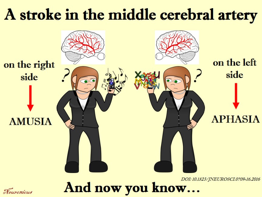
104-copy
unless you’re left-handed, then things are reversed.
References:
1. Sihvonen AJ, Ripollés P, Leo V, Rodríguez-Fornells A, Soinila S, & Särkämö T. (24 Aug 2016). Neural Basis of Acquired Amusia and Its Recovery after Stroke. Journal of Neuroscience, 36(34):8872-8881. PMID: 27559169, DOI: 10.1523/JNEUROSCI.0709-16.2016. ARTICLE  | FULLTEXT PDF
2.Bates E, Wilson SM, Saygin AP, Dick F, Sereno MI, Knight RT, & Dronkers NF (May 2003). Voxel-based lesion-symptom mapping. Nature Neuroscience, 6(5):448-50. PMID: 12704393, DOI: 10.1038/nn1050. ARTICLE
By Neuronicus, 9 November 2016


Tuesday, February 11, 2014

The First Map Showing All the Connections in Your Brain's White Matter

Gray matter damage is much easier to find, it shows up easily in external affects, hemiparesis, aphasia, etc. This is where I think our doctors are really failing us, they have not mapped 3d damage  to locations in either gray or white matter. I'm sure that my Middle cerebral artery blockage killed off a lot of  my white matter, but there is no known way to even identify the size of the damage. This might give some idea of the effects but it will be 50 years before it becomes standard practice unless a great stroke association directs and plans research to make it useful.
http://io9.com/the-first-map-showing-all-the-connections-in-your-brain-1520834855
map from the link, ask your doctor to decipher it compared to your damage.

Tuesday, August 13, 2013

Motor outcomes of patients with a complete middle cerebral artery territory infarct

I wonder if my doctor compares my infarct and subsequent damage to the ones  from this study. Nevermind he will never do anything so smart or work-related.  I have no idea of the exact location of my infarct. A 3d mapping of infarct locations and damage should have been completed however many years ago they had a 3 dimensional map of the arteries of the brain. But laziness and stupidity ruled.
http://124.16.31.131:8080/Jweb_sjzs/CN/article/downloadArticleFile.do?attachType=PDF&id=660

Research Highlights
(1) This study, for the first time, reported the motor outcomes of upper and lower extremities in patients with a complete middle cerebral artery territory infarct.
(2) After receiving rehabilitation treatments for 3–6 months, about 70% of patients were able to walk independently, but no patient achieved functional hand recovery.
(3) Results from this study will provide supporting evidence for developing rational rehabilitation strategies and establishing proper goals for stroke patients.(proper goal is simple - 100% recovery, nothing less)
Abstract
Detailed knowledge of motor outcomes enables to establish proper goals and rehabilitation strategies for stroke patients. Several previous studies have reported functional or motor outcomes in patients with a middle cerebral artery territory infarct. However, little is known about motor outcome in patients with a complete middle cerebral artery territory infarct. In this study, we investigated the motor outcomes in 23 patients with a complete middle cerebral artery territory infarct. All of these patients received comprehensive rehabilitative management, including movement therapy and neuromuscular electrical stimulation of the affected finger extensors and ankle dorsiflexors, for more than 3 months. Motor outcomes were measured at 6 months after stroke onset using the Medical Research Council, Motricity Index, the modified Brunnstrom Classification, and Functional Ambula-tion Category scores. The motor function of the lower extremities was found to be better than that of the upper extremities. After receiving rehabilitation treatments for 3–6 months, about 70% of these patients were able to walk independently (Functional Ambulation Category scores > 3), but no patient achieved functional hand recovery.