Changing stroke rehab and research worldwide now.Time is Brain! trillions and trillions of neurons that DIE each day because there are NO effective hyperacute therapies besides tPA(only 12% effective). I have 523 posts on hyperacute therapy, enough for researchers to spend decades proving them out. These are my personal ideas and blog on stroke rehabilitation and stroke research. Do not attempt any of these without checking with your medical provider. Unless you join me in agitating, when you need these therapies they won't be there. Did your doctor somehow miss the recovery protocol that Pedro Bach-y-Rita used to recover fully back in 1958 with only a partial brain?

What this blog is for:

My blog is not to help survivors recover, it is to have the 10 million yearly stroke survivors light fires underneath their doctors, stroke hospitals and stroke researchers to get stroke solved. 100% recovery. The stroke medical world is completely failing at that goal, they don't even have it as a goal. Shortly after getting out of the hospital and getting NO information on the process or protocols of stroke rehabilitation and recovery I started searching on the internet and found that no other survivor received useful information. This is an attempt to cover all stroke rehabilitation information that should be readily available to survivors so they can talk with informed knowledge to their medical staff. It lays out what needs to be done to get stroke survivors closer to 100% recovery. It's quite disgusting that this information is not available from every stroke association and doctors group.

Showing posts with label nanosensors. Show all posts

Friday, July 16, 2021

Scientists Create Injectable Swarm of Brain Reading Nanosensors

I like this one, seems easier to implement that some of the earlier ones.  But WHOM is going to determine the best method? I want to know what signals are being sent when neuroplasticity occurs. Why does a neuron give up its current job and take on a neighboring function? If we don't know that, neuroplasticity will never be repeatable on demand.

2. Use nanowires to listen in on single neurons
3. Or lay a grid across the cortex to listen in.

Scientists Create Injectable Swarm of Brain Reading Nanosensors

They say it could diagnose neurological disorders or allow for powerful brain-computer interfaces.

Fakurian Design via Unsplash / Futurism

A team of scientists has developed a new kind of biosensor that can be injected straight into the bloodstream, and will then travel to your brain, where they will — according to the scientists behind the project — monitor your neural activity and even potentially thoughts.

The cell-sized nanosensors, aptly named NeuroSWARM3, can cross the blood-brain barrier to the brain, where they convert neural activity into electrical signals, allowing them to be read and interpreted by machinery, according to work by a team of University of California, Santa Cruz scientists that will be presented next week at a virtual Optical Society conference.

The tech could, the researchers say, help grant extra mobility to people with disabilities in addition to helping scientists understand human thought better than before. However, they haven’t yet been tested on humans or even animals.

“NeuroSWARM3 can convert the signals that accompany thoughts to remotely measurable signals for high precision brain-machine interfacing,” lead study author A. Ali Yanik said in a press release. “It will enable people suffering from physical disabilities to effectively interact with the external world and control wearable exoskeleton technology to overcome limitations of the body. It could also pick up early signatures of neural diseases.”

It’s also a notably different approach to the problem of brain-computer interfaces from most high profile attempts, including Elon Musk’s Neuralink, which are working on implant-based solutions instead of nanosensor swarms.

During tests, the team found that their nanosensor swarm is sensitive enough to pick up on the activity of individual brain cells. Single-neuron readings aren’t new, but the ability to detect them with free-floating sensors, and especially the ability to wirelessly broadcast them through a patient’s thick skull, is an impressive technological development. If further tests continue to pan out, those capabilities could make real-time neuroscientific research simpler and neurological medicine more sophisticated.

“We are just at the beginning stages of this novel technology, but I think we have a good foundation to build on,” Yanik added. “Our next goal is to start experiments in animals.”


As a Futurism reader, we invite you join the Singularity Global Community, our parent company’s forum to discuss futuristic science & technology with like-minded people from all over the world. It’s free to join, sign up now!


Thursday, May 21, 2020

Tiny implanted sensors monitor brain injuries, then dissolve away

You'll have to ask your doctor and stroke hospital if this ever made it to human testing. Because with just a few modifications with nanosensors we could listen in on individual neuron signals and figure out EXACTLY what happens during neuroplasticity.  Assuming of course that there is anyone in stroke research that has two neurons to rub together for a spark of innovation.


Tiny implanted sensors monitor brain injuries, then dissolve away


A tiny implantable brain sensor could someday monitor conditions within the skull before dissolving away. Currently being tested in rats, this technology tracks temperature and pressure levels, potentially offering a new option for how brain injuries are studied and treated. By melting away when no longer useful, these micro-sized sensors remove the risks associated with current technology used to monitor brain injuries, according to a University of Illinois press release.
The study, led by John A. Rogers, professor of material sciences and engineering at the University of Illinois at Urbana-Champaign, and Wilson Ray, professor of neurological surgery at the Washington University School of Medicine in St. Louis, is published in the journal Nature.
"This is a new class of electronic biomedical implants," Rogers said in the release. "These kinds of systems have potential across a range of clinical practices, where therapeutic or monitoring devices are implanted or ingested, perform a sophisticated function, and then resorb harmlessly into the body after their function is no longer necessary."
Rogers stressed that the technology now used for monitoring after traumatic brain injuries or brain surgery can be dangerous for patients. Systems involve bulky wires that restrict patients' movements, and invasive implants may lead to brain hemorrhages, allergic reactions, and infections.
By contrast, the tiny dissolvable silicon devices that Rogers' team developed are smaller than a grain of rice. They are naturally biodegradable -- built up on incredibly thin silicon sheets -- and dissolve away after a few weeks of monitoring brain activity. They melt harmlessly into the human body's own fluids, the researchers say.
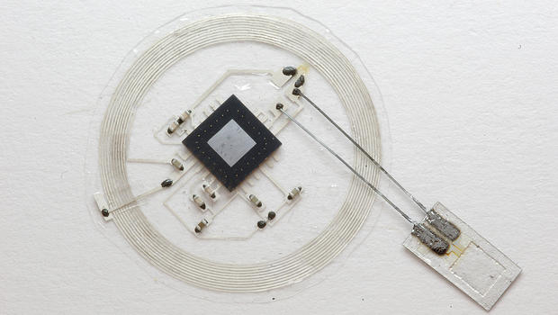
These devices are sensitive to pressure levels in the intracranial fluid that surrounds the brain, and have a temperature sensor synced to a wireless postage stamp-sized transmitter that is placed on top of the skull.
sensor-transmitter.jpg
The small sensor connects to an embeddable wireless transmitter that lies on top of the skull. Courtesy of John A. Rogers
"If you simply could throw out all the conventional hardware and replace it with very tiny, fully implantable sensors capable of the same function, constructed out of bioresorbable materials in a way that also eliminates or greatly miniaturizes the wires, then you could remove a lot of the risk and achieve better patient outcomes," Rogers added. "We were able to demonstrate all of these key features in animal models, with a measurement precision that's just as good as that of conventional devices."
The lab tests with rats were to see how compatible these tiny devices would be with a living organism's body.
So, what's the next phase? The team hopes to move toward human trials soon.
"The ultimate strategy is to have a device that you can place in the brain -- or in other organs in the body -- that is entirely implanted, intimately connected with the organ you want to monitor and can transmit signals wirelessly to provide information on the health of that organ, allowing doctors to intervene if necessary to prevent bigger problems," Rory Murphy, a neurosurgeon at Washington University who was a co-author of the paper.


Friday, November 3, 2017

Nanosensors Demystify Brain Chemistry

With these we should be able to answer the question of how exactly neuroplasticity works and make it repeatable on demand. But only if we destroy the existing fucking failures of stroke associations and get RFPs written and funded with foundation grants. Stroke is so fucking easy to solve  if you put your mind to it. I can do this and I'm stroke-addled.
https://www.rdmag.com/news/2017/11/nanosensors-demystify-brain-chemistry?
Fri, 11/03/2017 - 2:09pm
by AVS: Science and Technology of Materials, Interfaces, and Processing
Near-infrared microscopy (top) enables imaging of single-walled carbon nanotube sensors (bottom left) to image dopamine neurotransmission in brain tissue (bottom right).
Nanosensors are incredible information-gathering tools for myriad applications, including molecular targets such as the brain. Neurotransmitter molecules govern brain function through chemistry found deep within the brain, so University of California, Berkeley researchers are developing nanosensors to gain a better understanding of exactly how this all plays out. 
During the AVS 64th International Symposium & Exhibition, being held Oct. 29-Nov. 3, 2017, in Tampa, Florida, Markita del Carpio Landry, a professor of chemical and biomolecular engineering, and Abraham Beyene, a doctoral candidate in the Landry lab, will describe their design and use of near-infrared optical nanosensors to image the neurotransmitter dopamine within the brain.
“Developing sensors for brain chemistry is an exciting area of research that could transform how we diagnose diseases based on imbalances in brain chemistry, such as depression and anxiety,” Landry said. 
These nanosensors are created by combining carbon nanotubes and biomimetic synthetic polymers with the assistance of sound waves to promote the recognition of a selected small-molecule target. The formed sensors produce a fluorescent signal in the presence of their specified neurotransmitter target. Landry and her team are then able to directly quantify the neurotransmitter levels using the fluorescence intensity as a function of time.
“These complexes form nanosensors that fluoresce only within the presence of dopamine, a key neurotransmitter implicated in psychiatric disorders and neurodegenerative diseases such as Parkinson’s and Alzheimer’s disease,” Landry said. “We then build microscopes to detect the fluorescent response of the nanosensor so that we can image the nanosensors in living brain tissue.” 
The researchers are already using their sensors to explore how brain chemistry reacts to antidepressants. “We’re seeing some interesting results of how the antidepressant drug Merital affects the way the brain handles dopamine-based neurotransmission,” Landry said. “These key insights may help us to understand how antipsychotics and antidepressants work, and their side effects as well.” 
A simple method to assess brain chemistry is highly desirable for both research and clinical applications. While diseases such as cancer or diabetes are often diagnosed via methods such as blood tests that provide quantitative measurements of imbalances in blood or tissue chemistry, it’s impractical to take a “brain sample” to assess brain chemistry.
“My lab’s research focuses on the very challenging task of imaging brain chemistry with nanosensors that can report on neurotransmitter concentrations from within the brain and transmit their signals through brain tissue and the cranium,” Landry said. 
Landry and her colleagues are now building a new microscope, a “double infrared excitation-emission” form of fluorescence microscopy for deep brain neurotransmitter imaging, to allow them to image dopamine neurotransmission within the brains of awake and active animals.
“This will provide us with the capability to determine how antidepressants are affecting brain chemistry and to validate their effectiveness in a living animal model,” Landry said.

Monday, December 22, 2014

A “turn-on” fluorescent microbead sensor for detecting nitric oxide

Since nitric oxide is so helpful for us it would be good to know that the breathing exercises we are doing to produce it actually works.

Breathing Exercises for Your Heart

How else is your doctor making sure that the breathing protocol that was prescribed for you is working the way it is supposed to?
http://www.dovepress.com/articles.php?article_id=19691
Authors Yang LH, Ahn DJ, Koo E
Published Date December 2014 Volume 2015:10 Pages 115—123
DOI http://dx.doi.org/10.2147/IJN.S74924
Received 25 September 2014, Accepted 18 November 2014, Published 19 December 2014
Approved for publication by Dr Thomas J. Webster
Lan-Hee Yang,1,2 Dong June Ahn,3 Eunhae Koo1

1Advanced Materials Convergence Division, Korea Institute of Ceramic Engineering and Technology, Seoul, Republic of Korea; 2Department of Biomicrosystem Technology, Korea University, Seoul, Republic of Korea; 3Departments of Biomicrosystem Technology, Chemical & Biological Engineering, KU-KIST Graduate School, Korea University, Seoul, Republic of Korea


Abstract: Nitric oxide (NO) is a messenger molecule involved in numerous physical and pathological processes in biological systems. Therefore, the development of a highly sensitive material able to detect NO in vivo is a key step in treating cardiovascular and a number of types of cancer-related diseases, as well as neurological dysfunction. Here we describe the development of a fluorescent probe using microbeads to enhance the fluorescence signal. Microbeads are infused with the fluorophore, dansyl-piperazine (Ds-pip), and quenched when the fluorophore is coordinated with a rhodium (Rh)-complex, ie, Rh2(AcO-)4(Ds-pip). In contrast, they are able to fluoresce when the transition-metal complex is replaced by NO. To confirm the “on/off” mechanism for detecting NO, we investigated the structural molecular properties using the Fritz Haber Institute ab initio molecular simulations (FHI-AIMS) package. According to the binding energy calculation, NO molecules bind more strongly and rapidly with the Rh-core of the Rh-complex than with Ds-pip. This suggests that NO can bond strongly with the Rh-core and replace Ds-pip, even though Ds-pip is already near the Rh-core. However, the recovery process takes longer than the quenching process because the recovery process needs to overcome the energy barrier for formation of the transition state complex, ie, NO-(AcO-)4-(Ds-pip). Further, we confirm that the Rh-complex with the Ds-pip structure has too small an energy gap to give off visible light from the highest unoccupied molecular orbital/lowest unoccupied molecular orbital energy level.

Tuesday, August 5, 2014

Nanobiosensors: the future for diagnosis of disease?

With appropriate sensors these could be used to determine the extent of the penumbra so we could then send in the cavalry to save those neurons.
http://www.dovepress.com/articles.php?article_id=17806
Authors Prasad S
Published Date August 2014 Volume 2014:3 Pages 1—10
DOI http://dx.doi.org/10.2147/NDD.S39421
Received 10 February 2014, Accepted 26 March 2014, Published 4 August 2014
Shalini Prasad

Department of Bioengineering, University of Texas, Dallas, TX, USA

Abstract: Nanotechnology is having a profound impact on the development of a new class of biosensors known as nanobiosensors. Nanobiosensors commonly comprise a biological recognition molecule immobilized onto the surface of a signal transducer. The reaction between the biorecognition molecule and the analyte is a heterogeneous reaction and therefore the design of the biosensing interface is important in determining the performance of the nanobiosensor. Nanobiosensors are being widely used for molecular detection of biomarkers associated with diagnosis of disease. The application of new nanomaterials in biosensing has influenced biosensing research. The use of high surface area nanomaterials has been important in producing nanobiosensors with greater sensitivity and shorter response times. This review summarizes the advances in disease diagnostics, primarily through the detection of molecular biomarkers, such as proteins and nucleic acids mediated by use of nanobiosensors.

Keywords: nanomaterial, biosensors, disease diagnosis, biomimetic, macromolecular crowding, performance metrics

Download Article [PDF] 

Monday, March 31, 2014

‘Electronic skin' equipped with memory

This would be perfect for monitoring our movements so we could precisely tell what's wrong with them. And every neurologist should have that exact same thought and create a project to implement this for their patients.
I know that is a stupid dream but somehow we have to prove that our neurologists know nothing about stroke recovery.
http://www.nature.com/news/electronic-skin-equipped-with-memory-1.14952

Thursday, August 1, 2013

Chemists develop innovative nano-sensors for multiple proteins

This is so simple. There must be some identifiable proteins that circulate in the blood post-stroke. This would be a fast objective way to then diagnose a stroke. And we can get away from the stupidity of the current, 'Is it a stroke or drunkenness?'.  Which stroke association is going to fail to recognize and promote this possibility? I bet all of them. 

Chemists develop innovative nano-sensors for multiple proteins


Chemists at Johannes Gutenberg University Mainz (JGU) have developed a new method for parallel protein analysis that is, in principle, capable of identifying hundreds or even thousands of different proteins. It could be used to detect the presence of viruses and identify their type in tiny samples. At the same time, it is very cost-effective and quick. "We see possible applications of this technique in medicine, where it could be used, for example, for the rapid diagnosis of a wide range of diseases. It would be almost as easy to use as a pregnancy test strip," said Professor Carsten Sönnichsen of the Institute of Physical Chemistry. The test involves placing a tiny drop of blood, saliva, or other bodily fluid on a small test strip, which is then placed in a device developed at the JGU Institute of Physical Chemistry. This device is able to identify the specific proteins in the fluid and thus allows to quickly and reliably differentiate between harmless microorganisms and dangerous pathogens.
In order to detect the many different substances present in a small sample, the sensors need to be as tiny as possible, preferably the size of nano-particles. Sönnichsen's team of scientists have designed a sensor no larger than the head of a pin but capable of performing a hundred different individual tests on a surface that is only of one-tenth of a square millimeter in area. The 'test strips' consist of glass capillary tubes that have gold nano-particles as sensor elements on their internal surfaces. "We first prepare our nano-particles using short DNA strands, each of which binds to a specific type of protein," explained Janak Prasad, who developed the functionalization method. When a protein docks with one of these special DNA strands, called aptamers, the corresponding nano-particle changes its color. The color changes can be detected with the aid of a spectrometer. For this purpose, the capillary tubes are placed under a microscope designed, constructed, and provided with the necessary software by the Mainz-based team of chemists.
"We demonstrate a new approach for a multiplexed assay that detects multiple proteins simultaneously by letting a fluid flow past the randomly positioned gold nano-rods," explained Christina Rosman, first author of the study. The team from JGU's Institute of Physical Chemistry used four different target proteins to demonstrate the viability of the new concept, its ability to detect concentrations in the nanomolar range, and the possibility to recycle the sensors for more than one analysis. "We see the potential to extend our method to the simultaneous detection of hundreds or even thousands of different target substances," assert the authors in their article published in the June 2013 issue of the journal Nano Letters. Low-cost serial production of the sensors is feasible if advanced nano-fabrication methods such as nano-printing or optical trapping are used.
There are manifold possible applications of a test for multiple targets in a single procedure. The low-cost sensors could be directly used by physicians in their practices in order to detect and discriminate various types of flu viruses with which their patients could be infected. In addition, the technique would also be suitable for detecting the presence of toxins in the environment or in food, particularly in liquids such as milk or baby food, or the presence of doping or other illicit drugs.
Support for the research on this novel multiplexed protein sensor was provided by the Graduate School of Excellence 'Materials Science in Mainz' (MAINZ) and the European Research Council (ERC) 'Single Sense' project.
http://www.uni-mainz.de/presse/16589_ENG_HTML.php