Showing posts with label oxygenation. Show all posts
Showing posts with label oxygenation. Show all posts
Wednesday, September 30, 2026
Pomegranate Compound Shows Potential to Improve Heart Function by 80 Percent: Study


Sunday, January 12, 2025
The impact of brain-systemic oxygenation coupling in sleep-disordered breathing on cognitive function in elderly
My doctors at the hospital never even looked for my sleep apnea. Since this was tested in healthy adults your competent? doctor needs to ensure such resesarch occurs in stroke survivors. I don't think your doctor can or will do that; because incompetence!
The impact of brain-systemic oxygenation coupling in sleep-disordered breathing on cognitive function in elderly
Scientific Reports volume 15, Article number: 1523 (2025)
Abstract
Background:
Intermittent hypoxia, a consequence of sleep-disordered breathing (SDB), may contribute to an increased risk of cognitive decline. However, the association between SDB and cognition remains highly variable.
Methods:
Fifty-two community-dwelling healthy older adults (28 women) were recruited. All participants underwent neuropsychiatric evaluations, simultaneous ambulatory polysomnography (PSG), and near-infrared spectroscopy (NIRS) recordings. We quantified the average coherence between oxy-Hb and SpO2 signals during SDB events to determine whether it could predict cognitive outcomes in healthy older adults, where higher coherence indicates reduced protection against systemic hypoxia.
Results:
The mean (SD) coherence of oxy-Hb and SpO2 was 0.16 (0.07). Linear regression analysis showed a significant association between mean coherence and worse Stroop Color Word Test scores (t=-0.304, p = .004). In contrast, oxy-Hb reduction alone and conventional SDB parameters did not show a significant association with cognition.
Conclusion:
This is the first report to demonstrate an association between a novel parameter of brain-systemic oxygenation coherence in SDB and cognition in older adults. A higher coherence rate of cortical oxy-Hb and systemic SpO2 during SDB may reflect a loss of compensatory mechanisms against systemic hypoxia and could help stratify older adults with a higher risk for cognitive decline.
Tuesday, March 7, 2023
Landmark discovery provides a new perspective on tissue oxygenation
If your doctors and hospital aren't breathlessly following this for stroke intervention, you don't have a functioning stroke doctor or hospital. Until this is available what is your doctor doing to increase the oxygen supply to the brain?
Here, check these out.
cerebral blood flow (29 posts to July 2016)
Cerebral blood flow autoregulation (1 post to July 2021)
Cerebral Blood Flow Velocity (1 post to Febraury 2020)
cortical oxygenation (1 post to November 2020)
oxygen delivery (20 posts to January 2020)
oxygen uptake (5 posts to August 2013)
Super Saturated Oxygen Therapy (1 post to May 2021)
Normobaric oxygen (10 posts to January 2020)
brain blood flow (3 posts to April 2019)
The latest here:
Landmark discovery provides a new perspective on tissue oxygenation
Cardiovascular medicine, hematology and pulmonary medicine may soon have the first-ever therapies to correct poor tissue oxygenation, a key driver of disease in millions, including peripheral artery disease, sickle cell disease, heart failure, stroke, emphysema and many others.
The breakthrough follows a landmark discovery from investigators at Harrington Discovery Institute at University Hospitals (UH) and Case Western Reserve University School of Medicine. The research team showed that a modified version of hemoglobin, termed S-nitrosohemoglobin, senses areas with insufficient oxygen, and then restores blood flow for oxygenation. The study recently published in PNAS.
Our textbooks focus on how much oxygen is carried in the blood. But this work reveals, for the first time in humans, that it's really not about oxygen, but about nitric oxide when it comes to delivering oxygen. Critically, the nitric oxide must be carried by hemoglobin, in the form of S-nitrosohemoglobin, to be effective. This opens up an entirely new line of drug development."
Jonathan S. Stamler, MD, senior author of the study, President, Harrington Discovery Institute at UH, Robert S. and Sylvia K. Reitman Family Foundation Distinguished Professor of Cardiovascular Innovation, and Professor of Medicine and Biochemistry at UH and Case Western Reserve School of Medicine
Correcting inadequate tissue oxygenation has been a longstanding goal of medicine. Scientists have known for some time that oxygen levels in the blood don't predict oxygen delivery to tissues, and that oxygen availability is almost never the problem.
"We have a major mystery in medicine," Dr. Stamler explained. "Even when people die from 'low oxygen' they still have lots of oxygen left in the bloodstream. Yet medications that increase blood flow are unable to increase oxygen delivery. There are millions of patients across both major and rare diseases that have disorders of oxygen delivery, and we have no way to improve that situation."
Cancer Research eBook
Compilation of the top interviews, articles, and news in the last year.
Drugs that improve blood flow have so far failed to improve oxygen delivery. So, increasing oxygen doesn't work and increasing blood flow hasn't worked.
In the new study, the researchers identify S-nitrosohemoglobin as a unique vasodilator that not only opens blood vessels but produces major increases in tissue oxygenation. Further, in both mice and humans--including patients with peripheral artery disease, sickle cell disease, heart failure, stroke and emphysema--impairments in tissue oxygenation were found to correlate with levels of S-nitrosohemoglobin. A low level resulted in poor tissue oxygenation.
"This study shows that you must vasodilate through hemoglobin if you want to get oxygen to tissues, because hemoglobin knows precisely where in the body oxygen is low, and then it adjusts itself to give off the vasodilator nitric oxide in proportion to need," Dr. Stamler said. "Other vasodilators dilate vessels everywhere, and steal blood flow from places that need it most."
Stamler and colleagues are currently working to develop drugs that modify hemoglobin with nitric oxide to form S-nitrosohemoglobin. These new drugs are increasing tissue oxygenation for the very first time, even when oxygen levels in the blood are low.
He emphasized, "It's almost never about amounts of oxygen in medical conditions. It's about blood flow in little blood vessels. If you don't have S-nitrosohemoglobin, you cannot oxygenate. This is an entirely new perspective on tissue oxygenation, and one that hopefully solves a major problem in medicine."
Source:
Journal reference:
Reynolds, J.D., et al. (2023) Control of tissue oxygenation by S-nitrosohemoglobin in human subjects. PNAS. doi.org/10.1073/pnas.2220769120.
Saturday, January 14, 2023
Long COVID: major findings, mechanisms and recommendations
You don't want the reduced cerebral blood flow which you already had as part of your stroke. You can ask your doctor what protocol they used to correct that reduced flow; if they did nothing you don't have a functioning stroke doctor. This is why I'm vaccinated up the wazoo.
This is what I'm going to tell my doctor to do; don't listen to me,I'm not medically trained.
If your doctor has DONE NOTHING on cerebral blood flow and oxygenation then you don't have a functioning stroke doctor or hospital.
cerebral blood flow (27 posts to July 2016)
oxygenation (5 posts to March 2013)
The latest here:
Long COVID: major findings, mechanisms and recommendations
Nature Reviews Microbiology (2023)
Abstract
Long COVID is an often debilitating illness that occurs in at least 10% of severe acute respiratory syndrome coronavirus 2 (SARS-CoV-2) infections. More than 200 symptoms have been identified with impacts on multiple organ systems. At least 65 million individuals worldwide are estimated to have long COVID, with cases increasing daily. Biomedical research has made substantial progress in identifying various pathophysiological changes and risk factors and in characterizing the illness; further, similarities with other viral-onset illnesses such as myalgic encephalomyelitis/chronic fatigue syndrome and postural orthostatic tachycardia syndrome have laid the groundwork for research in the field. In this Review, we explore the current literature and highlight key findings, the overlap with other conditions, the variable onset of symptoms, long COVID in children and the impact of vaccinations. Although these key findings are critical to understanding long COVID, current diagnostic and treatment options are insufficient, and clinical trials must be prioritized that address leading hypotheses. Additionally, to strengthen long COVID research, future studies must account for biases and SARS-CoV-2 testing issues, build on viral-onset research, be inclusive of marginalized populations and meaningfully engage patients throughout the research process.
Introduction
Long COVID (sometimes referred to as ‘post-acute sequelae of COVID-19’) is a multisystemic condition comprising often severe symptoms that follow a severe acute respiratory syndrome coronavirus 2 (SARS-CoV-2) infection. At least 65 million individuals around the world have long COVID, based on a conservative estimated incidence of 10% of infected people and more than 651 million documented COVID-19 cases worldwide1; the number is likely much higher due to many undocumented cases. The incidence is estimated at 10–30% of non-hospitalized cases, 50–70% of hospitalized cases2,3 and 10–12% of vaccinated cases4,5. Long COVID is associated with all ages and acute phase disease severities, with the highest percentage of diagnoses between the ages of 36 and 50 years, and most long COVID cases are in non-hospitalized patients with a mild acute illness6, as this population represents the majority of overall COVID-19 cases. There are many research challenges, as outlined in this Review, and many open questions, particularly relating to pathophysiology, effective treatments and risk factors.
Hundreds of biomedical findings have been documented, with many patients experiencing dozens of symptoms across multiple organ systems7 (Fig. 1). Long COVID encompasses multiple adverse outcomes, with common new-onset conditions including cardiovascular, thrombotic and cerebrovascular disease8, type 2 diabetes9, myalgic encephalomyelitis/chronic fatigue syndrome (ME/CFS)10,11 and dysautonomia, especially postural orthostatic tachycardia syndrome (POTS)12 (Fig. 2). Symptoms can last for years13, and particularly in cases of new-onset ME/CFS and dysautonomia are expected to be lifelong14. With significant proportions of individuals with long COVID unable to return to work7, the scale of newly disabled individuals is contributing to labour shortages15. There are currently no validated effective treatments.
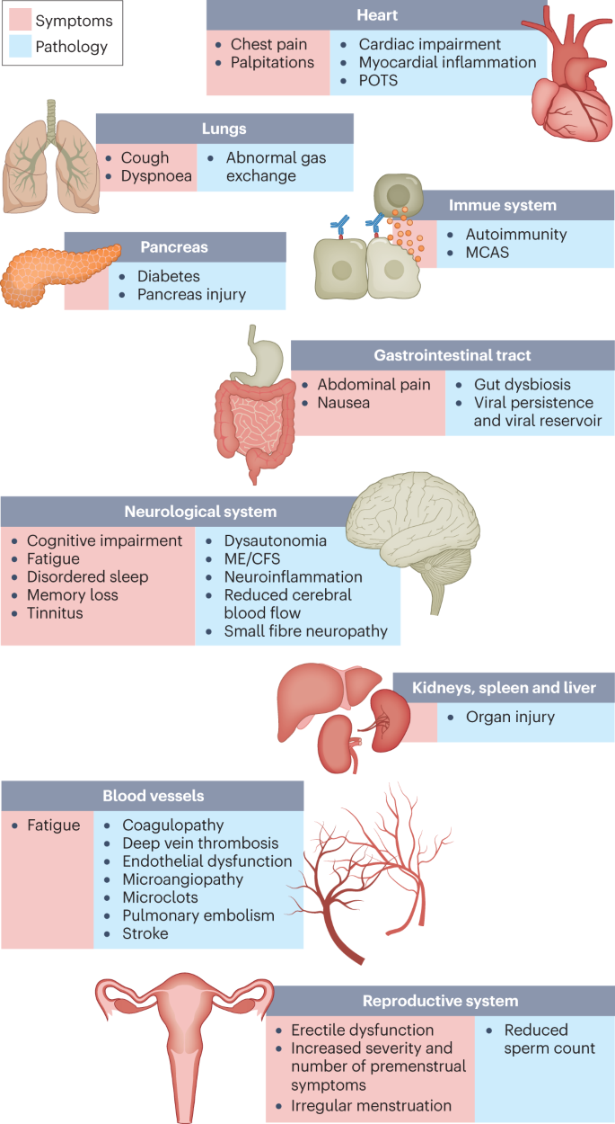
The impacts of long COVID on numerous organs with a wide variety of pathology are shown. The presentation of pathologies is often overlapping, which can exacerbate management challenges. MCAS, mast cell activation syndrome; ME/CFS, myalgic encephalomyelitis/chronic fatigue syndrome; POTS, postural orthostatic tachycardia syndrome.
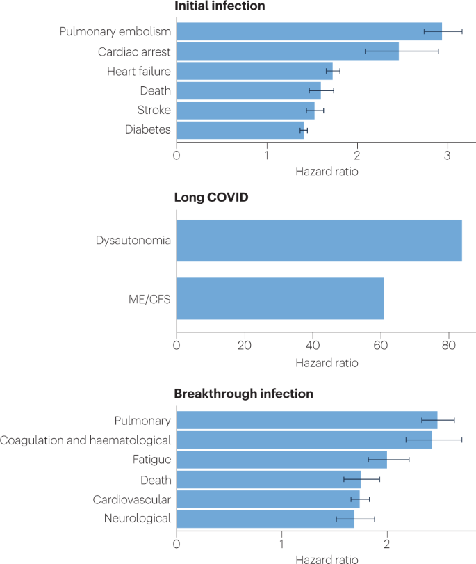
Because diagnosis-specific data on large populations with long COVID are sparse, outcomes from general infections are included and a large proportion of medical conditions are expected to result from long COVID, although the precise proportion cannot be determined. One year after the initial infection, severe acute respiratory syndrome coronavirus 2 (SARS-CoV-2) infections increased the risk of cardiac arrest, death, diabetes, heart failure, pulmonary embolism and stroke, as studied with use of US Department of Veterans Affairs databases. Additionally, there is clear increased risk of developing myalgic encephalomyelitis/chronic fatigue syndrome (ME/CFS) and dysautonomia. Six months after breakthrough infection, increased risks were observed for cardiovascular conditions, coagulation and haematological conditions, death, fatigue, neurological conditions and pulmonary conditions in the same cohort. The hazard ratio is the ratio of how often an event occurs in one group relative to another; in this case people who have had COVID-19 compared with those who have not. Data sources are as follows: diabetes9, cardiovascular outcomes8, dysautonomia8,201, ME/CFS10,202 and breakthrough infections4.
There are likely multiple, potentially overlapping, causes of long COVID. Several hypotheses for its pathogenesis have been suggested, including persisting reservoirs of SARS-CoV-2 in tissues16,17; immune dysregulation17,18,19,20 with or without reactivation of underlying pathogens, including herpesviruses such as Epstein–Barr virus (EBV) and human herpesvirus 6 (HHV-6) among others17,18,21,22; impacts of SARS-CoV-2 on the microbiota, including the virome17,23,24,25; autoimmunity17,26,27,28 and priming of the immune system from molecular mimicry17; microvascular blood clotting with endothelial dysfunction17,29,30,31; and dysfunctional signalling in the brainstem and/or vagus nerve17,32 (Fig. 3). Mechanistic studies are generally at an early stage, and although work that builds on existing research from postviral illnesses such as ME/CFS has advanced some theories, many questions remain and are a priority to address. Risk factors potentially include female sex, type 2 diabetes, EBV reactivation, the presence of specific autoantibodies27, connective tissue disorders33, attention deficit hyperactivity disorder, chronic urticaria and allergic rhinitis34, although a third of people with long COVID have no identified pre-existing conditions6. A higher prevalence of long Covid has been reported in certain ethnicities, including people with Hispanic or Latino heritage35. Socio-economic risk factors include lower income and an inability to adequately rest in the early weeks after developing COVID-19 (refs. 36,37). Before the emergence of SARS-CoV-2, multiple viral and bacterial infections were known to cause postinfectious illnesses such as ME/CFS17,38, and there are indications that long COVID shares their mechanistic and phenotypic characteristics17,39. Further, dysautonomia has been observed in other postviral illnesses and is frequently observed in long COVID7.
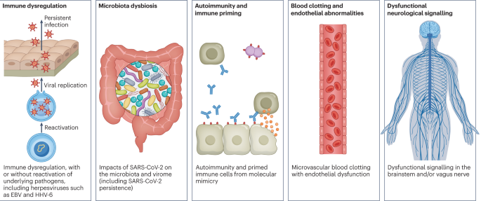
There are several hypothesized mechanisms for long COVID pathogenesis, including immune dysregulation, microbiota disruption, autoimmunity, clotting and endothelial abnormality, and dysfunctional neurological signalling. EBV, Epstein–Barr virus; HHV-6, human herpesvirus 6; SARS-CoV-2, severe acute respiratory syndrome coronavirus 2.
In this Review, we explore the current knowledge base of long COVID as well as misconceptions surrounding long COVID and areas where additional research is needed. Because most patients with long COVID were not hospitalized for their initial SARS-CoV-2 infection6, we focus on research that includes patients with mild acute COVID-19 (meaning not hospitalized and without evidence of respiratory disease). Most of the studies we discuss refer to adults, except for those in Box 1.
More at link.
Friday, October 28, 2022
High arterial oxygen levels and supplemental oxygen administration in traumatic brain injury: insights from CENTER-TBI and OzENTER-TBI
What will your doctor takeaway from this and all this other research on what the proper protocol is for cerebral blood flow and oxygenation? If your doctor has DONE NOTHING on cerebral blood flow and oxygenation then you don't have a functioning stroke doctor or hospital.
cerebral blood flow (27 posts to July 2016)
oxygenation (5 posts to March 2013)
High arterial oxygen levels and supplemental oxygen administration in traumatic brain injury: insights from CENTER-TBI and OzENTER-TBI
Intensive Care Medicine (2022)
Abstract
Purpose
The effect of high arterial oxygen levels and supplemental oxygen administration on outcomes in traumatic brain injury (TBI) is debated, and data from large cohorts of TBI patients are limited. We investigated whether exposure to high blood oxygen levels and high oxygen supplementation is independently associated with outcomes in TBI patients admitted to the intensive care unit (ICU) and undergoing mechanical ventilation.
Methods
This is a secondary analysis of two multicenter, prospective, observational, cohort studies performed in Europe and Australia. In TBI patients admitted to ICU, we describe the arterial partial pressure of oxygen (PaO2) and the oxygen inspired fraction (FiO2). We explored the association between high PaO2 and FiO2 levels within the first week with clinical outcomes. Furthermore, in the CENTER-TBI cohort, we investigate whether PaO2 and FiO2 levels may have differential relationships with outcome in the presence of varying levels of brain injury severity (as quantified by levels of glial fibrillary acidic protein (GFAP) in blood samples obtained within 24 h of injury).
Results
The analysis included 1084 patients (11,577 measurements) in the CENTER-TBI cohort, of whom 55% had an unfavorable outcome, and 26% died at a 6-month follow-up. Median PaO2 ranged from 93 to 166 mmHg. Exposure to higher PaO2 and FiO2 in the first seven days after ICU admission was independently associated with a higher mortality rate. A trend of a higher mortality rate was partially confirmed in the OzENTER-TBI cohort (n = 159). GFAP was independently associated with mortality and functional neurologic outcome at follow-up, but it did not modulate the outcome impact of high PaO2 levels, which remained independently associated with 6-month mortality.
Conclusions
In two large prospective multicenter cohorts of critically ill patients with TBI, levels of PaO2 and FiO2 varied widely across centers during the first seven days after ICU admission. Exposure to high arterial blood oxygen or high supplemental oxygen was independently associated with 6-month mortality in the CENTER-TBI cohort, and the severity of brain injury did not modulate this relationship. Due to the limited sample size, the findings were not wholly validated in the external OzENTER-TBI cohort. We cannot exclude the possibility that the worse outcomes associated with higher PaO2 were due to use of higher FiO2 in patients with more severe injury or physiological compromise. Further, these findings may not apply to patients in whom FiO2 and PaO2 are titrated to brain tissue oxygen monitoring (PbtO2) levels. However, at minimum, these findings support the need for caution with oxygen therapy in TBI, particularly since titration of supplemental oxygen is immediately applicable at the bedside.
Take-home message
| In two large prospective multicenter cohorts of traumatic brain injured patients, arterial and supplemental oxygen levels varied widely across centers during the first seven days after admission to the intensive care unit. |
| Exposure to high arterial blood oxygen or high supplemental oxygen—a therapeutic gas immediately titratable at the bedside—was independently associated with 6-month mortality, regardless of brain injury severity. |
Introduction
In patients with traumatic brain injury (TBI), hypoxemia is a major predictor of hospital and 6-month mortality [1]. Oxygen supplementation aims to reverse tissue hypoxia and, thus, improve cell viability, organ function, and survival in critically ill patients [2]. However, this may lead to administering more oxygen than needed to patients admitted to the intensive care unit (ICU) [3].
While hyperbaric oxygen is known to be neurotoxic [4], it is not clear whether high normobaric oxygen levels may play a detrimental role in the brain [5]. Hyperoxia, i.e., high inspiratory oxygen fraction, may be associated with excitotoxicity in severe TBI [6]. Furthermore, hyperoxemia, i.e., high blood oxygen partial pressure levels, may potentially worsen organ injury and impact the case fatality rate of critically ill patients with TBI [7, 8]. Therefore, not only too low but even extreme hyperoxemia might cause injury in TBI patients, as David et al. showed [9]. Data on more than 36,000 mixed ICU patients mechanically ventilated with early arterial partial pressure of oxygen (PaO2) suggested an independent U-shape association with hospital mortality [10]. A recent metanalysis of 32 studies in acute brain-damaged patients highlighted that hyperoxemia, differently defined across studies, was associated with an increased risk of poor neurological outcomes [11]. Patients with a poor neurological outcome also had a significantly higher maximum PaO2 and mean PaO2. These associations were present, especially in patients with subarachnoid hemorrhage and ischemic stroke, but not in traumatic brain injured.
Currently, there is no evidence to support the role of hyperoxemia or hyperoxia in a large real-world dataset of critically ill patients admitted to ICU with severe TBI [12,13,14].
Therefore, we described variability across centers in the blood oxygen levels (i.e., PaO2) and oxygen supplementation distributions (i.e., inspiratory oxygen fraction, FiO2) and investigated whether high PaO2 and FiO2 levels are associated with worse 6-month outcomes. We validated our findings in the multicenter Australian OzENTER-TBI database [15]. Finally, we explored whether PaO2 and FiO2 levels may contribute differently to outcomes in the presence of increasing levels of glial fibrillary acidic protein (GFAP), a biomarker of brain injury severity.
The aims of this study are to:
-
1.
Describe the values and the differences in PaO2 and FiO2 in the first week from ICU admission in mechanically ventilated TBI patients across centers in CENTER-TBI;
-
2.
assess whether high levels of PaO2 or FiO2 are independently associated with 6-month mortality and unfavorable neurologic outcome in CENTER-TBI;
-
3.
evaluate whether the impact of high levels of oxygen exposure or high levels of supplemental oxygen on 6-month outcome could be worsened by increasing brain injury severity, as assessed by acute (first 24 h) serum levels of GFAP in the CENTER-TBI cohort.
All these objectives (except the last one) were subsequently validated in an external cohort of patients with traumatic brain injury from OzENTER-TBI. Hypotheses of the current analyses were that exposure to high oxygen and FiO2 levels in TBI patients mechanically ventilated and admitted to ICU may promote brain injury and have a negative impact on both functional neurological disability and survival.
Thursday, January 21, 2021
Blood Pressure Management Before, During, and After Endovascular Thrombectomy for Acute Ischemic Stroke
Since no factual basis exists for blood pressure management post stroke, your doctor is just guessing. Hope she guesses right. Too high blood pressure and you could create a hemorrhage, too low means you aren't getting enough oxygen to those neurons in the penumbra that need it before they completely die.
Although there might be other ways to get more oxygen to your brain, don't listen to me, I'm not medically trained.
Does your doctor know about them?
Possible solutions: Obviously not vetted coming from me. Don't do them.
Normobaric oxygen (10)
How to Improve Your Brain Function with An Oxygen Concentrator April 2018
Or is it more important to increase the loading ability of red blood cells to carry more oxygen?
Like this?
University of Glasgow Study Demonstrates the Ability of Oxycyte® to Supply Oxygen to Critical Penumbral Tissue in Acute Ischemic Stroke August 2012
Or like this?
chronic cannabis users have higher cerebral blood flow and extract more oxygen from brain blood flow than nonusers. August 2017
Vinpocetine increases cerebral blood flow and oxygenation in stroke patients: a near infrared spectroscopy and transcranial Doppler study
The latest here:
Blood Pressure Management Before, During, and After Endovascular Thrombectomy for Acute Ischemic Stroke

Abstract
There is an absence of specific evidence or guideline recommendations on blood pressure management for large vessel occlusion stroke patients. Until randomized data are available, the periprocedural blood pressure management of patients undergoing endovascular thrombectomy can be viewed in two phases relative to the achievement of recanalization. In the hyperacute phase, prior to recanalization, hypotension should be avoided to maintain adequate penumbral perfusion. The American Heart Association guidelines should be followed for the upper end of prethrombectomy blood pressure: ≤185/110 mm Hg, unless post–tissue plasminogen activator administration when the goal is <180/105 mm Hg. After successful recanalization (thrombolysis in cerebral infarction [TICI]: 2b–3), we recommend a target of a maximum systolic blood pressure of < 160 mm Hg, while the persistently occluded patients (TICI < 2b) may require more permissive goals up to <180/105 mm Hg. Future research should focus on generating randomized data on optimal blood pressure management both before and after endovascular thrombectomy, to optimize patient outcomes for these divergent clinical scenarios.
Publication History
Publication Date:
20 January 2021 (online)
© 2021. Thieme. All rights reserved.
Thieme Medical Publishers, Inc.
333 Seventh Avenue, 18th Floor, New York, NY 10001, USA
Thursday, December 3, 2020
Living into your 90s; Leisure World Cohort Study
I will have to regularly follow this up since the study was extended again. At the 19 minute mark they talk about hypertension preventing micro-infarcts that lead to dementia. In my opinion this makes sense since you are forcing more oxygenated blood thru your brain. The con which your doctor will use is that high blood pressure could lead to a hemorrhage. Although there might be better ways to get more oxygen to your brain, don't listen to me, I'm not medically trained.
Possible solutions: Obviously not vetted coming from me. Don't do them.
Normobaric oxygen (10)
How to Improve Your Brain Function with An Oxygen Concentrator April 2018
Or is it more important to increase the loading ability of red blood cells to carry more oxygen?
Like this?
University of Glasgow Study Demonstrates the Ability of Oxycyte® to Supply Oxygen to Critical Penumbral Tissue in Acute Ischemic Stroke August 2012
Or like this?
chronic cannabis users have higher cerebral blood flow and extract more oxygen from brain blood flow than nonusers. August 2017
Vinpocetine increases cerebral blood flow and oxygenation in stroke patients: a near infrared spectroscopy and transcranial Doppler study
May 2015
The latest here:
Living into your 90s; Leisure World Cohort Study
Wednesday, November 25, 2020
Dietary flavanols improve cerebral cortical oxygenation and cognition in healthy adults
Does your stroke hospital have enough intelligence to read between the lines and recognize that greater brain oxygenation is what is needed immediately post stroke? Even if this research was done in healthy adults? Of course your stroke hospital will have to contact the researchers to get the protocol. Not doing any of that and your board of directors needs to be fired for not setting proper goals for the stroke unit. 100% RECOVERY FOR ALL!
Dietary flavanols improve cerebral cortical oxygenation and cognition in healthy adults
Scientific Reports volume 10, Article number: 19409 (2020)
Abstract
Cocoa flavanols protect humans against vascular disease, as evidenced by improvements in peripheral endothelial function, likely through nitric oxide signalling. Emerging evidence also suggests that flavanol-rich diets protect against cognitive aging, but mechanisms remain elusive. In a randomized double-blind within-subject acute study in healthy young adults, we link these two lines of research by showing, for the first time, that flavanol intake leads to faster and greater brain oxygenation responses to hypercapnia, as well as higher performance only when cognitive demand is high. Individual difference analyses further show that participants who benefit from flavanols intake during hypercapnia are also those who do so in the cognitive challenge. These data support the hypothesis that similar vascular mechanisms underlie both the peripheral and cerebral effects of flavanols. They further show the importance of studies combining physiological and graded cognitive challenges in young adults to investigate the actions of dietary flavanols on brain function.
Introduction
Lifespan wear and tear of the vascular system due to poor nutrition and lack of fitness, among other factors, can accelerate cognitive aging and lead to dementia. There is epidemiological evidence suggesting that flavonoids, a group of small molecules present in fruits and vegetables, can protect against vascular disease and cardiovascular-related mortality1,2,3,4. In particular, cocoa flavanols, a sub-group of flavonoids (also present in berries, grapes, apples and tea) have been shown to improve endothelial function in humans quite rapidly (within 1–2 h) by enhancing vasodilatory properties of peripheral arteries5,6. Acute benefits translate effectively into short-term (2–8 weeks) clinically relevant improvements in blood pressure and endothelial function (as measured by brachial flow-mediated dilatation, FMD)7,8, comparable to those of drugs, such as statins9,10. Mechanistically, the beneficial effects of cocoa flavanols on endothelial function have been linked to increases in bioavailability of nitric oxide (NO)6, which is known to be affected in the earliest stages of vascular disease11.Whilst the acute effects of flavanols have been mainly attributed to phase I/II-derived (−)-epicatechin metabolites, the short to long term benefits may be also driven by gut-derived metabolites12,13, although this remains to be established. Another emerging line of research further suggests that this class of plant-derived compounds may protect against cognitive decline in aging14,15,16 and cognitive resilience to neuropsychiatric disorders and stress17,18. Yet, the extent to which increases in circulatory levels of NO by flavanols can translate into benefits in the brain vasculature, and effectively influence cognitive performance in humans, is poorly understood.
Cerebral blood flow is controlled by neuronal activity but also by levels of arterial blood gases, in particular carbon dioxide (CO2)19. Relevant to our hypothesis is the fact that NO is known to contribute to CO2-dependent increases in cerebral blood flow in humans (hypercapnia)20. Furthermore, cerebrovascular reactivity to CO2 is widely accepted as a key biomarker of cerebrovascular health, and has been closely associated with cognitive function in health and disease states21,22,23,24,25. Hence, hypercapnia represents a robust model to test whether flavanol-mediated increases in endothelial function (as assessed by gold-standard FMD) mediate benefits in cerebrovascular and cognitive function.
Only a handful of studies have previously reported effects of flavanols on the human cerebral vasculature, both in a resting state26,27,28,29 and in response to cognitive challenges30,31,32, albeit in opposite directions (increase/decrease in blood flow/velocity). Further, modulation of cerebral physiological outcomes by flavanols in the context of neuronal/cognitive challenges frequently and surprisingly fail to translate into cognitive benefits26,30,31,32. A possibility is that the benefits of flavanols may only be visible at high levels of task difficulty. This highlights that, whilst some of these studies could provide ecological validity (as they target aging adults with cognitive and/or vascular problems), they were not designed in a manner that allows for an evaluation of the underlying physiological effects of these compounds in the human brain. This leaves some uncertainty about whether flavanols’ benefits in peripheral vascular function are reflected by similar effects on cerebrovascular reactivity, and whether the cognitive and vascular benefits are related.
There is a need to determine whether the peripheral vascular benefits of flavanols extend to the cerebral vasculature. This requires well-controlled experiments to demonstrate that flavanols (a) can modulate the brain’s vasculature; (b) that these effects, similarly to those found in the periphery, are revealed during physiological challenges likely involving the NO pathway; (c) that they affect cognitive performance, at least in challenging conditions; and (d) that cerebrovascular and cognitive benefits are linked.
In the current study, we employed separate physiological and cognitive challenges in a double-blind, within-subject, placebo-controlled acute (2 h) study to assess the underlying physiological actions of cocoa flavanols on cerebral and peripheral vascular and cognitive function. To measure cerebrovascular reactivity, we employed a CO2-breathing challenge (hypercapnia) before and after intake of either a high- or low-flavanol intervention. During hypercapnia, we measured cortical haemoglobin concentration using functional near-infrared spectroscopy (fNIRS)33, which allowed us to finely quantify the dynamics of cerebrovascular reactivity, providing information not only about the amplitude of this response, but also about its time course. Similar to peripheral measures, cerebral CO2-reactivity has been shown to be mediated by the NO pathway20, which is the hypothesized mechanism underlying the beneficial effects of flavanols on peripheral endothelial function6. To measure the cognitive effects of flavanols, we employed tasks with escalating levels of difficulty34, which could inform us of the level at which the cognitive benefits of flavanols may emerge.
Subscribe to:
Posts (Atom)