Changing stroke rehab and research worldwide now.Time is Brain! trillions and trillions of neurons that DIE each day because there are NO effective hyperacute therapies besides tPA(only 12% effective). I have 523 posts on hyperacute therapy, enough for researchers to spend decades proving them out. These are my personal ideas and blog on stroke rehabilitation and stroke research. Do not attempt any of these without checking with your medical provider. Unless you join me in agitating, when you need these therapies they won't be there. Did your doctor somehow miss the recovery protocol that Pedro Bach-y-Rita used to recover fully back in 1958 with only a partial brain?

What this blog is for:

My blog is not to help survivors recover, it is to have the 10 million yearly stroke survivors light fires underneath their doctors, stroke hospitals and stroke researchers to get stroke solved. 100% recovery. The stroke medical world is completely failing at that goal, they don't even have it as a goal. Shortly after getting out of the hospital and getting NO information on the process or protocols of stroke rehabilitation and recovery I started searching on the internet and found that no other survivor received useful information. This is an attempt to cover all stroke rehabilitation information that should be readily available to survivors so they can talk with informed knowledge to their medical staff. It lays out what needs to be done to get stroke survivors closer to 100% recovery. It's quite disgusting that this information is not available from every stroke association and doctors group.

Showing posts with label blue light. Show all posts

Friday, February 21, 2020

Hospital: Lighting helps stroke patients deal with fatigue, depression

Maybe you also want this even though it is in mice.

Moderate ultraviolet light exposure boosts the brainpower of mice thanks to increased production of the neurotransmitter glutamate May 2018 

Or all these:


The latest here:

Hospital: Lighting helps stroke patients deal with fatigue, depression

Copenhagen study shows that circadian-tuned LED brights and spectrum can help, although it might depend on how you measure things.
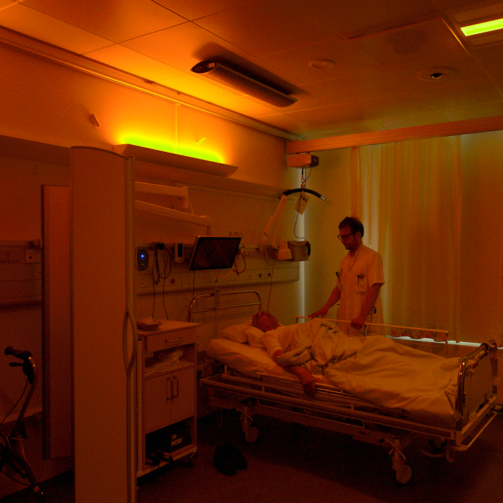
A stroke patient under soft warm nighttime lighting at Righospitalet, with neurologist Anders West of the University of Copenhagen. (Photo credit: Image courtesy of Chromaviso.)
Studies of 90 stroke patients in a Danish hospital indicate that LED lighting tuned to mimic the patterns of daylight can have a positive effect on depression, fatigue, anxiety, and wellbeing, although there was some amount of inconclusiveness, and lighting made no difference in cognition improvement.
The results of the observations at the Stroke Rehabilitation Unit in Copenhagen's Righospitalet from May 2014 through June 2015 were only recently published in the journal NeuroRehabilitation.
Righospitalet is part of the Copenhagen University Hospital. The studies were conducted by the the University of Copenhagen's departments of neurology and neurophysiology and by the hospital's department of ophthalmology, using specialty lighting from Chromaviso, based in Aarhus, Denmark.
The scientists generally tuned lighting in patient rooms to deliver the brighter and more blue-rich makeup associated with natural daylight during the mornings and afternoons. They toned down the brightness and delivered warmer spectral content in the evenings.
At the same time they delivered ordinary hospital lighting to control groups.
Read more like this: New studies show cycles of blue and red light promote circadian entrainment
The team worked on the hypothesis that the tuned lighting would support human circadian rhythms associated with the 24-hour cycle of day and night, whereas ordinary hospital lighting and indoor confinement deprives patients of circadian normalities and can thus cause physiological disturbances.
All patients were hospitalized for over two weeks.
In one study focused on fatigue, they concluded that “fatigue was significantly reduced in rehabilitation patients exposed to naturalistic lighting during admission.”  They based their conclusion on a couple of questionnaires, one called the Multidimensional Fatigue Inventory questionnaire, and the other the Rested Statement, noting that the experimental groups reported less fatigue compared to the control group.
However, by another measurement, the Pittsburgh Sleep Quality Index, “no differences were detected between groups in sleepiness or subjective sleep quality,” wrote the authors, led by Anders West from the neurology department.
Read more like this: Blue-light study shows promise in healing mild TBI
If those results were encouraging but mixed, the same could be said of a separate set of observations looking at a combination of depression, anxiety, wellbeing, and cognition.
“Depressive mood and anxiety was reduced, and wellbeing was increased in the (experimental) group at discharge compared to the (control group),” the authors reported.
However, “no difference was found in cognition,” the authors stated.
The team applied the Hamilton Depression Scale, the Major Depression Inventory, the WHO-Five Well-being Index, the Hospital Anxiety and Depression Scale, and the Montreal Cognitive Assessment to determine the results.
“This study is the first to demonstrate that exposure to naturalistic light during admission may significantly improve mental health in rehabilitation patients,” they concluded, adding that “further studies are needed to confirm these findings.”
Chromaviso has been very active in lighting tuned to health and recovery, in areas including dementia, psychiatric disorders, brain trauma, surgery, and others.

MARK HALPER is a contributing editor for LEDs Magazine, and an energy, technology, and business journalist (markhalper@aol.com).

Sunday, January 19, 2020

Blue light reduces organ injury from ischemia and reperfusion

Would wearing blue light googles immediately upon entering the ambulance or hospital prevent some of the reperfusion injury? We'll never know because we have NO leadership and NO strategy in stroke.

Blue light reduces organ injury from ischemia and reperfusion

Significance

It is well established that light regulates mammalian biology. And yet, we have been unable to define and thus harness the underlying mechanisms so as to apply them to alter the course of human disease. In this study we determine that the spectrum of light is a critical determinant of its effect on critical illness. We show that an acute and short (24 h) exposure to high-illuminance (1,400 lx) blue spectrum (peak 442 nm) light prior to ischemia/reperfusion (I/R) significantly attenuates the degree of organ injury. Our characterization of the biological mechanisms through which blue light beneficially alters the cellular response to I/R provides an opportunity to develop novel therapeutics for the prevention and treatment of many diseases.
Keywords: blue light, ischemia, reperfusion, organ injury, circadian rhythms

Abstract

Evidence suggests that light and circadian rhythms profoundly influence the physiologic capacity with which an organism responds to stress. However, the ramifications of light spectrum on the course of critical illness remain to be determined. Here, we show that acute exposure to bright blue spectrum light reduces organ injury by comparison with bright red spectrum or ambient white fluorescent light in two murine models of sterile insult: warm liver ischemia/reperfusion (I/R) and unilateral renal I/R. Exposure to bright blue light before I/R reduced hepatocellular injury and necrosis and reduced acute kidney injury and necrosis. In both models, blue light reduced neutrophil influx, as evidenced by reduced myeloperoxidase (MPO) within each organ, and reduced the release of high-mobility group box 1 (HMGB1), a neutrophil chemotactant and key mediator in the pathogenesis of I/R injury. The protective mechanism appeared to involve an optic pathway and was mediated, in part, by a sympathetic (β3 adrenergic) pathway that functioned independent of significant alterations in melatonin or corticosterone concentrations to regulate neutrophil recruitment. These data suggest that modifying the spectrum of light may offer therapeutic utility in sterile forms of cellular injury.

Blue Light Can Help Heal Mild Traumatic Brain Injury

Your doctor is incompetent if these earlier ones on blue light were not put into stroke protocols for your use. Go ahead and ask your doctor why they are so incompetent. Can't your doctor translate TBI interventions to stroke?

Laziness? Incompetence? Or just don't care? No leadership? No strategy? Not my job?  The board of directors didn't tell them that totally solving stroke was their job, not just lazily relying on the status quo?

After Stroke, 'Blue' Light May Help Beat the Blues  February 2017 


Blue light reduces organ injury from ischemia and reperfusion  December 2016


Could blue light reduce blood pressure?  November 2018 

Daily Blue Light Therapy Reduces Post-Concussion Symptoms After Mild Traumatic Brain Injury

June 2019

The Effects Of Naturalistic Light On Diurnal Plasma Melatonin And Serum Cortisol Levels In Stroke Patients During Admission For Rehabilitation: A Randomized Controlled Trial January 2019


The latest here:

Blue Light Can Help Heal Mild Traumatic Brain Injury

Early morning blue light exposure therapy can aid the healing process of people impact by mild traumatic brain injury, according to new research from the University of Arizona.
"Daily exposure to blue wavelength light each morning helps to re-entrain the circadian rhythm so that people get better, more regular sleep. This is likely true for everybody, but we recently demonstrated it in people recovering from mild traumatic brain injury, or mTBI. That improvement in sleep was translated into improvements in cognitive function, reduced daytime sleepiness and actual brain repair," said William D. "Scott" Killgore, psychiatry professor in the College of Medicine - Tucson and lead author on a new study published in the journal Neurobiology of Disease.
Mild traumatic brain injuries, or concussions, are often the result of falls, fights, car accidents, and sports participation. Among other threats, military personnel can also experience mTBI from exposure to explosive blasts: Shockwaves strike the soft tissue of the gut and push a burst of pressure into the brain, causing microscopic damage to blood vessels and brain tissue, Killgore said.
"Your brain is about the consistency of thick Jell-O," he said. "Imagine a bowl of Jell-O getting hit from a punch or slamming against the steering wheel in a car accident. What's it doing? It's absorbing that shock and bouncing around. During that impact, microscopic brain cells thinner than a strand of hair can easily stretch and tear and rip from the force."
Those with a concussion or mTBI might can momentarily seen stars, become disoriented, or even briefly lost consciousness following the injury; however, loss of consciousness doesn't always happen and many people who sustain a concussion are able to walk it off without realizing they have a mild brain injury, according to Killgore. Headaches, attention problems and mental fogginess are commonly reported after head injuries and can persist for weeks or months for some people.
Few, if any, effective treatments for mTBI exist. The US Army Medical Research and Development Command funded the research to find alternatives to medicinal methods of mTBI recovery.
"About 50 percent of people with mTBI also complain that they have sleep problems after an injury," Killgore said.
Recent research has shown that the brain repairs itself during sleep, so Killgore and his co-authors—John Vanuk, Bradley Shane, Mareen Weber, and Sahil Bajaj, all from the Department of Psychiatry—sought to determine if improved sleep led to a faster recovery.

In a randomized clinical trial, adults with mTBI used a cube-like device that shines bright blue light (with a peak wavelength of 469 nm) at participants from their desk or tables for 30 minutes early each morning for six weeks. Control groups were exposed to bright amber light.
"Blue light suppresses brain production of a chemical called melatonin," Killgore said. "You don't want melatonin in the morning because it makes you drowsy and prepares the brain to sleep. When you are exposed to blue light in the morning, it shifts your brain's biological clock so that in the evening, your melatonin will kick in earlier and help you to fall asleep and stay asleep."
People get the most restorative sleep when it aligns with their natural circadian rhythm of melatonin—the body's sleep-wake cycle associated with night and day.
"The circadian rhythm is one of the most powerful influences on human behavior," Killgore said. "Humans evolved on a planet for millions of years with a 24-hour light/dark cycle, and that's deeply engrained in all our cells. If we can get you sleeping regularly, at the same time each day, that's much better because the body and the brain can more effectively coordinate all these repair processes."
As a result of the blue light treatment, participants fell asleep and woke an average of one hour earlier than before the trial and were less sleepy during the daytime. Participants improved their speed and efficiency in brain processing and showed an increase in volume in the pulvinar nucleus, an area of the brain responsible for visual attention. Neural connections and communication flow between the pulvinar nucleus and other parts of the brain that drive alertness and cognition were also strengthened.
"We think we're facilitating brain healing by promoting better sleep and circadian alignment, and as these systems heal, these brain areas are communicating with each other more effectively. That could be what's translating into improvements in cognition and less daytime sleepiness," Killgore said.
Blue light from computers, smartphones and TV screens often gives blue light a bad rap. But according to Killgore, "when it comes to light, timing is critical. Light is not necessarily good or bad in-and-of-itself. Like caffeine, it all comes down to when you use it. It can be terrible for your sleep if you're consuming coffee at 10 o'clock at night, but it may be great for your alertness if you have it in the morning."
He and his team plan to continue their research to see if blue light improves sleep quality and how light therapy might affect emotional and psychiatric disorders. Killgore believes that most people, whether injured or healthy, could benefit from correctly timed morning blue light exposure, a theory he hopes to prove for certain in future studies

Thursday, June 13, 2019

Daily Blue Light Therapy Reduces Post-Concussion Symptoms After Mild Traumatic Brain Injury

Your doctor has some analysis to do. Is daytime sleepiness better treated with this or sex? 

Your doctor is incompetent if these earlier ones on blue light were not put into stroke protocols for your use. Go ahead and ask your doctor why they are so incompetent. 

Laziness? Incompetence? Or just don't care? No leadership? No strategy? Not my job?  The board of directors didn't tell them that totally solving stroke was their job, not just lazily relying on the status quo?

After Stroke, 'Blue' Light May Help Beat the Blues  February 2017 


Blue light reduces organ injury from ischemia and reperfusion  December 2016


Could blue light reduce blood pressure?  November 2018 

The latest here:

Daily Blue Light Therapy Reduces Post-Concussion Symptoms After Mild Traumatic Brain Injury

By Matt Silver

SAN ANTONIO, Tex -- June 11, 2019 -- Daily blue light therapy effectively reduces daytime sleepiness and post-concussion symptoms related to mild traumatic brain injury (TBI), according to a study presented here at SLEEP 2019, the 33rd Annual Meeting of the Associated Professional Sleep Societies (APSS).

“An estimated 30% to 80% of individuals report long-term sleep disruption following a mild traumatic brain injury,” said Adam Raikes, MD, University of Arizona, Tucson, Arizona. “Given the critical role of high-quality sleep on optimal cognitive, emotional, sport, and physical performance, ameliorating these adverse effects is essential.”

For the current randomised study, the researchers compared the effects of daily blue light therapy with those of amber light therapy in 27 individuals who have suffered mild TBI, hypothesising that subjects receiving blue light would self-report improved sleep outcomes.

All subjects underwent 6 weeks of daily light treatment for 30 minutes, with each treatment taking place in the morning, lasting 30 minutes, and consisting of direct exposure via light box.

Daytime sleepiness was measured using the Epworth Sleepiness Scale (ESS) and post-concussion symptoms were measured using the Rivermead Post-concussion Symptom Questionnaire (RPCSQ).

Individuals in the blue light group showed significantly improved daytime sleepiness (P = .025) and somatic (P = .053), and cognitive symptoms (P = .082) than those in the amber light group.

Bivariate correlations indicated that improvements in ESS scores were significantly associated with improvements in somatic symptoms (P = .014).

“These improvements are likely to have significant positive effects on individuals’ daytime functioning (cognitive, physical, emotional, and sport) and overall quality of life,” the authors concluded.

They noted that more research is needed to investigate the causality of these findings and the neurological mechanisms underpinning them.

[Presentation title: Daily Blue Light Therapy Reduces Daytime Sleepiness and Post-Concussion Symptoms After Mild Traumatic Brain Injury. Abstract 0935]

Tuesday, January 1, 2019

The Effects Of Naturalistic Light On Diurnal Plasma Melatonin And Serum Cortisol Levels In Stroke Patients During Admission For Rehabilitation: A Randomized Controlled Trial

Weren't these earlier ones enough to get your doctor to set up a stroke protocol?

After Stroke, 'Blue' Light May Help Beat the Blues  February 2017 


Blue light reduces organ injury from ischemia and reperfusion  December 2016

Could blue light reduce blood pressure?  November 2018 

Oh well, incompetence reigns supreme in stroke.  This repeat research is a direct result of not having a database of stroke research and protocols. Which our

fucking failures of stroke associations should have set up decades ago.  You're screwed along with your children and grandchildren that have strokes.


The Effects Of Naturalistic Light On Diurnal Plasma Melatonin And Serum Cortisol Levels In Stroke Patients During Admission For Rehabilitation: A Randomized Controlled Trial

Abstract

Background: Stroke patients admitted for rehabilitation often lack sufficient daytime blue light exposure due to the absence of natural light stimulation and are often exposed to light at unnatural time points.
We hypothesized that artificial light imitating daylight, termed naturalistic light, would stabilize the circadian rhythm of plasma melatonin and serum cortisol levels among long-term hospitalized stroke patients.  
Methods: A quasi-randomized controlled trial. Stroke patients in need of rehabilitation were randomized between May 1, 2014, and June 1, 2015 to either a rehabilitation unit equipped entirely with always on naturalistic lighting (IU), or to a rehabilitation unit with standard indoor lighting (CU). At both inclusion and discharge after a hospital stay of at least 2 weeks, plasma melatonin and serum cortisol levels were measured every 4 hours over a 24-hour period. Circadian rhythm was estimated using cosinor analysis, and variance between time-points.  
Results: A total of 43 were able to participate in the blood collection. Normal diurnal rhythm of melatonin was disrupted at both inclusion and discharge. In the IU group, melatonin plasma levels were increased at discharge compared to inclusion ( 2.9; IQR: −1.0 to 9.9, p = 0.030) and rhythmicity evolved p = 0.007). In the CU group, melatonin plasma levels were similar between discharge and inclusion and rhythmicity evolved. Overall, both patient groups showed normal cortisol diurnal rhythms at both inclusion and discharge.  
Conclusions: This study is the first to demonstrate elevated melatonin plasma levels and evolved rhythmicity due to stimulation with naturalistic light.    
Key words: Stroke; Rehabilitation; Circadian rhythm; Light; Melatonin; Cortisol
MANUSCRIPT
Introduction
Interventional uses of light have attracted growing interest since the recent discovery of the blue light absorbing Melanopsin-expressing photosensitive ganglion cells (ipRGCs) in the retinal ganglion cell layer. Especially a subtype of ipRGCs (M1) pass the highest amount of light stimulation through the optic nerve and retinohypothalamic tract to the master circadian clock system in the suprachiasmatic nucleus (SCN). Several studies indicate that sunlight is the strongest entrainment for the circadian rhythm because of the sensitivity for short-wavelength blue light [1].
Light stimulation to the SCN also happens through the intergeniculate leaflet (IGL), which appears to be an important secondary route for sunlight entrainment [2].
The SCN affects melatonin and cortisol in a manner involving the oscillation system within the SCN and its direct autonomic connection with peripheral tissue.
            Melatonin is produced from serotonin in the pineal gland, and its circuitous pathway is regulated by the SCN. Light normally inhibits melatonin secretion, such that it is low during the day and peaks late at night, and this temporal pattern is relatively unaltered by changes in sleep habits
[3]. During hospitalization, critically ill patients reportedly exhibit low melatonin levels and a disrupted diurnal melatonin rhythm [4,5]. Patients with cortical stroke also show decreased melatonin secretion [6-8] and a disturbed diurnal rhythm [9]. Although the physiological explanation of this phenomenon is unknown. It is possible that the initial edema and widespread cortical lesions may affect areas projecting to the IGL, impairing light perception to the SCN, and through that disrupting circadian rhythm regulation [6].
Another well-known circadian-regulating hormone, cortisol, synchronizes peripheral circadian oscillators and controls 60% of the circadian transcriptome [10]. Cortisol secretion is controlled by the SCN, where neuronal projections signal directly to the paraventricular hypothalamic nucleus (PVH) and dorsomedial hypothalamus (DMH). Cortisol levels normally rise around midnight, peak in the early morning, and decrease again around 9 a.m. Cortisol is reportedly elevated in response to external stimulus, such as hospital admission and surgery [11,12]. However, it seems likely that cortisol is more stable than melatonin in critically ill patients exposed to diurnal disruption [13].
            Hospitalization and circadian rhythm disruption reportedly have negative consequences [14]. Patients admitted for post-stroke rehabilitation carry a high risk of circadian disruption due to the duration of hospitalization and immobilization. This combination deprives patients of natural light from the sun, subjects them to many hours of artificial light from the evening and nighttime indoor hospital lighting.
            LED (light-emitting diode) technologies support the development of artificial light with specific wavelengths. Together with computerized technology, this enables the production of lamps that can imitate the natural sunlight spectrum and rhythm—termed naturalistic light, circadian light, or dynamic lighting. Melatonin levels are influenced by light interventions [15], and several studies show that short-wave light is an isolated melatonin manipulator [16-19]. Previously tested light interventions have not detectably altered melatonin levels in patients in real-hospital settings [20,21]. However, no studies have investigated the influence of naturalistic light on melatonin levels and its diurnal rhythm.
            In the present study, we aimed to determine whether naturalistic light could stabilize the circadian rhythm of melatonin and cortisol, and increase the expected low plasma melatonin levels in stroke patients admitted for rehabilitation.
Materials and Methods
Study design and Participants
This study was performed in the Stroke Rehabilitation Unit, Department of Neurology, Rigshospitalet, Copenhagen. The methods have been previously described in detail [14]. Briefly, the study included stroke patients who required over 2 weeks of in-hospital rehabilitation during the period from May 1st of 2014 to June 1st of 2015. Patients were excluded if they were unable to give consent due to their awareness status, severe aphasia, or less than 2 weeks of hospitalization in the rehabilitation unit. We conducted a parallel randomized controlled trial with two arms: an intervention group admitted to a rehabilitation unit equipped with naturalistic light (IU), and a control group admitted to a rehabilitation unit with standard indoor lighting (CU). No safety precautions were necessary regarding assessments and interventions. The study was approved by the Danish scientific ethics committee (H-4-2013-114) and the Danish Data Protection Agency (2007-58-0015), and is registered at ClinicalTrials.gov (Identifier: NCT02186392).

Randomization
Randomization was performed by non-blinded stroke nurses (quasi-randomization) at the acute stroke unit (with normal standard light conditions). The nurses were not involved in the study and were simply following normal procedure regarding the relocation of patients to the two rehabilitation units.

Naturalistic light intervention
In all areas of the intervention rehabilitation unit, a 24-hour naturalistic lighting scheme was implemented using multi-colored LED-based luminaires (lamps) managed by a centralized lighting controller according to the lighting scheme (Chromaviso, Denmark). The lighting was dim in the morning (from 7 am), increased to reach maximum illuminance around betweennoon and 3 pm with strong inclusion of the blue light spectrum, and then dimmed again throughout the evening with diminishment of the blue light spectrum, ensuring no IpRGC stimulation during nighttime. The luminaires were located in the ceiling and at the wall behind the beds, and theThis naturalistic lighting scheme ran constantly throughout the inclusion period.
Due to the complexity and the need for comprehensive technical description of the light, the light intervention ispresented in details in the method description paper [14]where the irradiance profiles can be found in figure 3a and 3b. The technical light description is produced in accordance with CIE TN 003 following the principles of Lucas et al. [22].Normal ceiling luminaries
were installed in the CU. They had new fluorescent tubes installed prior to the inclusion in order to uniform the light in all areas of the CU. The technical light description regard the irradiance profiles for the IU can be found in figure 3a and for CU in 3b in West et al.[14]

Measurements
All acute stroke patients underwent standard initial examinations. Additionally, the Morningness-Eveningness Questionnaire (MEQ) was performed at both inclusion and discharge to determine the distribution of circadian classes. Daily life in the patient ward was best suited to morning types, such that evening-type circadian class could potentially interfere with outcome for these patients. The MEQ is validated for determining individual circadian rhythm [23], and divides patients into five types: Definitely Evening Type, Moderately Evening Type, Neither Type, Moderately Morning Type, and Definitely Morning Type. The highest scores indicate the morning type.

Blood samples
Blood samples were collected at both inclusion and discharge
(hospital treatment complete/done) for measurement of melatonin and cortisol levels at 4-hour intervals, seven times over a 24-hour period: 08 a.m., noon, 04 p.m., 08 p.m., midnight, 04 a.m., and again at 08 a.m. To prevent external factors other than light from influencing plasma melatonin and serum cortisol levels, the participants were asked to avoid parameters which could influence the blood levels [14] (Table S1). Travel to different time zones and regular night work within the last 14 days were registered. The instructions were given both verbally and in writing. To avoid circadian stimulation, blood collection was performed in dim lighting from an old incandescent bulb, which has very low emission of the blue light spectrum. During collection, the lamp was pointed towards the arm, away from the patient. Blood samples were centrifuged directly after collection, and plasma and serum were separated. Samples were immediately stored at −50°C, and within 30 hours were stored at −80°C until further analysis.
Biochemical analysis
Plasma melatonin concentrations were analyzed by use of a Melatonin Direct Radioimmunoassay (LDN Labor Diagnostika Nord GmbH and Co. Nordhorn) according to the kit instructions. The limit of detection was 2,3 pg/mL,the measuring range was 2.3 - 1000 pg/mL and the analytical between-run coefficient of variations were 19,6% at 24 pg/mL and 14% at 70 pg/mL. 
Serum cortisol concentrations were determined on a Cobas e 411 analyzed (Roche Diagnostics, Basel, Switzerland) by an electro-chemiluminescence immunoassay.The limit of detection was 0.5 nmol/L, the measuring range was 2 - 17500 nmol/L and the analytical between-run coefficient of variation was 3% at 330 nmol/L.


MRI radiological classification
MRI sequences were performed, and brain lesions were classified according to volume and anatomic localization by a neuro-radiologist. The infarction volume (in cm3) was calculated by measuring the infarction size in the coronal, transversal, and sagittal planes. All scans were performed using a 1.5 Tesla MR scanner (Siemens, General Electrics), and included the following sequences: a sagittal T2-weighted turbo spin echo sequence (FSE), an axial T2-weighted FSE, an axial fluid attenuation inversion recovery (FLAIR) sequence, an axial 3 scan trace diffusion-weighted imaging sequence, a sagittal 3D T1WI sequence, and an axial susceptibility-weighted imaging sequence.

Outcomes
This study was part of a larger investigation of the effects of light on human rehabilitation patients health as measured by psychological parameters, biochemical parameters, fatigue, and sleep. As this subject is a relatively new scientific field, the study was considered an exploratory investigational study. We chose five primary endpoints, including melatonin and cortisol levels and rhythmicity in the present study.


Statistical analysis
All analyses were performed using SAS (SAS Inst. Inc., Cary, NC USA, 9.4). A p value of <0.05 was considered significant. Between-group differences regarding basic demographic parameters were calculated using the t-test for continuous variables, and chi-square-test for categorical variables. Normally distributed continuous variables were expressed as mean ± standard deviation (SD). The melatonin plasma levels and cortisol serum levels were not normally distributed; therefore, these data were expressed as median and interquartile range (IQR).
            Data were logarithmically transformed prior to mixed model analysis, and were subsequently transformed back to empirical fractiles to achieve parametric distribution, which were then converted to percentage variance ((x−1) * 100). The deviation of calculated cosinor rhythmicity was expressed as standard error (SE). Cosinor rhythmicity was analyzed assuming a 24-hour time-period [24]. The data were fitted to a combined cosine and sine function: y = M + k1COS(2pt/24) + k2SIN(2pt/24). The 24-hour rhythms of each group were further characterized by the following rhythm parameters: mesor (rhythm-adjusted average about which oscillation occurs), amplitude (difference between the highest and lowest values of the fitted cosinor curve), and times of peak and nadir [24,25]. Data analyses were performed using the GPLOT procedure in SAS. Mixed model analysis was performed in SAS to describe the variance between time-points of the diurnal rhythm of melatonin and cortisol at inclusion and discharge in each unit.
            Infarction size was correlated to melatonin and cortisol mean values using regression analysis. Infarction location was included as a confounding element by analysis of covariance. The Wilcoxon signed-rank test was used to describe within-group changes from inclusion to discharge. The mMelatonin mean plasma values were calculated from all time-points together (24 h). Due to the preserved diurnal rhythm, cortisol mean serum values were further divided into day (high-secretion phase; 24–12 h) and night (low-secretion phase; 12–24 h) values. Melatonin plasma levels did not show a diurnal rhythm in either unit; thus, the division of mean melatonin values into further stages was not relevant.

Results
Among 256 screened patients who required in-hospital neurorehabilitation, 90 met our inclusion criteria, of whom 73 avoided meeting exclusion criteria, death, and severe illness until discharge. Of these 73 included patients, 30 dropped out before discharge, while the remaining 43 patients completed the study (Figure 1). The main reasons for missed blood collection were the patient’s discomfort with the procedure, and technical complications with the first 9 included patients. Patients were also excluded from blood collection due to fragile veins and low hemoglobin concentration. Melatonin data from one patient were excluded due to prescribed melatonin treatment. Cortisol data were excluded due to very high cortisol values resulting from respiratory distress in one patient who unexpectedly died a few hours after the last blood sampling. NIHSS (Included N=43; 5.0 (±4.2); excluded N=30; 7.8 (±6.4): p=0.04) and Barthel (Included N=43; 56.9 (±30.0); excluded N=30; 39.1 (±31.2): p=0.02) scores were calculated in the group of excluded patients and indicated significant worse disability scores compared to the included participants (table S2).
[Figure 1]

            A total of 33 patients were willing and able to sufficiently answer the MEQ. The two groups did not significantly differ in circadian class distribution (chi-square test) (Table 1). Table 1 presents the demographic data. The two groups were well matched, except regarding the number of smokers (IU 13, CU 16, p = 0.02). Pre-analytical variability was estimated to be equal among the patients based on the information collected before blood sampling, and was therefore not included as a confounding or interaction element.
[Table 1]

Circadian rhythm of melatonin and cortisol
At both inclusion and discharge, both patient groups lacked a normal diurnal rhythm of plasma melatonin. Melatonin plasma levels did not follow a cosinor rhythmicity in either group, at either time-point (Table 2). Regarding the variance between time-points, the CU group appeared to have an abnormal but diurnal melatonin rhythmicity at inclusion (Table 3). However, this rhythmicity was absent at discharge which is also illustrated by Figure 2,b. In the IU group at inclusion, melatonin plasma levels only significantly differed between 08 p.m. and at discharge, melatonin levels significantly differed between each time-point (Table S3), with elevated levels from 08 a.m. to noon and from 08 p.m. to midnight illustrated by Figure 2,a. In the IU group, we detected significant changes over time between inclusion and discharge. Such differences were not evident in the CU group (Table 3).
            A significant cortisol cosinor rhythm (p < 0.0001) was detected in both patient groups at both inclusion and discharge (Table 2, Figure 2,
ca,
db). The CU group showed a significant amp/peak difference in cortisol values between inclusion and discharge (p = 0.005) which illustrate the decrease in cortisol levels between inclusion and discharge (Figure 2,d). Cosinor analysis and the calculated variance between time-points revealed that both groups showed a significant cortisol rhythm at both inclusion and discharge but that the diurnal rhythm also changes in the variance of the rhythmic pattern between inclusion and discharge at both unit (Table 3, Table S4), which 3,b also illustrate. The curves in Figure 3,b illustrate that the largest discrepancy between groups was during the first part of the day, when the CU group showed decreasing levels and the IU group showed stable levels.
[Table 2]
[Table 3]
 [Figure 2]
[Figure 3]

Mean levels of plasma melatonin and serum cortisol
Table 4 summarizes the differences in melatonin and cortisol levels between inclusion and discharge for all patients.
 [Table 4]

Melatonin plasma values significantly increased from inclusion to discharge in the IU group (n = 23; median diff, 2.9; IQR: −1.0 to 9.9; p = 0.030), but not in the CU group (n = 19; median diff, −1.5; IQR: −7.0 to 6.3; p = 0.418) (Table 4). Figure 3,a shows the melatonin delta-curve, illustrating the melatonin level changes between inclusion and discharge in the IU and not in the CU groups, and supporting a 24-hour increase.
            The mean day cortisol serum levels significantly decreased from inclusion to discharge in the CU group (n = 20; median diff, −59.6; IQR: −129.4 to 13.2; p = 0.003), but did not significantly change in the IU group (n = 22; median diff, 5.6; IQR: −68.7 to 59.7; p = 0.945). During the admission time-period, cortisol night values increased in the IU group, and decreased in the CU group which is illustrated by Figure 3,b. However, these changes were not statistical significant (Table 4).
            Analysis of covariance was performed to investigate cortical, striatocapsular, and large infarcts as confounding factors for the influence on melatonin and cortisol levels. Cortisol and melatonin levels were not significantly associated with these infarction types. Regression analysis revealed that lesion size was also not significantly correlated with melatonin (n = 27; Estimate, −0.03; 95% CI: −1.4, 0.09; p = 0.62) or cortisol values (n = 26; Estimate, 0.027; 95% CI: −0.34, 0.88; p = 0.38) (Estimate = diff. lesion size mm3). Regression analysis also showed that length of hospitalization was not significantly correlated with melatonin or cortisol levels.


Discussion
This study is the first to investigate the Our present results show for the first time that exposureeffect of a to a naturalistic light environment exposure on melatonin and cortisol levels in stroke patients during at least 2 weeks of hospitalization. can stimulate rhythmicity and increase melatonin plasma levels in stroke patients.
            At the time of inclusion in our study, the stroke patients in both groups exhibited an eradicated normal diurnal pattern of melatonin, with the lack of a normal peak. At discharge, the IU group exhibited significantly increased plasma melatonin levels and a present but abnormal diurnal rhythmicity. Conversely, the CU group exhibited significant but abnormal diurnal rhythmicity at inclusion, which was absent at discharge. The absent peak levels and disrupted diurnal rhythm of melatonin in our cohort is in line with the impaired melatonin secretion and disturbed rhythmicity commonly reported after stroke.
Since melatonin is synthetized from serotonin, it is reasonable to believe that melatonin production could be affected by the known reduction/disturbances of serotonin synthesis after stroke [26,27]. This could explain the absence of a melatonin secretion peak in our study. Furthermore, it has been suggested that widespread cortical lesions could affect areas projecting to the intergeniculate leaflet (IGL), potentially impairing light perception to the SCN and the pineal gland, and disrupting circadian rhythm regulation and melatonin secretion [6]. However, we did not find that melatonin and cortisol values were significantly correlated with lesion size, or with cortical and striatum infarcts. Notably, not all patients underwent MRI scanning; thus, the correlation was only calculated in a subgroup of patients, potentially influencing the results.
            Blue light exposure during the day reportedly increases nightly melatonin secretion [28,29] and prevents the melatonin suppression caused by light exposure at night [30]. This may explain the high melatonin secretion in the IU group compared to the CU group. The increased melatonin levels in the IU group appeared to persist over the 24-hour measurement period (Figure 3,a) despite the high exposure to the blue light spectrum at the start of the day. Although the physiological explanation is not immediately evident, it may be related to the disturbed diurnal rhythm. The CU group had reduced exposure to blue light during the daytime, which could make the melatonin suppression more sensitive to light [31] and inhibit melatonin secretion [32]. This might result in the CU group having lower melatonin levels than the IU group during the daytime, as well as at nighttime since the CU group was frequently exposed to blue light-emitting ward lights in their rooms at night. Beta-blockers have been shown to reduce the production of melatonin[33]. Beta-blockers are widely used in stroke prevention and therefore in our patient cohort. However, the distribution of beta-blockers between the two units was unequal, as there was a greater prescription at the IU (Inclusion: IU; N=12, CU; N=6. Discharge: IU; N=18, CU; N=8). This unequal distribution may have hypothetically decreased the melatonin production at the IU compared with the CU.
            Compared to melatonin, less is understood about cortisol’s response to light. We found no change in cortisol levels in the IU group, but significantly reduced cortisol levels in the CU group. The higher cortisol levels in the IU group compared to the CU group may be correlated with positive health effects, such as improved cognition, mood, and well-being [18]. However, these correlations could also be related to the light-enhanced cortical activity [34].
            Unlike the melatonin rhythm, the human cortisol rhythm does not seem to be associated with day and night. However, cortisol secretion is dependent on the phase of light, particularly transition periods from dark to light and, to a lesser extent, from light to dark. The IU and CU groups showed the greatest difference in cortisol serum levels during the first part of the day period (Table 4 and Figure 3,b). This corresponds well with previous findings that cortisol levels increase in response to the change from dim light to bright light exposure in the morning, but not in the afternoon or night [18,35,36]. However, it would also be expected that bright light would not affect cortisol levels during the afternoon or nighttime, since cortisol production is usually low at those times.
            Our results showed a discrepancy between the circadian rhythms of melatonin and cortisol. While the normal 24-h rhythm of melatonin secretion was eradicated, the normal 24-h rhythm of cortisol was preserved. This preserved cortisol rhythmicity is not evident for a normal preserved SCN function. Even in the absence of a functional SCN pacemaker, the adrenal gland and its own clock system can still be light-entrained by gating the sensitivity of the adrenal to ACTH via modulation of circadian corticosterone rhythms [37]. Although stroke hypothetically leads to IGL destruction, cortisol may be less sensitive to reduced IGL function and impaired serotonin levels than melatonin, due to its different approaches to light and its secondary circadian control. It remains uncertain whether this persists throughout a patient’s hospitalization. It is possible that the preserved 24-h cortisol rhythm resulted from a combination of the HPA axis and the autonomic nervous system, and their activation and inhibition from the SCN.

Limitations and strengths
Patients were randomly allocated following the normal procedure for an equal distribution of patients to the two rehabilitations units (quasi-randomization). The conditions in the two rehabilitation units were equal with regards to size, form, and staff professions. The impact of daylight on the facade of the two units was not completely identical, since the angle of sun exposure differed between the two wings during all four seasons. However, measurement of the incoming sunlight revealed no significant differences between the two units [14], assuming that levels above 200 lux were required to stimulate the circadian center [38]. As illustrated in West et al.[14] (figure 4a and 4b), there was no appreciable difference between units in daylight exposure at the window side bed across the year other than the use of curtains in the IU. Figure 4a illustrate that there was a difference in daylight exposure between IU and CU at the bed nearest to the door, but all illuminance levels fall below the required level of 200 lux D55 equivalent light to generate a diurnal stimulation of the circadian center [38].Thus, we do not view this difference as clinically important. Furthermore, it does not favor the IU.  
The intervention unit had blackout curtains that went up at 08 a.m. and down at 08 p.m. during all four seasons. Furthermore, the naturalistic light had much higher lux levels than the incoming natural sunlight, eliminating the influences of natural light in the IU and of differing light exposure between beds.
Therefore, variations in light exposure between bed positions (two beds: window side and away-from-window side) were only relevant in the CU. It was estimated that the lux values
lightsignificantly differed between beds during 40% of the meteorological time, over a five-hour period, during the peak summer season, and this difference disappeared outside the summer period. During the study period, information was collected on all bed positions, and all patients were placed near the window at the end of their stay due to the natural rotation in the units. Overall, we found no differences in bed positions between patients; thus, bed positions were excluded from the calculations.Artificial light sources at the control unitwerenormal indoor ceiling luminaries and a bedside lamp. The use of these light sources could not be measured due to the random use seen in a normal ward and >because of the absent in manipulation of the light sources due to the control setup. The technical light description regarding the ceiling light at the control unit is described in the method description
paper [14]. 
            Blood testing could only be performed for 43 participants. The two units significantly differed with regards to smoking (p = 0.02), which we considered to be a random finding. NIHSS and Barthel scores significantly differed between the included and excluded participants, which was expected since the most severely impaired patients had the most difficulties participating in blood collection. At the start of the study period, saliva collection was tested as a method; however, our the stroke patients showed a lack of saliva production, making this method unusable. Due to the RCT study design, all participants were equally disturbed during blood collection, for example, by waking for evening sampling.
            Strengths of this study include the power of having two comparable units, and the ability to include data for all four seasons, since sunlight exposure in Denmark significantly changes throughout the year. This study was performed in a real-hospital setting; therefore, the results reflect the real-life situation in a rehabilitation hospital ward. However, this study was part of an exploratory investigational study in a relatively new scientific area. Thus, more specific studies are needed to further address the effects of naturalistic light on the levels and rhythmicity of melatonin and cortisol.


Conclusions

The present results indicate a physiologically influence of naturalistic light on melatonin<
and cortisol levels in patients hospitalized more than 2 weeks<
reflect the impact of naturalistic light in a real-world clinical setting. There exists a need for clinical trials in circadian rhythm research with real patients in a real-world clinical settingthe field of circadian rhythm research, and our study addresses that need. These findingsdemonstrates a rationale for further investigations on  demonstrate that it is possible to increase plasma melatonin levels and to alter the circadian rhythm by using naturalistic light in a real-hospital setting. Further trials are needed to investigate the exact implications of the observed circadian rhythm alterations, and to examine the long-term effects of the circadian light intervention.


Acknowledgments
We are deeply grateful to the stroke patients for their participation in this study. We thank service manager Svend Morten Christiansson and architect Maj Lis Brunsgård Seligmann from the Service Center, Rigshospitalet Glostrup, for their interest in naturalistic light, and for making it possible to install naturalistic lighting throughout an entire hospital ward. We thank the company ChromaViso
especially Masterin optical engineeringTorben SkovHansenfor always being available for technical questions and assistance regarding the light set-up
and light description. We thank Nina Vindegaard Grønberg, MD, who was a great help in collecting data during periods of high work pressure. Finally, we are grateful to the health staff of the entire stroke department, Rigshospitalet Glostrup, for their engagement and professionalism as they provided support and logistical assistance during the project period. The last gratitude goes to The Market Development Foundation Denmark for financing the project.

Competing interest
The authors have declared that no competing interest exists.


Friday, November 23, 2018

Could blue light reduce blood pressure?

Weren't these earlier ones enough to get your doctor to set up a stroke protocol?

After Stroke, 'Blue' Light May Help Beat the Blues  February 2017 

Blue light reduces organ injury from ischemia and reperfusion  December 2016

Oh well, incompetence reigns supreme in stroke. 


Could blue light reduce blood pressure?


Healthline/Medical News Today | November 12, 2018
Exposure to blue light may help combat hypertension, according to a recent study. If the findings are replicated in a larger sample, blue light could provide a cost-effective and side effect-free intervention.
The results of an investigation into the effects of blue light on high blood pressure were recently published in the European Journal of Preventative Cardiology.
High blood pressure, or hypertension, is widespread in the US and many other Western countries.
According to the Centers for Disease Control and Prevention (CDC), high blood pressure is a primary or contributing cause of almost 1,000 deaths every day in the U.S.
Hypertension is also a risk factor for more serious conditions, such as heart attack, stroke, and kidney disease. For these reasons, managing blood pressure effectively is a high priority.
Currently, doctors usually prescribe medications such as angiotensin converting enzyme inhibitors and calcium channel blockers to help manage hypertension. Although these can be effective, side effects are common and include a persistent dry cough and dizziness.
Because high blood pressure is so prevalent, a great deal of research has investigated its causes. The importance of lifestyle, including diet and exercise, cannot be overstated, while other risk factors play smaller roles.
For instance, studies have demonstrated that exposure to sunlight influences hypertension. Blood pressure is, on average, lower during the summer months, and some research has linked high levels of long-term sunlight exposure to a reduced risk of dying from cardiovascular disease.

The bittersweet symphony of sunlight

Exposure to excessive levels of sunlight damages the skin, causing premature aging. Research has also established that ultraviolet (UV) light is a carcinogen — excessive exposure increases the risk of developing skin cancer.
On the other hand, as sunlight hits our skin, it drives a chemical reaction that produces vitamin D, which plays a range of essential roles in the body.
UV light also affects nitric oxide (NO) levels, and NO is an important signaling molecule in humans and virtually all other organisms.
The inner lining of blood vessels—called the endothelium—releases NO. This causes the smooth muscles around the blood vessels to relax, thereby lowering blood pressure.
When sunlight hits the skin, compounds such as nitrites break down to produce NO, which then diffuses into the deeper tissues and, finally, enters the blood vessels.
The interaction between sunlight, NO, and the smooth muscles of the blood vessels may explain why sunlight has a hypertension-minimizing effect.

A bolt from the blue

Finding a way to harness the benefits of UV light while removing the negative effects would be a significant discovery. Enter: blue light.
Recently, researchers from the University of Surrey in the UK and Heinrich Heine University Dusseldorf in Germany joined forces to investigate whether blue light without UV wavelengths has the potential to ease hypertension.
Earlier studies demonstrated that visible blue light, in a range of 420–453 nanometers in wavelength, can induce the production of NO without damaging DNA.
The latest study goes one step further. For the first time, researchers assessed whether blue light exposure could produce enough NO to significantly reduce high blood pressure.
Their proof-of-concept study involved 14 male participants aged 30–60, none of whom had preexisting cardiovascular issues, hypertension, or diabetes.

Blue light benefits

Each participant visited the testing facility twice, with 1 week between sessions. During one visit, they were exposed to 30 minutes of whole-body blue light. At the other, they received 30 minutes of "control light."
Most importantly, the scientists looked for changes in blood pressure. They also assessed other parameters, including heart rate, blood flow in the forearm, circulating levels of NO, and pulse wave velocity, a measure of how stiff the arteries are.
Members of the team took measurements before, during, and after the light exposure. They found that 30 minutes of whole-body blue light significantly lowered systolic blood pressure—the pressure in blood vessels as the heart contracts—by 8 millimeters of mercury.

According to the authors, this reduction was equivalent to, or more significant than, "the blood pressure-lowering effect sizes observed by typical blood pressure-lowering medications."

The other parameters also improved significantly under blue light, including pulse wave velocity. As expected, serum NO levels also increased. The authors conclude:
"Our present study demonstrates for the first time that whole-body blue light exposure at doses that are comparable to daily sunlight exposure decreases systolic blood pressure [...] in young, healthy male volunteers."
Of course, this pilot study included a very small number of participants, and it will need to be replicated on a much larger scale.
If the findings are duplicated, however, this could be a game-changer for the management of hypertension in some populations.
As one of the study's authors, Christian Heiss, a doctor and a professor at the University of Surrey, explains, "Exposure to blue light provides an innovative method to precisely control blood pressure without drugs. Wearable blue light sources could make continued exposure to light possible and practical."
Although blue light therapy could never replace changes in diet and lifestyle, it may provide additional benefits for certain people. As Dr. Heiss notes, "This would be particularly helpful to those whose blood pressure is not easily controlled by medication, such as older people."
To read more, click here.

Sunday, November 26, 2017

The Colour That Reduces Stress 3 Times Faster


For all the stress caused by your doctor in not doing anything about stopping the neuronal cascade of death or having any plan to get to 100% recovery.  Your doctor should have a plan to handle your stress, although I bet you have to tell her/him about this research.  Do not assume your blue light from your smartphone is appropriate.

The Colour That Reduces Stress 3 Times Faster

Lighting of this colour helps people relax three times quicker.

Blue light helps people to relax more quickly, new research finds.

Compared with regular white light, people are able to recover from a stressful experience three times quicker when sitting under blue light.

Regular stresses such as that caused by arguing with a friend or having a close deadline at work could be best tackled with blue light.

In the small study, 12 people were first stressed and then performed a relaxation session under either blue or white light.

The study’s authors explain their conclusions:

“…blue lighting accelerates the relaxation process after stress in comparison with conventional white lighting.
The relaxation time decreased by approximately three-fold (1.1 vs. 3.5 minutes).
We also observed a convergence time (3.5–5 minutes) after which the advantage of blue lighting disappeared.

This is not the first time studies have found that coloured lighting can influence people’s emotions.

The study’s authors explain:

“A study about the influence of color of walls in learning environments proved that pale colors caused more relaxation than vivid colors, and that heart rate decreased with short-wavelength colors (e.g., violet, blue and green) in comparison with longer-wavelength (e.g., yellow and red).
In addition, a few authors have successfully treated people with behavior disorders by influencing their emotional states (e.g., causing mental calm) by color lighting.
For instance, pink light was successfully utilized to reduce aggressiveness of delinquents in prison.
Furthermore, another color-lighting-based method with blue light have been used for disruptive behavior disorders…”


The study was published in the journal PLOS ONE (Minguillon et al., 2017).

Thursday, November 23, 2017

Blue-Light Therapy following Mild Traumatic Brain Injury: Effects on White Matter Water Diffusion in the Brain

Would this help stroke patients? I don't think they are referring to the blue light of your smartphones or iPads. Anything would have been helpful to counteract the massive fatigue I had post-stroke.  Or you can ask your doctor about the effectiveness of near-infrared light since it is only two years old and your doctor and stroke hospital are so efficient they already have incorporated infrared into their stroke protocols. (sarcasm)

New Study Discovers Near-Infrared Light Therapy (NILT) Effectively Treats Traumatic Brain Injury (TBI) Patients  Aug. 2015

Blue-Light Therapy following Mild Traumatic Brain Injury: Effects on White Matter Water Diffusion in the Brain

  • Social, Cognitive and Affective Neuroscience Laboratory (SCAN Lab), Department of Psychiatry, College of Medicine, University of Arizona, Tucson, AZ, United States
Mild traumatic brain injury (mTBI) is a common and often inconspicuous wound that is frequently associated with chronic low-grade symptoms and cognitive dysfunction. Previous evidence suggests that daily blue wavelength light therapy may be effective at reducing fatigue and improving sleep in patients recovering from mTBI. However, the effects of light therapy on recovering brain structure remain unexplored. In this study, we analyzed white matter diffusion properties, including generalized fractional anisotropy, and the quantity of water diffusion in isotropic (i.e., isotropic diffusion) and anisotropic fashion (i.e., quantitative anisotropy, QA) for fibers crossing 11 brain areas known to be significantly affected following mTBI. Specifically, we investigated how 6 weeks of daily morning blue light exposure therapy (compared to an amber-light placebo condition) impacted changes in white matter diffusion in individuals with mTBI. We observed a significant impact of the blue light treatment (relative to the placebo) on the amount of water diffusion (QA) for multiple brain areas, including the corpus callosum, anterior corona radiata, and thalamus. Moreover, many of these changes were associated with improvements in sleep latency and delayed memory. These findings suggest that blue wavelength light exposure may serve as one of the potential non-pharmacological treatments for facilitating structural and functional recovery following mTBI; they also support the use of QA as a reliable neuro-biomarker for mTBI therapies.


Introduction

Mild traumatic brain injury (mTBI) is a common and often unobtrusive wound that occurs when kinetic energy is transferred to the brain through some form of traumatic event, such as a fall, blow to the head, or blast wave. While there are typically no exceptionally conspicuous physical or neuroimaging signs of mTBI, the mechanical trauma to the brain leads to a mild temporary disruption of consciousness or other alteration of ongoing cognition. Also commonly known as “concussion,” mTBI can further lead to persistent alterations in neuropsychological functions, including changes in mood (e.g., depression), poor attention and concentration, and memory problems (1, 2). Importantly, sleep deprivation is also known to produce many of these same symptoms (3, 4). It is therefore possible that sleep disturbances following mTBI may cause, or at least exacerbate, ongoing post-concussion symptoms. However, the nature of these complaints and their contribution to the experience of daytime sleepiness is not well understood (5). An objective measure of daytime sleepiness is the multiple sleep latency test (MSLT), which is used to determine the time it takes an individual to fall asleep (sleep onset latency) when given the opportunity to take a nap. Following a head trauma, symptoms are believed to result from neuronal damage in the form of diffuse axonal injury (6, 7), leading to the release of specific proteins that in turn promote maladaptive functional and structural changes within the brain (8). Identifying neuro-markers of these changes remains an important challenge in ongoing attempts to understand and treat mTBI and post-concussive symptoms.
A very limited number of treatment options for mTBI have been proposed and experimentally validated. Available treatments include cognitive behavior therapy (9), neuropsychological rehabilitation (10), educational intervention (11), and pharmacological intervention (12). Although the effects are small, some intervention studies report reliable reductions in post-concussion symptoms, including sleep problems, following successful treatment (13). Considering a range of post-concussion symptoms can also occur as the result of sleep loss, it is likely that improving sleep quality in particular would also lead to improvements of other post-concussion symptoms, such as attention, concentration, memory, and mood disturbances. While improving sleep makes sense, this is often easier said than done. A natural and potentially powerful method for regulating the sleep–wake cycle is through targeted exposure to bright light in the morning hours. Exposure to short wavelength light (~430–475 nm; blue wavelength light) has been demonstrated as an alternative to pharmacological treatment methods that focus on improving alertness, concentration, daytime sleepiness, as well as sleep quality (14, 15). Intrinsically photosensitive retinal ganglion cells are particularly responsive to light within the short wavelengths. These cells transmit signals to hypothalamic nuclei, which in turn regulate the production of melatonin (16, 17). Morning exposure to blue wavelength light leads to a suppression of melatonin production, which contributes to a phase delay and stabilization of the circadian rhythm (18), increases daytime alertness and vigilance, and earlier onset of evening sleep (19, 20). Interestingly, a recent clinical trial showed that 4 weeks of 45 min of morning blue-light therapy (BLT) in comparison to longer wavelength placebo light was effective at reducing self-rated fatigue and daytime sleepiness among individuals recovering from TBI (21). However, the extent to which these behavioral changes correspond to structural changes within the brain has not been explored.
When considering the potential influences of BLT on mTBI, it is important to consider that mTBI is associated with microscopic changes in brain structure, particularly within the white matter axonal tracts. Abnormalities in fractional anisotropy (FA) in the brain following an mTBI have been studied extensively using diffusion tensor imaging (DTI), a method that allows high-resolution imaging of the directional movement of water molecules along axonal fiber tracts (i.e., how fast water molecules move along fiber tracts). Abnormalities in FA in individuals with an mTBI are reported in areas such as uncinate fasciculus (UF) (22), superior longitudinal fasciculus (SLF) (23), anterior corona radiata (ACR) (22), corpus callosum (CC) (24), and thalamus (25). Alterations in FA within (a) UF are reported to be associated with changes in Mini-Mental State Examination (MMSE) scores (cognitive function) and specifically, memory performance (22, 26); (b) SLF and CC are reported to be associated with executive function (attention and memory) (27); (c) ACR changes are correlated with changes in attention (22); and (d) anterior thalamic nucleus changes are also linked to changes in executive function, memory, and attention (25). In addition, studies have found that individuals with mTBI show alterations in white matter within the frontal lobe (frontal cortex/dorsolateral prefrontal cortex, DLPFC), and that these alterations are correlated with lower executive control and related cognitive functions (28). Also, compared to healthy controls (HCs), there are multiple studies that have reported abnormally high FA values in individuals with mTBI within several areas, including the genu and splenium of CC, ACR (bilaterally), lUF, and internal capsule (IC; bilaterally) (29, 30). Recently, new diffusion measures—quantitative anisotropy (QA), isotropic diffusion (ISO), and generalized fractional anisotropy (GFA)—were introduced to the field of DTI for the analysis of diffusion properties of white matter (31). QA and ISO represent how much water diffuses (i.e., density) in a specific/restricted direction and in an isotropic fashion (i.e., total isotropic component), respectively. In contrast, GFA, which is calculated from an orientation distribution function, is a measure of how fast water diffuses (i.e., diffusivity) in an anisotropic fashion, i.e., it represents degree to which diffusion is anisotropic (31, 32). Highly significant correlations between FA and GFA were reported in the past (33). In addition, the difference between QA and GFA pertains to the fact that QA is a measure of water diffusion along each fiber orientation, whereas GFA/FA is defined for each voxel. Compared to GFA/FA, QA is also reported to have lower susceptibility to partial volume effects of crossing-fibers, free-water diffusion in ventricles, and non-diffusive particles (31). Moreover, normalization of QA helps to stabilize the spin-density measurement across subjects. In this study, we investigated multiple diffusion measures (i.e., diffusivity as well as density measures) simultaneously to better characterize the white matter properties; therefore, in conjunction with GFA, we also estimated normalized QA (NQA) and ISO measures. To the best of our knowledge, no study to date has used these metrics simultaneously to examine the effect of light exposure treatment on the brain following mTBI.
In individuals with mTBI, how changes in post-concussion symptoms following an exposure to BLT may correspond to structural changes within the brain has not yet been explored. Recent evidence suggests that sleep is important for clearing the neurotoxins that build up throughout the day (34) and increases the production of oligodendrocyte progenitor cells that contribute to myelin formation (35), which could conceivably facilitate repair of axonal damage. Based on this, we hypothesized that 6 weeks of daily morning BLT, compared to a placebo condition with an amber-light therapy (ALT) device, would improve sleep and, consequently, lead to changes in white matter water diffusion, improvements in cognitive abilities such as attention and memory, and daytime sleepiness. To this end, we investigated whether individuals in the BLT and ALT groups showed significant changes in diffusion (i.e., GFA, NQA, and ISO), cognitive, and sleep measures. Furthermore, we examined the correlations between changes in diffusion measures from pre- to post-treatment and changes in neuropsychological performance and sleep onset latency.