Use the labels in the right column to find what you want. Or you can go thru them one by one, there are only 32,925 posts. Searching is done in the search box in upper left corner. I blog on anything to do with stroke. DO NOT DO ANYTHING SUGGESTED HERE AS I AM NOT MEDICALLY TRAINED, YOUR DOCTOR IS, LISTEN TO THEM. BUT I BET THEY DON'T KNOW HOW TO GET YOU 100% RECOVERED. I DON'T EITHER BUT HAVE PLENTY OF QUESTIONS FOR YOUR DOCTOR TO ANSWER.
Changing stroke rehab and research worldwide now.Time is Brain!trillions and trillions of neuronsthatDIEeach day because there areNOeffective hyperacute therapies besides tPA(only 12% effective). I have 523 posts on hyperacute therapy, enough for researchers to spend decades proving them out. These are my personal ideas and blog on stroke rehabilitation and stroke research. Do not attempt any of these without checking with your medical provider. Unless you join me in agitating, when you need these therapies they won't be there.
What this blog is for:
My blog is not to help survivors recover, it is to have the 10 million yearly stroke survivors light fires underneath their doctors, stroke hospitals and stroke researchers to get stroke solved. 100% recovery. The stroke medical world is completely failing at that goal, they don't even have it as a goal. Shortly after getting out of the hospital and getting NO information on the process or protocols of stroke rehabilitation and recovery I started searching on the internet and found that no other survivor received useful information. This is an attempt to cover all stroke rehabilitation information that should be readily available to survivors so they can talk with informed knowledge to their medical staff. It lays out what needs to be done to get stroke survivors closer to 100% recovery. It's quite disgusting that this information is not available from every stroke association and doctors group.
Showing posts with label Dr. Steven Cramer. Show all posts
Showing posts with label Dr. Steven Cramer. Show all posts
Because it's possibly quite dangerous; AND YOU DON'T KNOW THAT? My God, don't even the smartest people in stroke know what's going on?
Your competent? doctor WILL 100% GUARANTEE that HIT will not cause a stroke? By verifying that your aneurysms will not blow out? Not just pooh poohing your question?
Do you really want to do high intensity training?
Because Andrew Marr blames high-intensity training for his stroke.
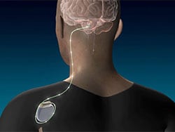
Stroke remains a significant cause of disability worldwide. In addition to multidisciplinary rehabilitation approaches, various forms of technology, including vagus nerve stimulation,
have emerged to facilitate neuroplasticity and, thereby, improve
functional status after stroke. Vagus nerve stimulation was recently
approved by the Food and Drug Administration, but questions remain
regarding its mechanism of action. Here, a potential role for
dopaminergic signaling is considered. This review first examines
evidence that dopamine
is important to neuroplasticity after stroke. Next, 2 different
dopaminergic pathways are considered potential mechanisms underlying
vagus nerve stimulation-related benefits after stroke, direct modulation
of brain dopaminergic pathways, and engagement of systemic dopaminergic
pathways such as those found in the gut-brain axis. A contribution of
dopamine signaling to vagus nerve stimulation efficacy could have
therapeutic implications that extend to a precision medicine approach to
stroke rehabilitation.
The first line in here should have been: 'Everything in stroke is a fucking failure, we can change that by following this strategy! But, no, the status quo remains! WHICH IS A COMPLETE FUCKING FAILURE!
Stroke is relatively common, being the third leading cause of death and disability globally (1). (1 in 4 per WHO will have a stroke!) Although few internists will direct the care of a patient with a recent
stroke, most will treat patients with a history of chronic stroke.
Furthermore, internists are often a patient’s lifelong primary contact
for stroke-related issues because many rehabilitation physicians and
stroke neurologists do not maintain long-term relationships with stroke
survivors.
Recent advances in the treatment
of an acute stroke have garnered considerable attention, but much of the
burden of long-term stroke disability nevertheless remains. Reperfusion
therapies, both intravenous and endovascular, can return patients …
Get full access to this article
View all available purchase options and get full access to this article.
Your doctors are responsible for your stress. You are under massive stress because your
doctor has nothing to get you 100% recovered.
Yeah, Dr. Cramer is a superstar stroke researcher but even he should have tried to solve the stress problem by getting 100% recovery protocols done.
Chronic stress(How am I going to recover?) is directly your doctor's responsibility to solve. THE SOLUTION IS 100% RECOVERY PROTOCOLS, not
guidelines or the crapola saying; 'All strokes are different, all
stroke recoveries are different'. If that saying comes out of your
doctor's mouth, you don't have a functioning stroke doctor, fire them.
Stroke is a leading cause of disability. Some people recover well after a stroke, and some do not. As a result, it’s crucial to understand the things that affect how well people bounce back after a stroke. Yet, until now, surprisingly little research has been done on the link between stroke recovery and certain types of stress.
A study published in the journal Stroke in November 2023 sheds new light on this subject. It looked at two specific kinds of stress:
Lifetime stress: Very stressful or traumatic events that people experienced in the past Poststroke acute stress: The sudden stress that people experience immediately after being diagnosed with a new stroke
“We found that higher lifetime stress predicted more poststroke acute stress. And this acute stress was a major player in stroke outcomes, even a year later,” says Steven Cramer, MD, a UCLA stroke neurologist and coauthor of the study.
Dr. Cramer is a nationally recognized expert on recovery after stroke. He was the recipient of the 2023 Outstanding Neurorehabilitation Clinician-Scientist Award from the American Society of Neurorehabilitation. What the study revealed
The new Stroke paper was based on data from the STRONG study, led by Dr. Cramer and UC Irvine researcher Alison Holman, PhD. The study included 763 patients from 28 stroke centers across the U.S. These patients took part in assessments at multiple time points, including:
Within a few days after a stroke diagnosis Three months after the stroke Twelve months after the stroke
The researchers found that having a history of trauma going as far back as childhood was tied to greater stress immediately after a stroke.
“People who have experienced a major life stressor may develop a different internal model for how they think about themselves and the world,” Dr. Cramer says. “If they have a stroke, they may tell themselves, ‘I can’t handle this.’ Or they may worry nonstop.”
In turn, being very stressed right after a stroke was tied to more impairment in movement and thinking skills at the three-month and 12-month assessments. Part of the reason may be that people who feel swamped by stress are unable to focus on their stroke recovery. They may turn down rehab therapy or plunge into depression. Less stress, better outcomes
Although this was a large study, further research is needed to confirm the findings. But these results could have implications for how stroke is treated.
“Our findings indicate that it is important to measure stress consistently and treat it early in patients with a new stroke,” Dr. Cramer says. Treatment options may include talk therapy, drug therapy or a combination of the two.
Dr. Cramer adds, “Measuring lifetime stress and acute stress at the time of stroke admission and initiating an appropriate treatment plan could profoundly change the trajectory of recovery after patients leave the hospital.” Take the Next Step
Learn more about the UCLA Health Comprehensive Stroke Center.
In here is one that is non-invasive, no surgery. Why was this research done with all this earlier stuff? Your mentors and senior researchers incompetently didn't know about it?
An early-stage clinical trial has shown that deep brain stimulation (DBS) applied to the cerebellum may aid the recovery of upper limb function after stroke.
Researchers
studied 12 people with moderate to severe upper extremity impairment
after stroke who received DBS to the dentate nucleus (DN) of the
cerebellum together with rehabilitation and found that 75% of the
participants had meaningful response in regaining some function of their
paralyzed arms.
PET revealed significant increments in brain metabolism around the part of the brain affected by the stroke.
"Our
findings support the safety and feasibility of deep brain stimulation
to the cerebellar dentate nucleus as a promising tool for modulation of
late-stage neuroplasticity for functional recovery," the authors write.
"The
results of the phase I study are promising; but more research is
needed. The next step is a phase II clinical trial that is already
enrolling patients at Cleveland Clinic," said senior author André
Machado, MD, PhD, chairman, Neurological Institute and Charles and
Christine Carroll Family Endowed Chair in Functional Neurosurgery,
Cleveland Clinic.
Machado
patented the DBS method in stroke recovery, a release from Cleveland
Clinic notes. Boston Scientific owns a license to those patents and
provided the Vercise DBS systems used in this trial. In 2010, Cleveland
Clinic Innovations established Enspire DBS Therapy, Inc, a Cleveland
Clinic portfolio company, and is commercializing technology developed at
Cleveland Clinic to commercialize the method and it co-funded the
study. Machado holds stock options and equity ownership rights with
Enspire and serves as the chief scientific officer.
"Stroke
is a leading cause of physical disability in the US and around the
world; and while physical and occupational therapy work and improve
function, about 50% of patients maintain severe levels of disability for
life," Machado said.
Neuroplasticity is a "well-documented
phenomenon that is associated with gradual spontaneous or therapy-driven
improvements in post-stroke motor function," the authors write. But the
extent of recovery "varies considerably across individuals" and depends
on a variety of factors, the authors write.
"Harnessing the
potential of neuroplasticity and modulating its extent and timing
remains a major frontier in medicine with vast upside and has been the
focus of our group," they continue.
The
researchers proposed a new surgical approach for "extending the degree
and temporal window of neuroplasticity after ischemic and traumatic
insults to the brain."
This approach involves continuous
stimulation of the cerebellar DN to "modulate neural activity and
ipsilesional cortical excitability through activation of the robust,
endogenous dentatothalamocortical pathway."
Machado explained that
cerebellar DN "promotes reorganization of the cerebral cortex around
the area affected by a stroke and increases the activity levels and
metabolism of the cerebral cortex." It also promotes new synapses and
the expression of long-term potentiation, a neuroplasticity process.
After
over one decade of preclinical studies, the researchers applied this
approach to humans for the first time, with the primary goal of
determining whether cerebellar DBS, in combination with rehabilitation,
is safe and feasible for post-stroke motor deficits.
To do so, they conducted the Electrical Stimulation of the Dentate Nucleus for Upper Extremity Hemiparesis Due to Ischemic Stroke
(EDEN) study, focusing on 12 individuals (mean age, 57.4 ± 6.5 years;
mean Upper-Extremity Fugl-Meyer Assessment [FM-UE] score, 22.9 ± 6.2
points) with chronic moderate to severe hemiparesis of the upper
extremity due to a unilateral middle cerebral artery stroke that took place between 12 and 36 months prior.
Each
participant underwent DBS surgery. After discharge and recovery from
the surgery, they had 3 months of rehabilitation before the device was
turned on and then 4-8 months of rehabilitation combined with DBS. They
continued with rehabilitation after discontinuation of stimulation
treatment.
Translational Potential
The researchers evaluated changes in motor impairment and function during five key intervals.
Pre-surgery to post-surgery
Rehab only (no DBS)
Experimental DBS plus rehab phase
2-month rehab-carryover phase in the absence of DBS
Long-term follow-up: 10 months after termination of the experimental treatment phase
In
addition, they used PET imaging to characterize metabolic changes
across ipsilateral perilesional cortex before and after the DBS plus
rehab phase, with the DBS turned off.
From pre- to post-surgery,
there was a median 0.5-point decrease in FM-UE score, which, although
not deemed significant, "supported the safety of the procedure."
There was a "modest" three-point median improvement across the pre-stimulation, rehab-only phase (P = .004). After that, an additional seven-point improvement was observed, when rehabilitation was combined with DN-DBS (P = .0005).
The
gain found during the DBS plus rehab phase was most pronounced in
participants who entered the study with some distal preservation of
motor function compared with those who entered without distal
preservation (P = .007).
During the 2-month,
rehab-carryover phase (continued rehabilitation as DBS was weaned by
weekly 25% amplitude reductions over the first month and then off during
the second month), no further change in FM-UE was observed.
The
median FM-UE score for the full cohort also remained unchanged at the
end of the long-term follow-up phase, "supporting the durability of the
previously realized treatment-related gains," the authors report.
Moreover,
the "robust functional gains were directly correlated with cortical
reorganization evidenced by increased ipsilesional metabolism," they
add.
Participants experienced no device failures and no study-related serious adverse events throughout the trial.
"This
emerging intervention has shown translational potential to modulate the
magnitude of neuroplastic reorganization toward recovery of function
and to extend its time window to late phases of disability," they
conclude.
Brooks Gross, PhD, program director, National Institute
of Neurological Disorders and Stroke (NINDS), agreed. "The safety and
feasibility data from this early study, combined with the potential
symptom improvements, certainly support the need for additional, larger
trials to see if cerebellar DBS is indeed a potential treatment for
post-stroke motor impairment," he stated in a news release.
'Exciting Data'
Commenting for Medscape Medical News,
Steven Cramer, MD, MSc, Susan and David Wilstein Endowed Chair in
Rehabilitation Medicine and professor, Department of Neurology, David
Geffen School of Medicine at UCLA, noted that patients with post-stroke
disability "have limited options in 2023, in terms of therapies to boost
stroke recovery."
This
study "provides exciting data on the safety and clinical efficacy of
cerebellar deep brain stimulation, along with favorable PET biomarker
data." But, as this is a "relatively small study with no control arm,
this approach should now proceed to testing in a phase II controlled
clinical trial," said Cramer, who is also the medical director of
research at the California Rehabilitation Institute in Los Angeles and
was not involved with the current study.
This study was
supported by the National Institutes of Health Brain Research Through
Advancing Innovative Neurotechnologies Initiative and by Enspire DBS, a
spin-off company of Cleveland Clinic. Machado serves as chief medical
officer and chair of the Scientific Advisory Board for Enspire DBS
Therapy and is paid with stock options. As the inventor, A.G.M. will
receive portions of commercialization and/or Cleveland Clinic Foundation
stock revenue and payments through Cleveland Clinic Foundation with
fees deducted. The other authors' disclosures are listed on the original
paper. Cramer serves as a consultant for AbbVie, Constant Therapeutics,
BrainQ, Myomo, MicroTransponder, Neurolutions, Panaxium, NeuExcell,
Elevian, Helius, Omniscient, Brainsgate, Nervgen, Battelle, and TRCare.
Nat Med. Published online August 14, 2023. Abstract
Steven C. Cremer, MD, (Really, you spelled his name wrong)stroke neurologist and professor of neurology at UCLA, talked about the 3
main types of rehab therapy given to poststroke patients and the
significant disparities in access.
Steven C. Cramer, MD
Known
as a leading cause for long-term disability, previous research has
shown that greater rehabilitation therapy poststroke can improve
functional outcomes.1 Therefore, the
delivery of effective rehabilitation programs with adequate resources,
dose, and duration is a critical aspect of care(Once again with 'care' NOT RESULTS OR RECOVERY!) poststroke, according to
guidelines from healthcare professionals affiliated with the American
Heart Association/American Stroke Association.2
Recently published in the journal Stroke, a study showed that rehabilitation therapy doses were low during the first year of recovery following stroke
and were also predicted by clinical factors.3 In 510 poststroke
patients, rehab therapy was mostly low and given in the first 3 months,
as 35.0% of patients had no physical therapy, 48.8% received no
occupational therapy, and 61.7% had no speech therapy. Also, discharge
destination was significantly related to cumulative therapy (for all
therapy types at visit 2, P <.05; occupational therapy visits 3 and 4, P <.05; speech therapy visits 3 and 4, P <.01) and across the variety of sites, as 0% to 71% of patients were discharged to an inpatient rehabilitation facility.
Steven C. Cramer, MD, stroke neurologist and professor of neurology at UCLA, sat down in an interview with NeurologyLive®
to discuss the 3 main classes of rehab therapy that were discussed in
the study. He also shared some of the surprising findings regarding
therapy utilization among poststroke patients. Cramer spoke about the
implications and potential impact on long-term function and therapy
utilization decline over time.
NeurologyLive®: Why did you choose those 3 specific therapies for this research?
Steven C. Kramer, MD:(Really, you spelled his name wrong again)
Those are probably the 3 main classes of rehab therapy that people get,
for better or for worse. Structurally, that's how it's broken down, and
historically, PTs (physical therapists) tend to be more involved with
walking and strength. OTs tend to be more involved with arms and
function. These are gross generalizations, and speech therapists tend to
be involved with language and swallowing. There are other entities such
as psychologists, social workers, and nutritionists, but the STRONG
study had a limited budget and there's a limited number of questions
that we could have.
There were 28 sites around the
country [involved in the study], each of which asked patients about
their rehab. We went with the 3 most common forms of rehab therapy that
people get after stroke. They correspond to 3 very common types of
things that go wrong with stroke: walking and strand, arm strength and
functional status, and then speech and swallowing.
Was there anything surprising that you found from the results and thought was quite notable?
Up
to the first 3 months [poststroke], a third of people never saw any
physical therapy and half never saw any occupational therapy. Also, 60%
never saw any speech therapy. We thought it was going to be barren and
we were surprised it was that barren. Hispanic patients got less
therapy, which was also a little bit surprising. It fits with other
reports that people of certain ethnicities and socioeconomic strata tend
to get less care. Maybe that wasn't totally surprising, but it
confirmed it for us. Then, maybe not surprise, but we were pleased that
even though some people were getting so little therapy, at least the
more severe cases tended to get more doses of therapy, even in the oddly
narrow spectrum.
Do you think there should be longer follow up study investigating the 3 types of therapies?
There
are 2 answers to that question. The narrower one is: should we follow
people for longer to see if they get more rehab therapy as time goes on?
This gets medium enthusiasm for it because what we saw and we described
is that with each success of 3-month quarter, the amount of new therapy
people was getting was smaller and smaller. By 1 year, fewer people got
any further rehab therapy. The yield would be modest because therapy
tends to stop over a matter of months, and for most people by a year.
This
is an offshoot of a larger study that looked at outcomes in relation to
genetics and that's not our focus here. We know that after stroke,
people have a real big sudden decline in function and things they can
do, and that there's some spontaneous recovery. For movement, it tends
to plateau around 3 months. For thinking and memory and language,
there's a big jump at 3 months and they often continue to improve in
little bits for maybe a year or sometimes longer. Somewhere around a
year, people started slow decline and the brain doesn't keep up.
Age-matched people with stroke showed faster decline after a year than
people who did not have a stroke who are showing a decline anyhow. The
average age of stroke is 70, and, by that age most people are losing a
little bit of function every year without stroke, but it’s even more for
patients poststroke.
I think it's important to
continue studies, examine how function declines beyond the year
poststroke, and look at the genetic correlates of that. Unfortunately, I
suspect we have doses given to patients even lower after a year, it's
more of the older populations that go to their physician and say, “I
want to walk better,” who are mostly people that have resigned to
whatever level of function they're left with at that point.
Transcript edited for clarity.
REFERENCES 1.
Young BM, Holman EA, Cramer SC; STRONG Study Investigators.
Rehabilitation Therapy Doses Are Low After Stroke and Predicted by
Clinical Factors. Stroke. 2023;54(3):831-839. doi:10.1161/STROKEAHA.122.041098 2.
Winstein CJ, Stein J, Arena R, et al. Guidelines for Adult Stroke
Rehabilitation and Recovery: A Guideline for Healthcare Professionals
From the American Heart Association/American Stroke Association. Stroke. 2016;47(6):e98-e169. doi:10.1161/STR.0000000000000098 3.
Many patients receive too little rehab therapy following stroke, study
finds. News Release. University of California, Los Angeles (UCLA),
Health Sciences. Published February 6, 2023. Accessed May 16, 2023.
https://www.newswise.com/articles/many-patients-receive-too-little-rehab-therapy-following-stroke-study-finds
Yeah, this is from Steven C. Cramer so you know it's going to be good, but come on, survivors want 100% recovery, not this tyranny of low expectations of 'improve' you're pushing on us. Will you be satisfied with not getting to 100% recovery when you're the 1 in 4 per WHO that has a stroke?
Interventions to improve stroke recovery
Steven C. Cramer KEY POINTS • Neural repair is a therapeutic strategy that is separate from acute stroke strategies such as reperfusion and neuroprotection, and has distinct biological targets, time windows for therapeutic efficacy, and issues to address in clinical trial design.• Many classes of therapy are under study to improve stroke recovery including small molecules, growth factors, monoclonal antibodies, stem cells, robotic devices, brain stimulation, activity-based therapies, telerehabilitation, and cognitive-based strategies.• Some repair-based therapies are introduced within days of stroke onset, in an attempt to amplify innate repair mechanisms, while other therapies are offered to patients from months to years after stroke onset, where the goal is to stimulate new forms of neural repair.• Restorative therapies improve behavioral outcomes on the basis of experience-dependent brain plasticity – a drug may galvanize the brain for repair, but behavioral reinforcement is also needed to achieve maximal gains. This is an important difference as compared to neuroprotective, reperfusion, and preventative stroke therapies, where the patient generally does not need to engage in any particular behavioral regimen to derive treatment benefit.• Several positive late-phase clinical trials of restorative therapies have been published, e.g., for activity-based therapies and for small molecules such as serotonergic drugs. BIOLOGY OF STROKE RECOVERY SUGGESTS THERAPEUTICS TARGETS A new stroke sets numerous biological pathways into motion. These include the ischemic cascade acutely, immunological events that evolve from pro-inflammatory systemic immuno-suppression, 1 and later a sequence of restorative events that support tissue repair and that also represent potential targets to improve stroke recovery. Animal studies indicate that an experimental stroke results in an ordered change in expression of numerous genes. Numer-ous growth-related events are seen, such as growth factor release, increased levels of growth inhibitors such as Nogo and MAG, capillary growth, axonal sprouting, synaptogenesis, glial cell activation, and changes in cortical excitability. These changes are seen both near and distant from injury, and gener-ally peak during the initial weeks post-stroke. 2–6 Studies of stroke recovery mechanisms in humans, informed by non-invasive neuroimaging and neurophysiolog-ical methods, are overall concordant with preclinical findings. 7 In parallel with behavioral improvement, cortical maps undergo reorganization. 8–10 compensatory changes in brain function and brain networks 11–13 arise, often bilaterally, 14–17 and are associated with changes in brain structure. 18,19 Un injured areas that are normally connected to the infarct region as part of a distributed network may show depressed function, a process known as diaschisis, 20,21 resolution of which may be linked with behavioral improvement. These restorative events represent potential therapeutic targets to promote brain repair.
NEURAL REPAIR IS DISTINCT FROM NEUROPROTECTION
A clear distinction must be made between therapeutic targets related to neuroprotection and those related to repair. These are parallel treatment strategies that have temporally distinct targets – a neuroprotective therapy is initiated from minutes to hours after stroke onset to salvage threatened tissue, while a repair therapy is typically introduced days after stroke onset or later. Brain repair aims to improve function by restoring normal patterns of brain structure and function, e.g., regaining voluntary control of arm reaching; this is distinguished from compensation, which aims to improve function by substitut-ing new patterns, e.g, teaching a patient to reach by swinging his torso to propel a paretic arm. 22 Many classes of restorative therapy are under study, using many different strategies. 6,23–27 These are listed in Box 59-1, which emphasizes interventions that have reached the point of human trials. Some repair-based therapies are introduced within days, or perhaps weeks, of stroke onset in an attempt to amplify innate repair mechanisms. For some restorative therapies, a critical period exists, whereby introduction within a specific time window provides a therapeutic benefit that is lost when treatment is delayed. 28–30 This is one of the many parallels between stroke recovery and normal brain develop-ment. 7,31 Other repair-based therapies are less limited by a time window and that may be offered to patients in the chronic phase, from months to years after stroke onset. Advan-tages of this approach include availability of a large enroll-ment pool and a stable baseline that is helpful to interpret treatment effects.
Patients
show substantial differences in response to rehabilitation therapy
after stroke. We hypothesized that specific genetic profiles might
explain some of this variance and, secondarily, that genetic factors are
related to cerebral atrophy post-stroke.
Methods
The
phase 3 ICARE study examined response to motor rehabilitation
therapies. In 216 ICARE enrollees, DNA was analyzed for presence of the
BDNF val66met and the ApoE ε4 polymorphism. The relationship
of polymorphism status to 12-month change in motor status (Wolf Motor
Function Test, WMFT) was examined. Neuroimaging data were also evaluated
(n=127).
Results
Subjects were 61±13 years old (mean±SD) and enrolled 43±22 days post-stroke; 19.7% were BDNF val66met
carriers and 29.8% ApoE ε4 carriers. Carrier status for each
polymorphism was not associated with WMFT, either at baseline or over
12 months of follow-up. Neuroimaging, acquired 5±11 days post-stroke,
showed that BDNF val66met polymorphism carriers had a
1.34-greater degree of cerebral atrophy compared to non-carriers
(P=.01). Post hoc analysis found that age of stroke onset was 4.6 years
younger in subjects with the ApoE ε4 polymorphism (P=.02).
Conclusion
Neither the val66met BDNF nor ApoE ε4 polymorphism explained inter-subject differences in response to rehabilitation therapy. The BDNF val66met
polymorphism was associated with cerebral atrophy at baseline, echoing
findings in healthy subjects, and suggesting an endophenotype. The ApoE
ε4 polymorphism was associated with younger age at stroke onset, echoing
findings in Alzheimer’s disease and suggesting a common biology.
Genetic associations provide insights useful to understanding the
biology of outcomes after stroke.
Good luck figuring out what a premotor therapy is and you need robotics besides. I believe most of my premotor cortex was destroyed so nothing here will help me even if I could figure it out.
and Steven C. Cramer a,b,∗ a Department of Anatomy & Neurobiology, University of California, Irvine, CA, USA b Department of Neurology, University of California, Irvine, CA, USA
* Address for correspondence: Steven C. Cramer, MD, University of California, Irvine Medical Center, 200S. Manchester Ave. Suite206, Orange, CA 92868, USA. Tel.: +1 714 456 6876; Fax: +1 714456 8805; E-mail: scramer@uci.edu
L. Dodakian et al. / Targeted dorsal premotor engagement Page,Szaflarski,Eliassen,Pan,&Cramer,2009;Strup-pler et al., 2007) after stroke. Evidence suggests that contralesional PMd might contribute to recovery, too,particularly in patients with more severe stroke (Bute-fisch et al., 2005; Kantak, Stinear, Buch, & Cohen,2012; Lotze et al., 2011; Rehme, Fink, von Cramon,&Grefkes,2011).Changes in PMd activity are thought to support behavioral gains through connections with ipsilesional primary motor cortex (M1), contralesional brain areas, spinal cord targets, and possibly reticulospinal brain stem neurons (Fregni & Pascual-Leone,2006; Fridman, et al., 2004; James et al., 2009; Kantak, et al., 2012). Together, these findings suggest that therapies that increase PMd activity could improve motor status after stroke. This idea was examined in the current study by testing two main hypotheses. The first is that practicing a motor behavior known to engage PMd circuitry will improve motor status after stroke to a greater extent than will practicing a repetitive motor behavior unrelated to PMd. These cond is that the extent to which such a PMd-based therapy provides superior gains in motor status will vary with the availability of PMd anatomically, and perhaps functionally–a therapy can not provide benefit if its biological target is excessively injured by stroke(Nouri & Cramer, 2011). These two hypotheses were examined in the cur-rent study, with a focus on the distal upper extremity. A Premotor Therapy was designed, the features of which emphasized normal functions of a PMd circuit.Key anatomical components of the PMd circuit include PMd, which processes novel external cues in order to guide the timing and the choice of voluntary movements(Askim,Indredavik,&Haberg,2010;Chouinard&Paus,2006;Geyer,Matelli,Luppino,&Zilles,2000;Koch et al., 2006; Kurata & Hoffman, 1994; O’Shea,Johansen-Berg,Trief,Gobel,&Rushworth,2007;Pass-ingham, 1993; Rushworth, Johansen-Berg, Gobel, &Devlin, 2003; Schluter, Rushworth, Passingham, &Mills, 1998), as well as M1 and the corticospinaltract, important for expressing the output of corticalcomponents of the circuit. A study of healthy control subjects (described below) confirmed that performing the timed movement tasks that constitute Premotor Therapy was associated with increased activity within a dorsal premotor circuit. A robotic device (Takahashi,Der-Yeghiaian, Le, Motiwala, & Cramer, 2008), found to improve post-stroke arm motor function in a prior study, served as an ideal vehicle for implementing the timed cues central to Premotor Therapy, and furthermore allowed inclusion of therapy in a gaming context, an approach useful to modulating the function of selected brain circuits (Bavelier, Levi, Li, Dan, &Hensch, 2010; Colzato, van den Wildenberg, Zmigrod,& Hommel, 2012). In the current study, this Premotor Therapy was contrasted with Motor Therapy , in which subjects performed the same distal arm movements as with Premotor Therapy but without cues or novelty. Patients with chronic stroke underwent a baseline example plus MRI for assessing anatomical and functional features of a PMd circuit, were randomized to two weeks of Motor Therapy vs. Premotor Therapy via the robotic device, and then had their motor outcome assessed 1 month after completion of therapy. These data were used to address the above two hypotheses.
Additions include includes Steven Cramer, M.D., Seth
Finklestein, M.D., Teresa Kimberley, Ph.D., P.T., Daniel Laskowitz,
M.D, MHS, David Lin, M.D., and Gary Steinberg, M.D.
ALLSTON, Mass., Feb. 9, 2021 /PRNewswire/ -- Elevian,
an emerging biotech company developing new medicines that target the
GDF11 pathway, announced the addition of a stroke clinical advisory team
to advance development of recombinant GDF11 (rGDF11) to promote
recovery post stroke. The team includes Steven Cramer, M.D., Seth Finklestein, M.D., Teresa Kimberley, Ph.D., P.T., Daniel Laskowitz, M.D, MHS, David Lin, M.D., and Gary Steinberg, M.D.
"We have assembled several of the leading experts in the emerging
field of stroke recovery, bringing together knowledge and experience
about the clinical implications of stroke and emerging therapies," said Mark Allen, M.D., CEO of Elevian. "Together we have mapped out a clinical strategy using rGDF11 to promote recovery post stroke."
"Stroke is a massive, unmet medical need. It is the second leading
cause of death worldwide and the number one cause of long-term
disability," said Seth Finklestein, MD, Neurologist at Massachusetts
General Hospital (MGH) and Chair of Elevian's Stroke Clinical Advisory
Board. "Elevian has produced exciting preclinical efficacy data
demonstrating that rGDF11 promotes motor function recovery post stroke.
These data, if translated to humans, could provide an important new
therapy for patients who have suffered a stroke."
Dr. Finklestein is currently a Neurologist at Massachusetts General Hospital (MGH), and Former Head of the CNS Growth Factor Research Laboratory at MGH and Associate Professor at Harvard Medical School
(HMS). He is also Former VP and Head of the Neuroscience Division at
Viacell, Inc., former CEO of Biotrofix, Inc., and current CEO at
Recovery Therapeutics, Inc. Dr. Finklestein is a graduate of Haverford College and Harvard Medical School. His major interest is brain repair and recovery after stroke.
Dr. Steven C. Cramer is a Professor of Neurology at the University of California, Los Angeles (UCLA).
He is also the Director of Research at California Rehabilitation
Institute, and co-PI of the NIH StrokeNet clinical trials network. Dr.
Cramer received his medical degree from University of Southern California, his Residency in Internal Medicine at UCLA, and completed his Residency in Neurology and a Fellowship in Cerebrovascular Disease at Massachusetts General Hospital. Dr. Cramer also earned a Master's degree in clinical investigation from Harvard Medical School.
His research focuses on neural repair after central nervous system
injury in humans, with an emphasis on stroke and on recovery of
movement, with a major emphasis is on translating new drugs and devices
to reduce disability after stroke, and on individualizing therapy for
each person's needs. Dr. Cramer has been awarded the Stroke
Rehabilitation Award from the American Heart Association and the Barbro B. Johansson Award in Stroke Recovery from the World Stroke Organization.
Teresa Jacobson Kimberley, Ph.D.,
P.T., is a professor and director of the Brain Recovery Lab, in the
department of Physical Therapy in the School of Health and
Rehabilitation Sciences at the MGH Institute of Health Professions. She
has an appointment as Research Staff at Massachusetts
General Hospital (MGH) Department of Neurology, and as Core Faculty in
the Center for Neurotechnology and NeuroRecovery. Kimberley received her
bachelor's in Physical Therapy and her doctorate in Rehabilitation
Science from the University of Minnesota-Twin Cities.
Her lab's focus is on understanding the pathophysiology of motor
impairment and develop novel rehabilitation interventions for neurologic
disorders, such as dystonia and stroke. Her research helped pioneer the
use of neuroimaging and non-invasive brain stimulation in the
investigation of rehabilitation-related areas.
Dr Laskowitz is a Professor and Vice Chair of Neurology at Duke University
where he serves as the Medical Director for the Neurovascular
Laboratories and leads the Neuroscience Medicine program at the Duke
Clinical Research Institute. He received his MD and his Master of Health
Science in clinical research from the Duke University School of Medicine and completed his neurology residency training at the University of Pennsylvania.
His perspective on drug development is shaped by the compelling unmet
needs in the care of his patients with acute and chronic brain injury.
His research focus is on the role of genetic influences on
neuroinflammatory responses, secondary neuronal injury, and recovery
from ischemic and traumatic brain injury. Dr. Laskowitz has been
involved with several translational trials evaluating new therapies in
stroke and acute brain injury. He is a fellow of the American Heart
Association and American Neurological Association and has authored or
co-authored more than 200 peer-reviewed articles.
Dr. Lin is a critical care Neurologist and Neurorehabilitation specialist at Massachusetts General Hospital. He is the Director of the MGH NeuroRecovery Clinic. He is also an Instructor in Neurology at Harvard Medical School.
In his clinical practice, Dr. Lin cares for patients with acute
neurologic injuries including stroke, brain hemorrhage, traumatic brain
injury, seizures, and spinal cord injury in the MGH Neurosciences
Critical Care Unit and he provides recommendations to facilitate best
possible recovery at the MGH NeuroRecovery clinic. Dr. Lin's research
involves understanding mechanisms of brain plasticity in patients order
to guide recovery after stroke and other acute brain injuries.
Dr. Steinberg is the Founder and Co-Director of the Stanford Stroke
Center, former Chair of Neurosurgery, and Director of the Stanford
Moyamoya Center. His 33 years of experience in basic and translational
neuroscience research has focused on hemorrhagic and ischemic stroke, as
does his neurosurgical clinical practice. Dr. Steinberg received his
medical degree from Stanford University and did residencies at Stanford University,
for General Surgery and Neurosurgery, and at Santa Clara Medical
Center. His lab investigates pathomechanisms of cerebral ischemia,
develops neuroprotective agents, and employs novel approaches to enhance
post-stroke functional recovery. He has successfully translated his
preclinical work into several stem cell clinical trials for stroke,
spinal cord injury and traumatic brain injury, as well as leading
numerous other clinical cerebrovascular trials.
About Elevian, Inc. Elevian is an emerging
biotech company developing medicines that target the GDF11 pathway, with
the potential to treat and prevent many age-related diseases.
Elevian's lead program uses recombinant GDF11 (rGDF11) to promote
recovery post stroke. The company has established additional programs
focused on the use of rGDF11 to treat diabetes and obesity, and the
regulation of GDF11 via novel molecules.
Stroke
recovery therapies promote favorable neural plasticity, both during
spontaneous recovery and the chronic phase. Activity-based therapies
based on intense practice, some aided by integration of computers and
telehealth, have shown promise. These studies emphasize key therapeutic
variables such as dose, intensity, and timing. Preclinical drug studies
have shown promise, but human translation has been challenged by
identifying the target patient subgroup, requirements for concomitant
training, and aligning biomarkers with preclinical evidence.
Footnotes
For Sources of Funding and Disclosures, see page 350.
The
opinions expressed in this article are not necessarily those of the
American Heart Association., Correspondence to: Lorie G. Richards, PhD,
520 Wakara Way, Salt Lake City, UT 84108. Email lorie.richards@hsc.utah.edu
Steven C. Cramer KEY POINTS • Neural repair is a therapeutic strategy that is separate from acute stroke strategies such as reperfusion and neuroprotection, and has distinct biological targets, time windows for therapeutic efficacy, and issues to address in clinical trial design.• Many classes of therapy are under study to improve stroke recovery including small molecules, growth factors, monoclonal antibodies, stem cells, robotic devices, brain stimulation, activity-based therapies, telerehabilitation, and cognitive-based strategies.• Some repair-based therapies are introduced within days of stroke onset, in an attempt to amplify innate repair mechanisms, while other therapies are offered to patients from months to years after stroke onset, where the goal is to stimulate new forms of neural repair.• Restorative therapies improve behavioral outcomes on the basis of experience-dependent brain plasticity – a drug may galvanize the brain for repair, but behavioral reinforcement is also needed to achieve maximal gains. This is an important difference as compared to neuroprotective, reperfusion, and preventative stroke therapies, where the patient generally does not need to engage in any particular behavioral regimen to derive treatment benefit.• Several positive late-phase clinical trials of restorative therapies have been published, e.g., for activity-based therapies and for small molecules such as serotonergic drugs.