Changing stroke rehab and research worldwide now.Time is Brain! trillions and trillions of neurons that DIE each day because there are NO effective hyperacute therapies besides tPA(only 12% effective). I have 523 posts on hyperacute therapy, enough for researchers to spend decades proving them out. These are my personal ideas and blog on stroke rehabilitation and stroke research. Do not attempt any of these without checking with your medical provider. Unless you join me in agitating, when you need these therapies they won't be there.

What this blog is for:

My blog is not to help survivors recover, it is to have the 10 million yearly stroke survivors light fires underneath their doctors, stroke hospitals and stroke researchers to get stroke solved. 100% recovery. The stroke medical world is completely failing at that goal, they don't even have it as a goal. Shortly after getting out of the hospital and getting NO information on the process or protocols of stroke rehabilitation and recovery I started searching on the internet and found that no other survivor received useful information. This is an attempt to cover all stroke rehabilitation information that should be readily available to survivors so they can talk with informed knowledge to their medical staff. It lays out what needs to be done to get stroke survivors closer to 100% recovery. It's quite disgusting that this information is not available from every stroke association and doctors group.

Showing posts with label hemiplegic gait. Show all posts

Saturday, January 18, 2025

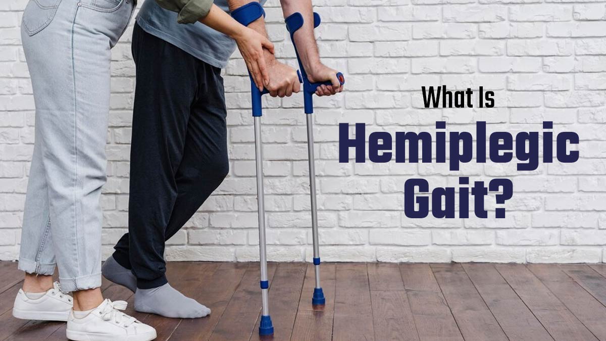
What Is Hemiplegic Gait? Exploring Its Symptoms, Causes and Treatment

 Absolutely nothing here will guarantee gait recovery! Why don't you have those protocols?

Laziness? Incompetence? Or just don't care? NO leadership? NO strategy? Not my job? Not my Problem?

What Is Hemiplegic Gait? Exploring Its Symptoms, Causes and Treatment

Learn about hemiplegic gait, a walking problem caused by a stroke or brain injury. Our expert explains the symptoms, causes, and treatments available to help people improve their movement and quality of life.

What Is Hemiplegic Gait? Exploring Its Symptoms, Causes and Treatment

Walking is something most people do every day without thinking. However, for those with hemiplegic gait, even taking a simple step can be very hard. Hemiplegic gait is a type of walking problem caused by weakness or paralysis on one side of the body. This condition often happens after a stroke or brain injury. People with hemiplegic gait may drag one leg or move it in a circular way to avoid tripping. 

To learn more about this condition, we spoke with Dr. Agnetia Vinoth, a consultant neurosurgeon at Apollo Cancer Centre in Chennai. He explained the challenges of hemiplegic gait and highlighted the symptoms, causes and treatments to improve the lives of those affected.

What Is Hemiplegic Gait? 

— Journal of Brown Hospital Medicine (@BrownJHM) January 3, 2023


Dr. Agnetia Vinoth explains that hemiplegic gait often happens after a stroke or brain injury. This condition makes it hard for a person to move, which can lead to less independence and a lower quality of life. Hemiplegic gait means that one arm or leg is weak or paralysed due to these injuries, causing unusual walking patterns.

Dr  Vinoth also notes that people with this condition may struggle to lift the affected limb, drag it, or swing it awkwardly while walking. While hemiplegic gait is most common in stroke patients, it can also occur in those with brain tumours, cerebral palsy, or multiple sclerosis.

What Are the Causes of Hemiplegic Gait?

1 (39)

According to our expert, hemiplegic gait results from an impairment in the brain's motor control pathways. Below are a few causes of the walking disorder. 

  • Stroke: This is the most common cause. A stroke happens when blood flow to the brain is blocked, leading to brain cell death and loss of movement abilities.
  • Head Injuries: Traumatic brain injuries can also damage the brain's motor control pathways, affecting how a person walks.
  • Multiple Sclerosis: This disease affects the nervous system and can lead to movement problems, including hemiplegic gait.
  • Cerebral Palsy: This condition occurs before or during birth and can cause weakness in one side of the body, resulting in walking difficulties.

The above-mentioned conditions disrupt the corticospinal tract, which is important for coordinating voluntary movements.

Symptoms of Hemiplegic Gait

Dr. Agnetia Vinoth claims that symptoms of hemiplegic gait can vary with the severity of the neurological damage, but common signs include the following. 

  • Weakness or paralysis on one side: The affected limb will have a weakened leg, causing an inability to lift, move, or bear weight.
  • Leg circumduction: A patient will drag his affected leg outward in a circular motion because of weakness; sometimes dragging along the floor. 
  • Foot drop: When muscles cannot be lifted from under the foot, they tend to drag on the ground.
  • Hip adduction: The involved limb can rotate medially sometimes causing scissoring of the limbs while walking.
  • Limited swing of arms: The limb on the side may be rigid and cannot swing from the body's side.

“The effects of hemiplegic gait can vary from mild to severe. Some people may have a significant disability that makes it very hard for them to walk, while others may only have mild difficulties,” said the expert. For those with milder symptoms, using walking aids like canes or walkers can help them balance and avoid falling. These tools allow them to support their weight on both sides, making it easier to move around safely.

ALSO READ: Cold Weather and Cigarettes: Heightened Risks for Heart Disease and Impaired Lung Health

Hemiplegic Gait Treatment

2 (11)

“Therapy for hemiplegic gait adopts an approach for mobilising the patient to overcome this disability maximally and obtain the highest possible level of function,” said Dr Agnetia Vinoth. He further detailed the treatment options in an interdisciplinary manner, which includes the following. 

(YOU'RE MISSING THE MOST IMPORTANT PROBLEM! CURE SPASTICITY! Are you that much of a blithering idiot?)

  • Physical Therapy: A big part of the treatment is physical therapy, which involves strengthening the muscles, flexibility, and coordination, and will include the return to the biomechanics of daily life. 
  • Occupational Therapy: Services would offer support to patients to adapt to their practical life despite the restrictions of their illness by improving activities in the activities of daily living such as dressing, eating, and bathing.
  • Assistive Devices: Assistive devices such as canes, walkers, or orthotic braces will offer stability and good support to patients who are severely impaired in their gait, thus reducing the chances of falls and average mobilization in some patients.
  • Botox injections: These patients also benefit a lot from Botox injections when the spasticity level is more than normal it decreases the stiffness and makes moving easier.

Conclusion

Hemiplegic gait is a complex and common problem that can happen after a stroke or brain injury. Many patients find it difficult to move after being diagnosed. However, with the right mix of medical care and physical therapy, people with hemiplegic gait can see significant improvements(But survivors want 100% recovery! Why are you OK with setting the tyranny of low expectations for your patients? They aren't OK with such crapola!). This tailored treatment can help reduce severe mobility issues and make it easier for them to move around.

Thursday, March 30, 2023

Spatio-temporal parameters and intralimb coordination patterns describing hemiparetic locomotion at controlled speed

I see nothing here that will help survivors recover.

Spatio-temporal parameters and intralimb coordination patterns describing hemiparetic locomotion at controlled speed

Abstract

Background

Comparison between healthy and hemiparetic gait is usually carried out while subjects walk overground at preferred speed. This generates bias due to the lack of uniformity across selected speeds because they reflect the great variability of the functional level of post-stroke patients. This study aimed at examining coordinative adaptations during walking in response to unilateral brain damage, while homologous participants walked at two fixed speeds.

Methods

Five patients with left and five with right chronic hemiparesis, characterized by similar level of motor functioning, were enrolled. Ten non-disabled volunteers were recruited as matched control group. Spatio-temporal parameters, and intralimb thigh-leg and leg-foot coordination patterns were used to compare groups while walking on a treadmill at 0.4 and 0.6 m/s. The likelihood of Continuous Relative Phase patterns between healthy and hemiparetic subjects was evaluated by means of the root mean square of the difference and the cross correlation coefficient. The effects of the group (i.e., healthy vs. hemiparetics), side (i.e., affected vs.unaffected), and speed (e.g., slow vs. fast) were analyzed on all metrics using the Analysis of Variance.

Results

Spatio-temporal parameters of all hemiparetic subjects did not significantly differ from those of healthy subjects nor showed any asymmetry between affected and unaffected limbs. Conversely, both thigh-leg and foot-leg coordination patterns appeared to account for pathology related modifications.

Conclusion

Comparisons between hemiparetic and healthy gait should be carried out when all participants are asked to seek the same suitable dynamic equilibrium led by the same external (i.e., the speed) and internal (i.e., severity of the pathology) conditions. In this respect, biomechanical adaptations reflecting the pathology can be better highlighted by coordinative patterns of coupled segments within each limb than by the spatio-temporal parameters. Accordingly, a deep analysis of the intralimb coordination may be helpful for clinicians while designing therapeutic treatments.

Background

Compensatory strategies adopted by post-stroke patients to increase stability and efficiency of locomotion have been widely described in literature by spatio-temporal parameters, kinematic, and kinetic measures referring to walking both overground [1–3] and on treadmill [4, 5]. On the whole, the gait of post-stroke patients is mainly characterized by reduced speed, stride length, and cadence, decreased angular excursions at leg joints, increased energetic cost, and asymmetry in kinematic and kinetic variables [3]. Furthermore, a recent study highlighted that post-stroke patients walk with different patterns between treadmill and overground and, in particular, the treadmill enhances the asymmetry between affected and unaffected limbs, decreases the self-selected speed and the step length, and increases stance and double support percentages [6].

A cerebrovascular accident has been also shown to significantly modify the coordinative relationship of segments within (i.e., intralimb coordination) and between (i.e., interlimb coordination) lower limbs [7–9]. Specifically, previous authors have noticed that the intralimb coordinative patterns of chronic hemiparetics walking overground at their preferred speed can be characterized by asymmetry between unaffected and affected sides [7], and that the botulinum toxin injections into the rectus femoris of the paretic side can improve the thigh-leg coordination patterns of both limbs [8]. In addition, it was observed that the increase in speed positively influences the interlimb coordination of both arm and lower limb movements of acute patients undergoing intense rehabilitative treatments [9].

One of the common features of all mentioned studies [4, 6–9] is that participants were asked to walk at their self selected speeds, that is – as acknowledged by some authors [3, 4, 10] – the walking speed was not uniform across subjects, basically due to the inherent variability related to the cerebrovascular accident (i.e., different clinical picture, rehabilitative training, neuromuscular adaptation across patients).

Actually, previous authors have already highlighted the importance of considering gait metrics in relation to the walking speed when attempting to classify gait abnormality [11, 12]. This is because the velocity can be viewed as a control parameter of the dynamic system that, when scaled, involves changes of coordination even within the gait of walking alone [7, 13]. Furthermore, in a previous study, Monaco and colleagues [14] demonstrated that the gait analysis, when carried out at controlled and comparable speed, can better pinpoint features between young and elderly healthy subjects than the comparison at self-selected pace.

Concerning post-stroke patients, it is well known that the self-selected speed reflects specific compensative strategies related to their own degree of impairment such that patients walking in a wide range of preferred velocities are characterized by very different functional deficit [15]. However, the speed, per se, affects in a significant fashion gait patterns inducing relevant modifications of the hemiparetic locomotion [12, 15, 16]. As matter of the fact, the inherent instability of the affected limb prompts the patients to an early shift of the weight on the unaffected one inducing an asymmetric duration of stance and swing phases [15, 16]. In spite of this, when patients are asked to walk at faster speeds, i.e., pace comparable to healthy subjects, they show a bilateral increment of joint angular excursions and muscle activation [17], an improved symmetry in double and single support portions [15, 17], and a significant modification of the coordination of pelvis and trunk rotations [12]. The speed-related improvements of the hemiparetic gait seem to be due to more appropriate timing of lower limb muscles, better suited movement coordination, and possibly facilitation of intralimb and interlimb energy transfers [17].

According to these results, it is possible to hypothesize that part of data variability leading previous authors to characterize motor performance of post-stroke patients may have been ascribed to the wide range of speeds rather than to the pathology itself. Specifically, metrics used to describe motor deficits of locomotion, may lack of a suitable sensitivity when different groups of homogenous subjects (e.g., similar age, anthropometric features, severity of the deficit) are observed while walking at the same pace.

This methodological study aimed at investigating the attitude of both spatio-temporal parameters and intralimb coordination patterns to capture some of the neuromuscular adaptations of the hemiparetic locomotion. This goal has been achieved by comparing gait patterns of affected and unaffected sides in post-stroke hemiparetic patients to those of healthy subjects while walking on a treadmill at the same speeds.

More at link.

Monday, August 24, 2015

Effects of handrail hold and light touch on energetics, step parameters, and neuromuscular activity during walking after stroke

A couple of important points from this, your doctor should  be able to determine which 60% successfully walk again in a community setting. 

Effects of handrail hold and light touch on energetics, step parameters, and neuromuscular activity during walking after stroke


T. IJmker12*, C. J. Lamoth3, H. Houdijk12, M. Tolsma2, L. H. V. van der Woude3, A. Daffertshofer1 and P. J. Beek1
1 MOVE Research Institute Amsterdam, Department of Human Movement Sciences, Faculty of Behavioural and Movement Sciences, VU University Amsterdam, van der Boechorststraat 9, Amsterdam, 1081 BT, The Netherlands
2 Heliomare Rehabilitation, Research and Development, Relweg 51, Wijk aan Zee, 1949 EC, The Netherlands
3 University of Groningen, University Medical Center Groningen, Center for Human Movement Sciences, Center for Rehabilitation, Antonius Deusinglaan 1, Groningen, 9713AV, The Netherlands
For all author emails, please log on.
Journal of NeuroEngineering and Rehabilitation 2015, 12:70  doi:10.1186/s12984-015-0051-3
The electronic version of this article is the complete one and can be found online at: http://www.jneuroengrehab.com/content/12/1/70

Received:5 February 2015
Accepted:26 June 2015
Published:23 August 2015
© 2015 IJmker et al.

Open Access This article is distributed under the terms of the Creative Commons Attribution 4.0 International License (http://creativecommons.org/licenses/by/4.0/), which permits unrestricted use, distribution, and reproduction in any medium, provided you give appropriate credit to the original author(s) and the source, provide a link to the Creative Commons license, and indicate if changes were made. The Creative Commons Public Domain Dedication waiver (http://creativecommons.org/publicdomain/zero/1.0/) applies to the data made available in this article, unless otherwise stated.

Abstract

Background

Holding a handrail or using a cane may decrease the energy cost of walking in stroke survivors. However, the factors underlying this decrease have not yet been previously identified. The purpose of the current study was to fill this void by investigating the effect of physical support (through handrail hold) and/or somatosensory input (through light touch contact with a handrail) on energy cost and accompanying changes in both step parameters and neuromuscular activity. Elucidating these aspects may provide useful insights into gait recovery post stroke.

Methods

Fifteen stroke survivors participated in this study. Participants walked on a treadmill under three conditions: no handrail contact, light touch of the handrail, and firm handrail hold. During the trials we recorded oxygen consumption, center of pressure profiles, and bilateral activation of eight lower limb muscles. Effects of the three conditions on energy cost, step parameters and neuromuscular activation were compared statistically using conventional ANOVAs with repeated measures. In order to examine to which extent energy cost and step parameters/muscle activity are associated, we further employed a partial least squares regression analysis.

Results

Handrail hold resulted in a significant reduction in energy cost, whereas light touch contact did not. With handrail hold subjects took longer steps with smaller step width and improved step length symmetry, whereas light touch contact only resulted in a small but significant decrease in step width. The EMG analysis indicated a global drop in muscle activity, accompanied by an increased constancy in the timing of this activity, and a decreased co-activation with handrail hold, but not with light touch. The regression analysis revealed that increased stride time and length, improved step length symmetry, and decreased muscle activity were closely associated with the decreased energy cost during handrail hold.

Conclusion

Handrail hold, but not light touch, altered step parameters and was accompanied by a global reduction in muscle activity, with improved timing constancy. This suggests that the use of a handrail allows for a more economic step pattern that requires less muscular activation without resulting in substantial neuromuscular re-organization. Handrail use may thus have beneficial effects on gait economy after stroke, which cannot be accomplished through enhanced somatosensory input alone.
Keywords:
Energy cost; Stroke; Neuromuscular function; Gait; Balance support

Background

Regaining the ability to walk independently is an important goal in the rehabilitation of stroke survivors. Only 60 % of all stroke survivors eventually attain this goal to the level of community walking [1]. An important limiting factor in this regard is the substantial metabolic cost of hemiparetic gait, which can be more than two times larger than in healthy subjects [2]–[4], and which is predictive of community ambulation [5]. We have previously shown that an increased (metabolic) effort to control balance contributes to this decreased gait economy [6], and that this cost can be reduced considerably by providing balance support in the form of a handrail or cane [7].
Using a handrail or cane may have biomechanical and/or somatosensory advantages that could facilitate balance control. Biomechanically, the use of a handrail or cane increases the base of support, resulting in greater margins of stability, and enables one to generate corrective forces via the hands to compensate for perturbations [8]. Apart from this biomechanical advantage, the use of a handrail or cane may provide additional somatosensory (tactile and proprioceptive) information about body orientation and movement relative to the point of contact [8], [9]. This may reduce sensory noise/uncertainty and might therefore lead to better balance control [9], [10]. There is experimental support that, even in the absence of additional biomechanical support, the mere contact of fingertips or hand with a stable support surface can decrease the excursion of the center of mass during standing and walking [9]–[13]. This decrease matched that observed with firm handrail hold in healthy participants and stroke survivors. This suggests that enhanced somatosensory information may add to the mechanical stabilization through holding a handrail, which in turn may result in a decreased energy cost of walking after stroke.
To unravel the factors underlying the differential effects of handrail hold and light touch on the energy cost of walking, it is imperative to investigate which gait parameters alter in line with metabolic changes and which neuromuscular modifications might engender these effects. In stroke survivors, handrail or cane use yields increased stride length and time as well as decreased cadence and step width and variability [14], [15]. These changes may be linked to an improved gait efficiency through a more optimal step length/frequency combination [16], [17], and lower step-to-step transition costs with a smaller step width [18]. Using a handrail or cane may also improve gait symmetry [19], [15], which may also contribute to enhanced gait economy [20].
Effects of holding a handrail or cane have also been examined in terms of changes in neuromuscular control as reflected in altered amplitude and timing of muscle activation. Some studies reported decreases in EMG burst duration and a decrease in amplitude of several lower limb muscles during cane use [21], [22]. Furthermore, a decrease in the variability of EMG profiles of the lower leg muscles has been found as a result of handrail support, which indicates a more consistent timing of muscle activity possibly relating to increased (lateral) gait stability [23], [24]. Reduced EMG amplitude and more accurate timing of muscle activity may reflect improved economy [25]. In contrast, other studies reported no effect of firm handrail hold or light touch contact with a cane on muscle activity [26], [27], while light touch contact has even been shown to result in higher activation amplitudes than force contact [12], [26].
As of yet, it is unclear whether somatosensory and/or biomechanical aspects of handrail or cane use affect the energy cost of walking after stroke, nor whether altered step parameters and/or altered neuromuscular control are responsible for this effect. Our research aims were therefore twofold: 1) to compare the effects of light touch contact with a handrail and firm handrail hold on the energy cost of walking, step parameters, and muscle activity (in terms of amplitude and timing) in stroke survivors, and 2) to examine which changes in step parameters and muscle activity are associated with the observed changes in energy cost. To evaluate changes in muscle activation amplitude and timing we used a principal component analysis (PCA), since this method allows for studying patterns of multivariate muscle activation instead of looking at isolated muscle activities alone. 

More at link.

Tuesday, October 7, 2014

Gait analysis assisted by robotic walker in patients with post-stroke hemiparesis

What a novel idea. Objective analysis of a post-stroke gait. Why hasn't this been done and taught in PT schools? If you don't objectively know what the problems are you can never tell if those problems are resolved and exactly what protocols corrected them. This is just a microcosm of all the problems in stroke rehab. 

Gait analysis assisted by robotic walker in patients with post-stroke hemiparesis


Flávia Loterio* and Teodiano Bastos-Filho
  • * Corresponding author: Flávia Loterio
Universidade Federal do Espírito Santo, Vitória, ES, Brazil
For all author emails, please log on.
BMC Proceedings 2014, 8(Suppl 4):P267  doi:10.1186/1753-6561-8-S4-P267

The electronic version of this article is the complete one and can be found online at: http://www.biomedcentral.com/1753-6561/8/S4/P267

Published:1 October 2014
© 2014 Loterio and Bastos-Filho; licensee BioMed Central Ltd.

This is an Open Access article distributed under the terms of the Creative Commons Attribution License (http://creativecommons.org/licenses/by/4.0), which permits unrestricted use, distribution, and reproduction in any medium, provided the original work is properly cited. The Creative Commons Public Domain Dedication waiver (http://creativecommons.org/publicdomain/zero/1.0/) applies to the data made available in this article, unless otherwise stated.

Poster presentation

Stroke is the leading cause of motor dysfunction in adults worldwide. The motor impairment, characteristic of a post-stroke patient, is the hemiparetic gait. This paper proposes a gait analysis assisted by a robotic walker in patients with post-stroke hemiparesis. The test group will have two categories: the first consisting of healthy subjects, and the second one by subjects with post-stroke hemiparesis. Both groups will perform an initial trajectory without the aid of a walker, and another one with its assistance. This route is a straight line, in a flat ground. After training with the walker, volunteers participating repeat the trajectory using the device. The robotic walker is equipped with 3D force sensors in the forearm support, which can confirm the correct weight applied to it. It also can deduce the subject's movement intentions, through the efforts made by his/her upper limbs. Spatio-temporal parameters of gait using inertial sensors will be analyzed. These sensors will be placed in specific points of the subject's legs, and a laser scanning sensor will be located in the center of the walker in order to get distances to the legs. Electromyography (EMG) electrodes will be also placed on the subject's legs to evaluate the muscle activity and energy consumption during the gaits. After analyzing the data, the results will be compared between the two groups and between the gaits before and after the training. The results might be useful for the process of rehabilitation of post-stroke patients, and in normal and hemiparetic gait.