Use the labels in the right column to find what you want. Or you can go thru them one by one, there are only 33,610 posts. Searching is done in the search box in upper left corner. I blog on anything to do with stroke. DO NOT DO ANYTHING SUGGESTED HERE AS I AM NOT MEDICALLY TRAINED, YOUR DOCTOR IS, LISTEN TO THEM. BUT I BET THEY DON'T KNOW HOW TO GET YOU 100% RECOVERED. I DON'T EITHER BUT HAVE PLENTY OF QUESTIONS FOR YOUR DOCTOR TO ANSWER.
Changing stroke rehab and research worldwide now.Time is Brain!trillions and trillions of neuronsthatDIEeach day because there areNOeffective hyperacute therapies besides tPA(only 12% effective). I have 523 posts on hyperacute therapy, enough for researchers to spend decades proving them out. These are my personal ideas and blog on stroke rehabilitation and stroke research. Do not attempt any of these without checking with your medical provider. Unless you join me in agitating, when you need these therapies they won't be there.
What this blog is for:
My blog is not to help survivors recover, it is to have the 10 million yearly stroke survivors light fires underneath their doctors, stroke hospitals and stroke researchers to get stroke solved. 100% recovery. The stroke medical world is completely failing at that goal, they don't even have it as a goal. Shortly after getting out of the hospital and getting NO information on the process or protocols of stroke rehabilitation and recovery I started searching on the internet and found that no other survivor received useful information. This is an attempt to cover all stroke rehabilitation information that should be readily available to survivors so they can talk with informed knowledge to their medical staff. It lays out what needs to be done to get stroke survivors closer to 100% recovery. It's quite disgusting that this information is not available from every stroke association and doctors group.
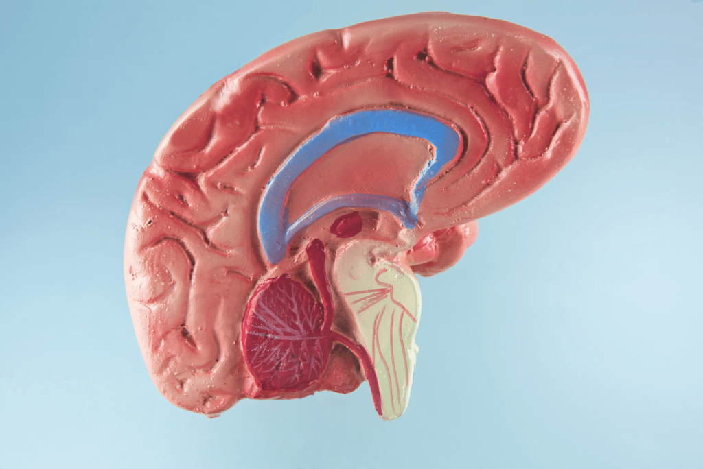
Once you identify the specific area where the damage is; WHAT IS THE EXACT REHAB PROTOCOL TO BE USED TO RECOVER FROM THAT DAMAGE? Totally incomplete research!
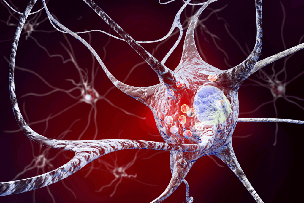
The study examined the potential of artificial intelligence (AI),
specifically the large language model GPT-4, to help locate brain
lesions resulting from strokes. These lesions play a crucial role in
predicting the long-term effects of a stroke and determining the
appropriate treatment and prognosis for affected individuals. The
research, conducted by Dr. Jung-Hyun Lee from SUNY Downstate Health
Sciences University, utilized text from health histories and neurologic
exams of 46 stroke patients to train GPT-4 in accurately identifying the
location and extent of lesions in the brain.
The findings of the study revealed that GPT-4 was successful in
locating lesions in the brains of many participants, determining the
side of the brain affected as well as the specific brain region, with
the exception of lesions in the cerebellum and spinal cord. The AI model
demonstrated a sensitivity of 74% and a specificity of 87% in
identifying the side of the brain with lesions, and a sensitivity of 85%
and a specificity of 94% in pinpointing the brain region involved.
Additionally, GPT-4 showed consistency in its results for the number of
brain lesions, side of the brain, and brain regions in a majority of
cases.
Although GPT-4 was able to provide accurate answers for 41% of
participants when combining responses to all three questions across all
three times, the study notes that further refinement and validation are
needed before its clinical use. A key limitation of the study is that
the accuracy of GPT-4 relies on the quality of information it receives,
and detailed health histories and neurologic exam information may not
always be available for all stroke patients. However, the potential of
AI models like GPT-4 to assist in locating brain lesions after a stroke
is seen as promising, particularly in underserved regions where access
to neurologic care is limited.
The study highlights the importance of accurate identification of
brain lesions following a stroke, as this information can significantly
impact the long-term outcomes and treatment strategies for affected
individuals. By leveraging the capabilities of AI models like GPT-4 to
analyze health histories and neurologic exam data, neurologists may be
able to streamline the diagnostic process and improve the efficiency of
lesion localization. This advancement has the potential to reduce
disparities in healthcare access and delivery, especially in regions
where neurologic care is scarce.
Moving forward, further research and validation are needed to enhance
the accuracy and reliability of AI models like GPT-4 in locating brain
lesions after a stroke. As technology continues to evolve, the
integration of AI in neurology practice holds promise for improving
patient outcomes and enhancing the accessibility and quality of
healthcare services worldwide. Continued collaboration between
neurologists, researchers, and AI experts will be crucial in harnessing
the full potential of artificial intelligence in advancing neurologic
care and addressing the global health challenges posed by strokes and other neurological conditions.
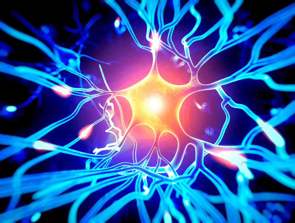
What Are Neuroplasticity Supplements, Herbs, Nootropics, and How Do They Work?
Omega-3 Fatty Acids
Omega-3s, found in fish and algae oil supplements, are important for brain health.
They can potentially increase neuroplasticity through several mechanisms:
Enhancing Neuronal Membrane Fluidity
Omega-3s, such as EPA (eicosapentaenoic acid) and DHA
(docosahexaenoic acid), are essential components of neuronal cell
membranes. They help maintain the fluidity and flexibility of these
membranes, which is crucial for efficient communication between neurons.
Flexible membranes allow for easier transmission of signals and the
formation of new connections, supporting neuroplasticity[1].
Promoting Synaptic Function
The proper functioning of synapses is essential for learning and
memory. Omega-3s may help optimize synaptic transmission and facilitate
the strengthening of synaptic connections, which are vital aspects of
neuroplasticity[2].
Supporting Healthy Inflammatory Response
Inflammation in the brain can hinder neuroplasticity by disrupting
communication between neurons. Omega-3 fatty acids support healthy
inflammatory responses in the nervous system. Therefore,omega-3s create a
more conducive environment for neural connections to form and adapt,
thereby supporting neuroplasticity[3].
BDNF is a protein that promotes the growth, survival, and maintenance
of neurons. It is also involved in the formation of new synapses and
the adaptation of existing ones, both of which are fundamental processes
in neuroplasticity.
Omega-3 fatty acids, particularly DHA, increase BDNF levels in the
brain, facilitating neural connectivity and enhancing the brain’s
adaptive capacity[4].
Modulating Neurotransmitters
Omega-3s can influence the production and release of
neurotransmitters like dopamine, serotonin, and glutamate. These
neurotransmitters play central roles in cognitive processes and mood
regulation. By helping regulate the balance of neurotransmitters,
omega-3s can support overall brain function and emotional well-being,
which are integral components of neuroplasticity[5].
Antioxidants
Antioxidants can potentially increase neuroplasticity through their
ability to neutralize oxidative stress, which can impair the brain’s
adaptive capacity. Antioxidants support neuroplasticity in the following
ways:
Reducing Oxidative Stress (ROS)
Oxidative stress occurs when there is an imbalance between harmful
ROS and the body’s antioxidant defenses. Excessive oxidative stress can
damage neurons and their components, including DNA, proteins, and lipids[6].
Antioxidants neutralize ROS and help protect neurons from this
damage, preserving their function and viability. Healthy neurons are
better equipped to engage in neuroplasticity.
Protecting Mitochondrial Function
Mitochondria are the energy-producing organelles within neurons.
Oxidative stress can impair mitochondrial function, leading to energy
deficits and decreased neuronal plasticity[7][8].
Antioxidants can help maintain mitochondrial health by mitigating
oxidative damage, ensuring neurons have the energy they need to support
neuroplasticity.
Inflammation Modulating Effects
Unmodulated Chronic inflammation in the brain can hinder
neuroplasticity by disrupting the normal functioning of neurons and
glial cells[9].
Antioxidants, by modulating inflammation, create a more favorable
environment for neural plasticity. They can modulate immune responses,
supporting overall brain health[10].
Promoting BDNF Production
As previously mentioned, BDNF is a protein that plays a crucial role
in neuroplasticity. Some antioxidants, such as flavonoids found in
certain fruits and vegetables, increase the production of BDNF[11].
Elevated BDNF levels facilitate the formation of new synapses and
strengthen existing ones, which are essential aspects of neuroplasticity[12].
B Vitamins
B vitamins play an important role in brain health and can support neuroplasticity through several mechanisms:
Neurotransmitter Production
B vitamins, particularly B6 (pyridoxine), B9 (folate), and B12
(cobalamin), are essential for the synthesis of neurotransmitters such
as serotonin, dopamine, and norepinephrine. Ensuring an adequate supply
of these B vitamins helps maintain neurotransmitter balance, which is
important for neuroplasticity[13].
Homocysteine Regulation
Elevated levels of homocysteine, an amino acid, are associated with
an increased risk of cognitive decline. B vitamins, specifically folate,
B6, and B12, help regulate homocysteine levels in the body. B vitamin
supplementation may help reduce the risk of cognitive impairment and
support overall brain health, including neuroplasticity[14].
DNA Methylation and Synaptic Plasticity
DNA methylation is an epigenetic process that can influence gene
expression. B vitamins, particularly folate, play a role in DNA
methylation, which can affect the expression of genes involved in
synaptic plasticity. Proper methylation patterns are necessary for the
formation and strengthening of synaptic connections, a fundamental
process in neuroplasticity[15].
Protection Against Oxidative Stress
B vitamins, including niacin (B3) and riboflavin (B2), are involved
in your body’s antioxidant defenses. Antioxidants help protect neurons
from oxidative stress, which can damage cell membranes, proteins, and
DNA. By reducing oxidative stress, B vitamins support the health and
function of neurons and facilitate neuroplasticity[16].
Mood Regulation
B vitamins are known to play a role in mood regulation, and mood is
closely linked to cognitive function and neuroplasticity. For example,
B6 is involved in the production of serotonin, a mood-enhancing
neurotransmitter. Balancing mood can indirectly support cognitive
processes and neuroplasticity[16].
A balanced diet with foods rich in B vitamins, such as leafy greens,
legumes, nuts, and fortified cereals, can provide the necessary
nutrients to support brain health and neuroplasticity. However, in cases
of deficiency or certain medical conditions, B vitamin supplementation
may be recommended under the guidance of a healthcare professional.
Lion’s Mane Mushroom (Hericium erinaceus)
Lion’s mane mushroom (Hericium erinaceus) is a natural dietary
supplement that has gained attention for its potential to support
neuroplasticity and overall brain health. While the exact mechanisms of
how lion’s mane mushroom influences neuroplasticity are still being
researched, there are several ways it may contribute to this process:
Nerve Growth Factor (NGF) and pro-BDNF Stimulation
Lion’s Mane Mushroom contains compounds known as hericenones and
erinacines, which have been shown in some studies to stimulate the
production of nerve growth factor (NGF). NGF is a protein essential for
the growth, maintenance, and survival of neurons.
By promoting NGF production, Lion’s Mane Mushroom may facilitate the
growth of new neurites (extensions of nerve cells) and the formation of
new synaptic connections, which are key aspects of neuroplasticity[17].
Lion’s Mane Mushroom supplements increase levels of pro-BDNF, which
is a precursor to BDNF. Pro-BDNF is synthesized and released by neurons
and is involved in synaptic pruning which is an important process of
neuroplasticity[18].
Enhanced Myelination
Myelin is the protective sheath that covers nerve fibers and enhances
the efficiency of signal transmission between neurons. Lion’s Mane
Mushroom may support myelination by promoting the growth and
differentiation of oligodendrocytes, the cells responsible for myelin
production.
Improved myelination can lead to faster and more efficient communication between neurons, potentially enhancing neuroplasticity[19].
Antioxidant Properties
Lion’s Mane Mushroom is rich in antioxidants, which help combat
oxidative stress and reduce damage to neurons caused by free radicals.
Oxidative stress can impair neuronal function and hinder
neuroplasticity. By reducing oxidative stress, Lion’s Mane Mushroom may
create a more favorable environment for synaptic plasticity and neural
adaptation[20].
Inflammation-Balancing Effects
Lion’s Mane Mushroom has immune-balancing properties, which may help
promote a healthy inflammatory response and thus a healthier environment
for synaptic remodeling and plasticity[21].
Improved Cognitive Function
Some studies suggest that lion’s mane mushroom supplementation can
lead to improved cognitive function, including memory and learning.
Enhanced cognitive abilities can indirectly support neuroplasticity by
facilitating the acquisition of new information and the formation of new
neural connections[22].
While there is promising research on the potential benefits of Lion’s
Mane Mushroom for neuroplasticity, more studies are needed to fully
understand its mechanisms and effects.
Individual responses to Lion’s Mane Mushroom may vary, and its use
should be approached with caution, especially if you have any underlying
medical conditions or are taking medications. As with any dietary
supplement, it’s advisable to consult with a healthcare professional
before incorporating lion’s mane mushroom into your routine.
Caffeine
Caffeine is a stimulant that primarily affects the central nervous
system, and while it is more commonly associated with increasing
alertness and concentration, it can also have some indirect effects on
neuroplasticity.
Enhanced Cognitive Function
Although there is no clear consensus, some studies suggest that
caffeine can temporarily improve cognitive function, including
attention, memory, and learning.
By increasing alertness and mitigating the sensation of fatigue,
caffeine may help individuals engage more effectively in cognitive tasks
that require neuroplasticity, such as learning new information or
adapting to changing circumstances. This enhanced cognitive function can
indirectly support neuroplasticity by facilitating the acquisition and
processing of new information[23].
Stimulation of Neurotransmitter Release
Caffeine stimulates the release of certain neurotransmitters,
including dopamine and norepinephrine, in the brain. These
neurotransmitters play roles in mood regulation, attention, and arousal.
Increased neurotransmitter activity can enhance alertness and focus,
potentially aiding in the engagement of cognitive processes associated
with neuroplasticity[24].
Improved Synaptic Transmission
Caffeine can enhance synaptic transmission, the process by which
signals are transmitted between neurons at synapses. It can increase the
release of neurotransmitters like glutamate, which is crucial for
synaptic plasticity and learning.
By promoting more efficient signaling between neurons, caffeine can
indirectly support the strengthening and formation of neural
connections, essential aspects of neuroplasticity[25].
Neuroprotective Effects
Some studies suggest that caffeine has neuroprotective properties,
which means it may help protect neurons from damage caused by factors
like oxidative stress and neuroinflammation. By preserving the health of
neurons, caffeine can create a more conducive environment for
neuroplasticity to occur[26].
By blocking adenosine receptors, caffeine enhances alertness,
potentially influencing neural activity and learning. This heightened
neuronal firing, along with potential BDNF elevation, might foster an
environment conducive to neuroplasticity. However, individual responses
to caffeine vary, and excessive intake can lead to negative effects[23].
The caffeine’s effect on neuroplasticity can vary among individuals
and depend on factors such as the dose of caffeine consumed and an
individual’s tolerance to caffeine.
While moderate caffeine consumption may have some potential cognitive
benefits, excessive caffeine intake can lead to side effects, including
anxiety, jitteriness, and disrupted sleep, which can ultimately impair
cognitive function and neuroplasticity[23].
Additionally, the long-term effects of chronic caffeine use on
neuroplasticity are still an active area of research, and more studies
are needed to fully understand the relationship between caffeine and
brain plasticity. It’s advisable to consume caffeine in moderation and
be mindful of its potential side effects, especially if you have
underlying medical conditions or are sensitive to caffeine[23].
Bacopa Monnieri
Bacopa monnieri, commonly known as Brahmi, is an herb used in
traditional medicine, particularly in Ayurveda, that is believed to have
several cognitive-enhancing properties, including the potential to
increase neuroplasticity[27].
Although no research has directly shown that Bacopa monnieri directly
causes neuroplasticity, here are some ways Bacopa monnieri may
influence neuroplasticity:
Enhanced Synaptic Transmission
Bacopa monnieri is thought to enhance synaptic transmission, which is
the process by which neurons communicate at synapses. It may increase
the release of neurotransmitters like acetylcholine, which is important
for learning and memory[28].
Improved synaptic transmission can facilitate the strengthening and
formation of neural connections, a key aspect of neuroplasticity.
Neuroprotective Effects
Bacopa monnieri is rich in antioxidants, which help protect neurons
from oxidative stress and damage caused by free radicals. By preserving
the health of neurons, Bacopa monnieri can create a more conducive
environment for neuroplasticity to occur[29][30].
Modulation of Neurotransmitters
Bacopa monnieri may influence the levels and activity of various
neurotransmitters, including serotonin, dopamine, and GABA
(gamma-aminobutyric acid). Modulating neurotransmitter activity can
indirectly support neuroplasticity by enhancing cognitive function and
emotional well-being[29][31].
Stress Reduction
Chronic stress can impair cognitive function and hinder neuroplasticity. Bacopa monnieri may promote calmness and enhance mood[28][32]. By mitigating stress, it can create a more favorable environment for the brain to engage in neuroplasticity processes.
Enhanced Memory Consolidation
Bacopa monnieri may help with improved memory retention and
consolidation. The formation of long-term memories involves synaptic
plasticity, and enhancing memory processes can indirectly support
neuroplasticity[28].
Neurotrophic Factor Modulation
Some research suggests that Bacopa monnieri may influence the
expression of brain-derived neurotrophic factor (BDNF), a protein
critical for the growth, survival, and adaptability of neurons. Elevated
BDNF levels facilitate the formation of new synapses and the
strengthening of existing ones, fundamental processes in neuroplasticity[29].
Supplements, herbs, and nootropics show promise in enhancing
neuroplasticity, which is crucial for brain adaptability and cognitive
health. Omega-3 fatty acids, antioxidants, B vitamins, Lion’s Mane
Mushroom, and caffeine can potentially support neuroplasticity through
various mechanisms.
These compounds aid in maintaining neuronal health, promoting
synaptic function, balancing inflammation, and enhancing
neurotransmitter activity.
While these supplements offer exciting potential, individual
responses vary, necessitating consultation with healthcare
professionals, especially for those with underlying health conditions. A
well-balanced diet rich in these nutrients can also contribute to brain
health and cognitive adaptability.
These supplements offer a promising avenue for individuals seeking to improve cognitive function and overall well-being.
The only outcome measure survivors care about is 100% recovery. don't you dare use the tyranny of low expectations to suggest to survivors anything else.
University of Nebraska Medical Center, stacie.christensen@unmc.edu Monica Dial
College of St. Mary, mdial@csm.edu Tell us how you used this information in this short survey. See link and comment Follow this and additional works at: https://digitalcommons.unmc.edu/cahp_pt_pres Part of the Physical Therapy Commons Recommended CitationRecommended Citation Christensen, Stacie Mae Larreau and Dial, Monica, "From Textbooks to Clinical Practice: Selecting and Implementing Outcomes Measures in Stroke Rehabilitation" (2024). Posters and Presentations: Physical Therapy. 44. https://digitalcommons.unmc.edu/cahp_pt_pres/44 This Presentation is brought to you for free and open access by the Physical Therapy at DigitalCommons@UNMC. It has been accepted for inclusion in Posters and Presentations: Physical Therapy by an authorized administrator of DigitalCommons@UNMC. For more information, please contact digitalcommons@unmc.edu.
The author is currently finalizing the slides for this presentation. Check back soon for the final work.
I'm sure your competent? doctor wants you to be cognitively aroused for better stroke recovery and s/he has had music protocols for all stroke patients for well over a decade.
Summary: A new study explores the influence of
personalized music on cognitive arousal and performance, drawing on the
Yerkes-Dodson law’s inverted-U theory. The study used participants’
physiological and behavioral signals to map arousal levels against
performance, revealing that music can significantly affect one’s
productivity by aligning arousal to an optimal level.
Exciting
music, in particular, was found to enhance performance, demonstrating
the potential of music as a simple, everyday tool to regulate cognitive
states. This research opens the door to personalized brain-computer
interfaces that adjust arousal for improved cognitive functioning in
daily tasks.
Key Facts:
The study
validates the Yerkes-Dodson law by showing an inverted-U relationship
between cognitive arousal and performance, with optimal outcomes
achieved at moderate arousal levels.
Participants exposed to exciting music performed better, highlighting music’s capacity to elevate arousal to a beneficial state.
The
research introduces a performance-based arousal decoder, offering
insights into tailoring interventions like music to individual cognitive
and physiological profiles for enhanced productivity.
Source: NYU
Human
brain states are unobserved states that can constantly change due to
internal and external factors, including cognitive arousal, a.k.a.
intensity of emotion, and cognitive performance states.
Maintaining
a proper level of cognitive arousal may result in being more productive
throughout daily cognitive activities. Therefore, monitoring and
regulating one’s arousal state based on cognitive performance via simple
everyday interventions such as music is a critical topic to be
investigated.
Researchers from NYU Tandon led by Rose Faghih—inspired by the
Yerkes-Dodson law in psychology, known as the inverted-U
law—investigated the arousal-performance link throughout a cognitive
task in the presence of personalized music.
The research is published in the IEEE Open Journal of Engineering in Medicine and Biology.
The
Yerkes-Dodson law states that performance is a function of arousal and
has an inverted-U shaped relationship with cognitive arousal, i.e., a
moderate level of arousal results in optimal performance, on the other
hand, an excessively high level of arousal may result in anxiety, while a
deficient level of arousal may be followed by boredom.
In this
study, participants selected music with calming and exciting music
components to mimic the low and high-arousing environment. To decode the
underlying arousal and performance with respect to everyday life
settings, they used peripheral physiological data as well as behavioral
signals within the Bayesian Decoders.
In particular, electrodermal
activity (EDA) has been widely used as a quantitative arousal index. In
parallel, behavioral data such as a sequence of correct/incorrect
responses and reaction time are common cognitive performance
observations.
The decoded
arousal and performance data points in the arousal-performance frame
depict an inverted U shape, which conforms with the Yerkes-Dodson law.
Also, findings present the overall better performance of participants
within the exciting background music.
Considering the Yerkes-Dodson law, the researchers develop a
performance-based arousal decoder that can preserve and account for the
cognitive performance dynamic. Such a decoder can provide a profound
insight into how physiological responses and cognitive states interplay
to influence productivity.
Although several factors, such as the
nature of the cognitive task, the participant’s baseline, and the type
of applied music, can impact the outcome, it might be feasible to
enhance cognitive performance and shift one’s arousal from either the
left or right side of the curve using music.
In particular, the
baseline of arousal level varies among humans, and the music may be
selected to set the arousal within the desired range.
The outcome
of this research can advance researchers closer to developing a
practical and personalized closed-loop brain-computer interface for
regulating internal brain states within everyday life activities.
About this music and cognitive performance research news
Author: Rose Faghih Source: NYU Contact: Rose Faghih – NYU Image: The image is credited to Neuroscience News
Ask your doctor why you didn't get 100% recovered like she did! Don't be polite about it, screaming may be required, and the competence of your doctor should be in question! In my stroke survivor opinion, all stroke doctors should have EXACT 100% RECOVERY PROTOCOLS! They've known since medical school that stroke rehab was a compete fucking failure and should have initiated research that solved that problem. At least leaders would do that. Isn't your doctor a leader?
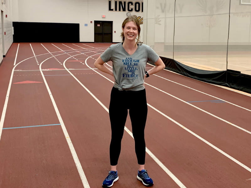
The only other stroke survivors that I know got back to running are these:
By Deborah Lynn Blumberg, American Heart Association News
Molly
Fitzgerald was 27 when she had a stroke. A month later, she had another
one that partially paralyzed her. Now 31, she runs and hikes. (Photo
courtesy of Molly Fitzgerald)
Molly
Fitzgerald woke up one fall morning with intense pain in her neck. It
was so bad she thought about going to the emergency room. But Molly
eventually pegged the pain to work stress. Maybe the migraines she'd had
lately were also to blame, she thought.
She took pain medicine
and used a heating pad, trying to ride things out. A week later, the
pain was still there, so she went to an urgent care facility near her
Minneapolis home. After doing an electrocardiogram, or EKG, to look at
her heart's electrical activity, the doctor said nothing was wrong.
"But I don't feel right," Molly said.
Worried she was being dramatic, Molly went home with a muscle relaxant.
She
woke up one day soon after feeling dizzy. She stood up from bed and
almost fainted. In the bathroom, her body kept leaning to the right. She
kept vomiting.
The word "stroke" popped into her head. She
dismissed it. She was only 27. Strokes were for older people, she
thought, and her face and speech were fine.
It was probably a
reaction from the muscle relaxant, she thought. She collapsed on the
couch to rest and called her mother, Karen Fitzgerald.
"Go to the hospital," Karen said.
In
the ER, Molly waited for hours. She got an anti-nausea medication. It
could be the flu, a doctor said, or maybe vertigo, a spinning sensation
that can be linked to issues with the inner ear. Ultimately, he proposed
a treatment that could help stop the spinning.
Molly had another idea. "Let's do some scans first," she said.
She had an MRI and CT scan to look at her brain activity. Soon after, the doctor came into her room.
"So," he said, "you had a little stroke."
She'd
had a cerebellar stroke, one that happens when blood is restricted to
the part of the brain that controls body and eye movement and balance.
The cause was vertebral artery dissection, when the wall of an artery in
the back of the neck tears and blood supply to the brain is blocked. It
wasn't clear why it happened. The doctor prescribed medicine to prevent
blood clots from forming.
"Hopefully this will never happen again," the doctor said.
Molly walked out of the hospital a few days later.
Over
the next few weeks, she had dizzy spells and headaches. Once, she felt a
cold sweat on one side of her body. She went to the ER.
"You're probably just healing," another ER doctor said.
A
month after her stroke, Molly was a few days from returning to her
marketing job at a local dairy company. She had brunch with a friend,
caught up with her parents in Chicago and went to take a nap.
Suddenly,
on the way to bed she felt an intense pain in her head. Her jaw locked
up. She looked at herself in the mirror. Her mouth wasn't drooping. But
Molly was nervous. What if it was another stroke? She called a friend
who lived five minutes away to come over.
Molly put on her shoes
and fed her cat, Gigi. Bending over, she got a huge headrush, then her
ears started ringing. Her vision was distorted; everything looked tinted
green.
"Oh no, it's another stroke," she told herself. Molly grabbed her phone to call 911. But she kept opening the calculator app.
Finally, she dialed.
"I think I'm having a stroke," she told the operator.
"How do you know?" the operator said.
"Because I've already had one," said Molly.
EMTs
and Molly's friend arrived at the same time. Molly couldn't talk. Her
left side was paralyzed. Paramedics asked if it was possible Molly had
taken any drugs.
"She doesn't even drink coffee right now," her friend said.
In the ambulance, Molly threw up while on her back, causing her to feel like she was suffocating.
"Stay with us," the EMT said as Molly's eyes fluttered closed. "Stay awake."
When
her parents, Donald and Karen, arrived at the hospital, Molly was in
bed, shaking. The left side of her face drooped. For days, she couldn't
walk.
An avid runner, she feared she'd never run again. When she
started therapy, she felt dizzy and sick her first time using a walker.
It didn't last long. She was disappointed.
"Why are you being so hard on yourself?" Donald asked.
"I thought I'd be a great student," Molly said.
Molly Fitzgerald and her mom, Karen, watching a movie in Molly's inpatient rehab room. (Photo courtesy of Molly Fitzgerald)
Soon,
though, she started seeing progress. Ten days later, she was walking
again. Then she went to stay with her parents in Chicago at her
childhood home.
It was Thanksgiving and she relished being with
her sisters, one older, one younger. To support Molly's recovery, Donald
set up an agility course in the basement with a reclining bike, a
soccer ball and an air hockey table.
"We always talk about grit," he said. "Molly's got grit and she just keeps pushing."
Molly
also did occupational therapy and physical therapy to get her strength
back. Months later, in the fall of 2020, when COVID-19 canceled all
marathons, Donald created his own 26.2-mile run in the Chicago suburbs
as a fundraiser for stroke research in Molly's honor.
Donald and
his brother-in-law, Jim Haack, wore red T-shirts that said, "Miles with a
Purpose." They raised nearly $7,000 for the American Heart Association.
Molly ran the last mile with her dad and uncle.
Molly
Fitzgerald (center) ran the final mile of the race her dad organized,
Miles with a Purpose. She crossed the finish line with her dad, Don
(left) and uncle, Jim Haack. (Photo courtesy of Molly Fitzgerald)
The following summer, Molly had recovered enough to fulfill a longtime dream. She moved to San Francisco.
Now,
five years after her strokes, Molly is 31. When she's not working doing
brand marketing for a California coffee manufacturer, she enjoys
running and hiking in various parts of California. The only off-limits
activities are things that could tear her artery again, such as water
skiing and riding a roller coaster.
She still has frequent
periods of fatigue, especially after intense physical activity or busy
workdays, and gets migraines every few months. "Fatigue from brain
damage isn't something you can muscle through," she said.
Also an
avid knitter, Molly got a tattoo of a knitting swatch with several
knitting errors in it. The image sends two messages: her challenges and
her triumphs.
"You do have certain deficits that you carry as
scars," she said. "But you learn to manage them, and today I'm pretty
good compared to where I was. I feel very lucky."
Molly Fitzgerald's knitting tattoo that symbolizes her stroke recovery journey. (Photo courtesy of Molly Fitzgerald)
Stories From the Heart chronicles the inspiring journeys of heart disease and stroke survivors, caregivers and advocates.
American Heart Association News Stories
American
Heart Association News covers heart disease, stroke and related health
issues. Not all views expressed in American Heart Association News
stories reflect the official position of the American Heart
Association. Statements, conclusions, accuracy and reliability of
studies published in American Heart Association scientific journals or
presented at American Heart Association scientific meetings are solely
those of the study authors and do not necessarily reflect the American
Heart Association’s official guidance, policies or positions.
Copyright is owned or held by the American Heart Association, Inc., and all rights are reserved. Permission
is granted, at no cost and without need for further request, for
individuals, media outlets, and non-commercial education and awareness
efforts to link to, quote, excerpt from or reprint these stories in any
medium as long as no text is altered and proper attribution is made to
American Heart Association News.
Other uses, including
educational products or services sold for profit, must comply with the
American Heart Association’s Copyright Permission Guidelines. See full terms of use. These stories may not be used to promote or endorse a commercial product or service.
HEALTH
CARE DISCLAIMER: This site and its services do not constitute the
practice of medical advice, diagnosis or treatment. Always talk to your
health care provider for diagnosis and treatment, including your
specific medical needs. If you have or suspect that you have a medical
problem or condition, please contact a qualified health care
professional immediately. If you are in the United States and
experiencing a medical emergency, call 911 or call for emergency medical
help immediately.
Time-restricted eating was associated with increased risk for death from heart disease.
This was a less than 8-hour eating window compared with a 12-to-16-hour window, which is the average in the U.S.
A time-restricted eating window of less than 8 hours was associated
with increased risk for cardiovascular death, according to study
findings presented at the Epidemiology, Prevention, Lifestyle &
Cardiometabolic Scientific Sessions.
“Restricting daily eating time to a short period, such as 8 hours per
day, has gained popularity in recent years as a way to lose weight and improve heart health,” Victor Wenze Zhong, PhD,
professor and chair of the department of epidemiology and biostatistics
at the Shanghai Jiao Tong University School of Medicine in Shanghai,
said in a press release. “However, the long-term health effects of
time-restricted eating, including risk of death from any cause or
cardiovascular disease, are unknown.”
Time-restricted eating was associated with increased risk for death from heart disease. Image: Adobe Stock
The present study included 20,078 participants aged 20 years or older
from the National Health and Nutrition Examination Survey from 2003 to
2018 who completed two valid 24-hour dietary recalls and provided self-reported usual dietary intake
(mean age, 49 years; 50% men; 73% white). Average time-restricted
eating, a form of intermittent fasting, was stratified by self-reported
duration: less than 8 hours, 8 to 10 hours, 10 to 12 hours, 12 to 16
hours or more than 16 hours.
Eating within a 12-to-16-hour window was identified as the mean U.S. eating duration and served as the reference group.
The median follow-up was 8 years.
Compared with the reference group, an eating duration of less than 8
hours was associated with increased risk for CV mortality (HR = 1.91;
95% CI, 1.2-3.03) and was also observed in subgroups of adults with CVD
(HR = 2.07; 95% CI, 1.14-3.78) and cancer (HR = 3.04; 95% CI,
1.44-6.41).
No other eating durations were associated with CV mortality, with the
exception 8 to 10 hours in adults with CVD compared with 12 to 16 hours
(HR = 1.66; 95% CI, 1.03-2.67), according to the presentation.
Moreover, Zhong and colleagues reported no significant associations
between eating duration and all-cause or cancer mortality in the overall
sample or either CVD or cancer subgroups, except for the eating
duration of more than 16 hours, which was associated with lower risk for
cancer mortality in adults with cancer (HR = 0.47; 95% CI, 0.23-0.95).
“It’s crucial for patients, particularly those with existing heart
conditions or cancer, to be aware of the association between an 8-hour
eating window and increased risk of cardiovascular death. Our study’s
findings encourage a more cautious, personalized approach to dietary
recommendations, ensuring that they are aligned with an individual’s
health status and the latest scientific evidence,” Zhong said in the
release. “Although the study identified an association between an 8-hour
eating window and cardiovascular death, this does not mean that
time-restricted eating caused cardiovascular death.”
Background
Early detection of large vessel occlusion (LVO) facilitates triage to
an appropriate stroke center to reduce treatment times and improve
outcomes. Prehospital stroke scales are not sufficiently sensitive, so
we investigated the ability of the portable Openwater optical blood flow
monitor to detect LVO.
Methods
Patients were prospectively enrolled at two comprehensive stroke
centers during stroke alert evaluation within 24 hours of onset with
National Institutes of Health Stroke Scale (NIHSS) score ≥2. A 70 s
bedside optical blood flow scan generated cerebral blood flow waveforms
based on relative changes in speckle contrast. Anterior circulation LVO
was determined by CT angiography. A deep learning model trained on all
patient data using fivefold cross-validation and learned discriminative
representations from the raw speckle contrast waveform data. Receiver
operating characteristic (ROC) analysis compared the Openwater
diagnostic performance (ie, LVO detection) with prehospital stroke
scales.
Results
Among 135 patients, 52 (39%) had an anterior circulation LVO. The
median NIHSS score was 8 (IQR 4–14). The Openwater instrument had 79%
sensitivity and 84% specificity for the detection of LVO. The rapid
arterial occlusion evaluation (RACE) scale had 60% sensitivity and 81%
specificity and the Los Angeles motor scale (LAMS) had 50% sensitivity
and 81% specificity. The binary Openwater classification
(high-likelihood vs low-likelihood) had an area under the ROC (AUROC) of
0.82 (95% CI 0.75 to 0.88), which outperformed RACE (AUC 0.70; 95% CI
0.62 to 0.78; P=0.04) and LAMS (AUC 0.65; 95% CI 0.57 to 0.73; P=0.002).
Conclusions
The Openwater optical blood flow monitor outperformed prehospital
stroke scales for the detection of LVO in patients undergoing acute
stroke evaluation in the emergency department. These encouraging
findings need to be validated in an independent test set and the
prehospital environment.
Data availability statement
Data
are available upon reasonable request. The de-identified data that
support the reported findings are available from the corresponding
author upon reasonable request.
But still not fast enough to get to 100% recovery and you're not even measuring that!
Survivors want to know how many got to
100% recovery! If you're not measuring that you'll never get there, and
just to make sure I'd have you all fired for incompetently not even
understanding the only goal in stroke for survivors is 100% recovery!
Accelerating
door‐in‐door‐out (DIDO) times at primary stroke centers (PSCs) for
patients with large vessel occlusion (LVO) acute ischemic stroke
transferred for possible endovascular stroke therapy (EVT) is important
to optimize outcomes. Here, we assess whether automated LVO detection
coupled with secure communication at non‐EVT performing PSCs improves
DIDO time and increases the proportion of patients receiving EVT after
transfer.
METHODS
From
our prospectively collected multicenter registry, we identified
patients with LVO acute ischemic stroke that presented to one of 7 PSCs
in the Greater Houston area from January 1, 2021, to February 27, 2022.
Noncontrast computed tomography and computed tomographic angiography
were performed in all patients at the time of presentation, per standard
of care. A machine learning (artificial intelligence [AI]) algorithm
trained to detect LVO (Viz.AI) from computed tomographic angiography was
implemented at all 7 hospitals. The primary outcome of the study was
DIDO at the PSCs and was determined using multivariable linear
regression adjusted for sex and on/off hours. Secondary outcomes
included likelihood of receiving EVT post‐transfer.
RESULTS
Among
115 patients who met inclusion criteria, 80 were evaluated pre‐AI and
35 post‐AI. The most common occlusion locations were middle cerebral
artery (51.3%) and internal carotid artery (25.2%). There were no
substantial differences in demographics or presentation characteristics
between the 2 groups. Median time from onset to PSC arrival was 117
minutes (interquartile range, 54–521 minutes). In univariable analysis,
patients evaluated at the PSCs after AI implementation had a shorter
DIDO time (median difference, 77 minutes; P<0.001). In
multivariable linear regression, patients evaluated with automated LVO
detection AI software were associated with a 106‐minute (95% CI, −165 to
−48 minutes) reduction in DIDO time but no difference in likelihood of
EVT post‐transfer (odd ratio, 2.13 [95% CI, 0.88–5.13).
CONCLUSION
Implementation
of a machine learning method for automated LVO detection coupled with
secure communication resulted in a substantial decrease in DIDO time at
non‐EVT performing PSCs.
Viz.ai has announced a strategic collaboration with Medtronic
to improve the coordination of cryptogenic stroke patient care between
neurology and cardiology teams.
For stroke patients who are at risk of atrial fibrillation (AF)
post-stroke and may need additional cardiac monitoring, stroke care
teams in the USA will have the opportunity to use the Viz Connect
solution—a software tool that automates the communication across
disciplines, including neurology and cardiology.
Recent clinical study results indicate that both community hospitals
and academic centres are in need of stronger, standardised care pathways
between neurology and cardiology to ensure that stroke patients receive
guideline-directed therapy, as stated in a recent press release. One
example the release cites is the DiVERT Stroke clinical study,
in which only 16% of stroke patients from community hospitals and 34%
of patients at academic centres received a cardiology consult.
“Through our collaboration with Medtronic, we have the opportunity to
bring cardiology and neurology closer together by using software tools
that help facilitate stroke patient care,” said Chris Mansi, chief
executive officer and co-founder at Viz.ai. “We are confident this
collaboration will help more patients get the continuity of care and
treatment they need to reduce secondary stroke recurrence.”
According to Viz.ai, Viz Connect has demonstrated impact on improving
patient access to cardiac care after a cryptogenic stroke—strokes with
an unknown cause, but that impact close to 800,000 people each year in
the USA, require cardiac workup, and are followed by a second stroke
within two years in roughly 20% of cases. Examples of Viz Connect’s
impact detailed in the release include an average increase of more than
50% in in-patient cardiology follow-up, and an average time of under
five minutes from when the notification is sent from neurology to when
it is reviewed by a cardiologist.
“We look forward to helping hospital care teams more easily get
patients to the right specialist at the right time,” said Stacey
Churchwell, vice president and general manager, Cardiovascular
Diagnostics and Services within the Cardiac Rhythm Management business,
which is part of the Cardiovascular Portfolio at Medtronic.
Good 8:29 video, not going to help you recover from your stroke but will make you more knowledgeable than your doctor!
Oops, I'm not playing by the polite rules of Dale Carnegie, 'How to Win Friends and Influence People'.
Telling supposedly smart stroke medical persons they know nothing about stroke is a no-no even if it is true.
Politeness
will never solve anything in stroke. Yes, I'm a bomb thrower and proud
of it. Someday a stroke 'leader' will try to ream me out for making them look bad by being truthful, I
look forward to that day.
Well, I'm not coupled, but that isn't stopping me from enjoying drinks with female friends listening to live jazz music.
And didn't your competent? doctor tell all the males in their patient load to drink with buddies twice a week? WHY NOT? Your doctor incompetently didn't know of the research or didn't follow thru? Either case is grounds for firing your doctor!
Summary: Couples with similar drinking habits,
specifically those who both consume alcohol, tend to live longer than
those who don’t share the same drinking patterns. This finding draws on
“the drinking partnership” theory, suggesting that shared alcohol
consumption correlates with improved marital outcomes and possibly,
greater longevity.
While the study stops short of endorsing
increased alcohol consumption among couples, it highlights the
significance of shared lifestyle habits on health and relationship
satisfaction. The research, part of the Health and Retirement study,
followed 4,656 couples over two decades, providing a comprehensive look
at the long-term implications of mutual drinking habits on life span.
Key Facts:
Shared Drinking Habits Linked to Longevity: Couples who both drink alcohol tend to live longer compared to those with discordant drinking habits or who abstain altogether.
Impact on Relationship Quality:
Concordant drinking couples report higher relationship satisfaction,
potentially due to increased intimacy and shared activities.
Groundbreaking Longitudinal Study:
The research analyzed data from the Health and Retirement study,
tracking 4,656 couples from 1996 to 2016, underscoring the robustness of
the findings.
Source: University of Michigan
In a recent study published in The Gerontologist, Kira
Birditt, research professor at the U-M Institute for Social Resarch’s
Survey Research Center, found that couples who are concordant in their
drinking behavior (that is, both members drink alcohol) tend to live
longer.
She says a
theory in alcohol literature called “the drinking partnership,” where
couples who have similar patterns of alcohol use tend to have better
marital outcomes (such as less conflict and longer marriages), was the
inspiration behind the study.
Birditt
would like to explore further questions related to couples’ alcohol
consumption and how it affects their relationship. Credit: Neuroscience
News
Although a great deal of research has
examined the implications of couples’ drinking patterns for marital
outcomes, the implications for health are less clear. Behaviors that are
good for marriage are not necessarily good for health, Birditt says.
“The
purpose of this study was to look at alcohol use in couples in the
Health and Retirement Study and the implications for mortality,” she
said.
“And we found, interestingly, that couples in which both
indicated drinking alcohol in the last three months lived longer than
the other couples that either both indicated not drinking or had
discordant drinking patterns in which one drank and the other did not.”
And while it may sound like that’s a recommendation to drink more with your spouse, Birditt cautions against that reading.
The study specifically looked at drinking patterns and defined
“drinking” very broadly, examining whether or not a participant had had a
drink within the last three months. However, it may suggest the
importance of remembering how spouses can impact each other’s health.
Drinking
concordance among couples may be a reflection of compatibility among
partners in their lifestyles, intimacy and relationship satisfaction.
“We’ve
also found in other studies that couples who drink together tend to
have better relationship quality, and it might be because it increases
intimacy,” Birditt said.
That impact might merit further study.
Birditt would like to explore further questions related to couples’
alcohol consumption and how it affects their relationship.
“We
don’t know why both partners drinking is associated with better
survival. I think using the other techniques that we use in our studies
in terms of the daily experiences and ecological momentary assessment
questionnaires could really get at that to understand, for example,
focusing on concordant drinking couples,” she said.
“What are their daily lives like? Are they drinking together? What are they doing when they are drinking?
“There
is also little information about the daily interpersonal processes that
account for these links. Future research should assess the implications
of couple drinking patterns for daily marital quality, and daily
physical health outcomes.”
The
Health and Retirement study is a nationally representative study of
adults aged 50 and older in the United States. It includes couples who
are interviewed every two years. Participants included 4,656
married/cohabiting different-sex couples (9,312 individuals) who
completed at least three waves of the HRS from 1996 to 2016.
About this longevity research news
Author: Morgan Sherburne Source: University of Michigan Contact: Morgan Sherburne – University of Michigan Image: The image is credited to Neuroscience News
A breathless tweet from @JNIS_BMJ: BREAKTHROUGH in Stroke Treatment!Meta-analysis: Mechanical Thrombectomy >> Medical Management for large infarct stroke! ++ functional recovery & quality-adjusted life-years PLUS more cost-effective over life.
You can decide how breakthrough it is; I don't see full 100% recovery for all!
Correspondence to
Dr Hugo H Cuellar, Department of Radiology and Interventional
radiology, Ochsner-Louisiana State University, Shreveport, LA 71104,
USA; hugo.cuellarsaenz@lsuhs.edu
Abstract
Background
Mechanical thrombectomy (MT) for acute ischemic stroke is generally
avoided when the expected infarction is large (defined as an Alberta
Stroke Program Early CT Score of <6).
Objective
To perform a meta-analysis of recent trials comparing MT with best
medical management (BMM) for treatment of acute ischemic stroke with
large infarction territory, and then to determine the cost-effectiveness
associated with those treatments.
Methods
A meta-analysis of the RESCUE-Japan, SELECT2, and ANGEL-ASPECT trials
was conducted using R Studio. Statistical analysis employed the weighted
average normal method for calculating mean differences from medians in
continuous variables and the risk ratio for categorical variables.
TreeAge software was used to construct a cost-effectiveness analysis
model comparing MT with BMM in the treatment of ischemic stroke with
large infarction territory.
Results
The meta-analysis showed significantly better functional outcomes, with
higher rates of patients achieving a modified Rankin Scale score of 0–3
at 90 days with MT as compared with BMM. In the base-case analysis
using a lifetime horizon, MT led to a greater gain in quality-adjusted
life-years (QALYs) of 3.46 at a lower cost of US$339 202 in comparison
with BMM, which led to the gain of 2.41 QALYs at a cost of US$361 896.
The incremental cost-effectiveness ratio was US$−21 660, indicating that
MT was the dominant treatment at a willingness-to-pay of US$70 000.
Conclusions
This study shows that, besides having a better functional outcome at
90-days' follow-up, MT was more cost-effective than BMM, when accounting
for healthcare cost associated with treatment outcome.


