Changing stroke rehab and research worldwide now.Time is Brain! trillions and trillions of neurons that DIE each day because there are NO effective hyperacute therapies besides tPA(only 12% effective). I have 523 posts on hyperacute therapy, enough for researchers to spend decades proving them out. These are my personal ideas and blog on stroke rehabilitation and stroke research. Do not attempt any of these without checking with your medical provider. Unless you join me in agitating, when you need these therapies they won't be there.

What this blog is for:

My blog is not to help survivors recover, it is to have the 10 million yearly stroke survivors light fires underneath their doctors, stroke hospitals and stroke researchers to get stroke solved. 100% recovery. The stroke medical world is completely failing at that goal, they don't even have it as a goal. Shortly after getting out of the hospital and getting NO information on the process or protocols of stroke rehabilitation and recovery I started searching on the internet and found that no other survivor received useful information. This is an attempt to cover all stroke rehabilitation information that should be readily available to survivors so they can talk with informed knowledge to their medical staff. It lays out what needs to be done to get stroke survivors closer to 100% recovery. It's quite disgusting that this information is not available from every stroke association and doctors group.

Friday, September 30, 2016

The Case for Cashews for depression

When I meet friends for wine and movies I bring cashews also.
http://www.mensjournal.com/health-fitness/nutrition/the-case-for-cashews-20140305
Two handfuls of cashews each day may keep depression at bay. A growing body of research has found that in lieu of taking a prescription drug, some people can turn to foods that are high in tryptophans, like cashews. Depressive episodes are often triggered when the body drops in serotonin and tryptophans can boost it again, but people tend to turn to nutrition as a last resort. One more natural source of tryptophan is cashews. "Several handfuls of cashews provide 1,000-2,000 milligrams of tryptophan, which will work as well as prescription antidepressants," says Dr. Andrew Saul, a therapeutic nutritionist and editor-in-chief of Orthomolecular Medicine News Service. The body turns tryptophan into serotonin, a major contributor to feelings of sexual desire, good mood, and healthy sleep.
The high levels of magnesium and vitamin B6 found in cashews may also help to stabilize mood. Approximately five ounces of cashews a day will provide a middle-aged man with his daily-required magnesium intake, a nutrient that, when low, can trigger mild depression. Vitamin B6 lends a hand to converting tryptophan into serotonin and helps magnesium enter into the body's cells. It's likely a trio of nutrients that help with depression. "You don't want to think that one individual nutrient is the magic bullet," says Saul.

Guthrie hospital earns national stroke accreditation - Sayre, PA

You'll notice that nowhere in here do they refer to RESULTS.  We don't give a shit about care, care and culture doesn't solve any of the problems in stroke. Call that hospital CEO(

Joseph A. Scopelliti, MD

)general number (1-888-448-8474 )  and demand to know what the RESULTS are; tPA efficacy, 30 day deaths, 100% recovery.
Double Big f*cking whoopee.
You can check out Joint Commission standards here:
 I saw absolutely nothing about what should be done the first week or anything about measuring 30-day deaths and 100% recovery.  God, these people are worse than worthless. Complacent good-for-nothings.


The puffery article here: They went for two certs, still nothing on results.
http://www.stargazette.com/story/news/local/2016/09/29/guthrie-hospital-earns-national-stroke-accreditation/91283754/
Guthrie's new accreditation will allow stroke sufferers to be treated locally.
Guthrie Robert Packer Hospital was recently named as a Primary Stroke Center by the Joint Commission and American Heart Association. The Sayre hospital also earned the Joint Commission's Gold Seal of Approval and the American Heart Association/American Stroke Association’s Heart-Check mark for Advanced Certification for Primary Stroke Centers.
Minutes matter when a person is experiencing a stroke, according to Guthrie Stroke Program Medical Director Dr. Matthew Quigley.
"A stroke can be fatal or have devastating, life-altering effects, and the sooner that treatment is started, the better the likelihood of a positive outcome," Quigley explained. "Being named as a Primary Stroke Center ensures that if a patient in the Twin Tiers is having a stroke, they will be quickly transferred to a local regional center, rather than having to travel hundreds of miles. Guthrie is proud to be able to now offer our communities this life-saving option.”
The accreditations and classifications were awarded following a "rigorous" onsite review in July 2016 by Joint Commission experts. Officials evaluated the hospital's compliance with stroke-related standards and requirements, including program management, the delivery of clinical care and performance improvement, according to a news release. (NOT RESULTS)
“Achieving this distinction is truly a team effort," said Guthrie Robert Packer Hospital’s Emergency Department chairman Dr. Marc Harris. "Hospitals named as Primary Stroke Centers must have a (highly) skilled, coordinated stroke team which involves clinicians in emergency services, radiology, surgery and the hospitalist program."
As well as being recognized as Primary Stroke Center, Guthrie has recently increased its capability to treat even the most severe strokes, the news release stated. The hospital introduced a Bi-plane to its Interventional Radiology Suite, which creates comprehensive three dimensional views that allow Guthrie neurosurgeons and interventional radiologists to administer medications, coil aneurysms or retrieve clots.
The accreditation remains active for two years, according to the release.
Strokes are the No. 4 cause of death and a leading cause of adult disability in the United States, according to the American Heart Association/American Stroke Association. On average, someone suffers a stroke every 40 seconds; someone dies of a stroke every four minutes; and 795,000 people suffer a new or recurrent stroke each year.

10 Things to Remember When It Seems Like Things Won’t Get Better

What psychological couseling are you getting in the hospital? All I got was the counselor trying to get me to agree that my body failed me by having a stroke. I was totally on my own to figure out how to stay positive. I did get the question; 'Are you having suicidal thoughts?' I was smart enough not to give any hint that I knew how to do it but would never take that step. The easiest way for me would have been to kneel in front of our 3-4 foot deep goldfish pond and just tip forward. But I was stubborn enough to know that I could get over this setback.

10 Things to Remember When It Seems Like Things Won’t Get Better

So far away that we can’t even see it, and it seems our flares and bad days will never end.
Whether it’s depressionanxiety, migraines, fibromyalgia or anything else you may have, here are ten things to remember when you think things will never get better.
1. Life is a cycle.
It’s a continuously shifting balance of up and down. If it’s down season right now, ride it out. The seasons will change again.
2. If you need help or if you’re a danger to yourself or others, go to the hospital — now.
It may seem like the worst place in the world, but the goal of hospitalization is to get stable in the least amount of time as possible.
3. Try mental health therapy, no matter what your diagnosis is.
We know our family and friends may be tired of hearing us complain, but we can’t keep it all in either. Find a therapist you connect with and share whatever you want. Therapy is a safe place with no judgment, and it’s all confidential. The National Alliance on Mental Illness (NAMI) even has a Crisis Text Line so you can be connected with a crisis counselor.
4. Ask for help. Don’t be stubborn.
I know we’re warriors and don’t want to ask for help, but when we’re hurting, we need help. Sometimes we don’t need help, and sometimes we need a lot of help. Don’t force yourself to go downstairs to get the water if you only end up falling down the stairs. Trust me, that only makes it worse.
5. You are not a burden.
Let me repeat that because so many of us feel that way: You are not a burden. You are a wonderful, loved, cherished person.
6. Listen to your body. 
Your body sends you signals for a reason. Pay attention to them. If all you want to do is sleep, then sleep. Sleep is restorative and healing. If you need medication, then take medication. It’s not weak. It’s taking care of ourselves.
7. Find people who understand.
Many of us aren’t able to leave our homes easily, and therefore, we might have to go online to meet some new people. Conduct some Facebook searches and find a good support group for your condition or read articles or find forums. That always helps me.
8. Share your story.
It externalizes it from yourself. You can do this by writing a journal, talking to people and starting a blog. Take your story and use it to do some good.
9. Don’t apologize.
This is not your fault. These things are out of your control, and it’s a disease, a disability or a sickness. You have no reason to apologize. (I’m still working on this one, too.)
10. Never give up.
Find something to hold on to. A pet that needs you take care of it, a family member to lean on and all the people in your corner. Music is also great. Find some empowering tunes that you can sing along to. Find music you love and put on some headphones. Jam out. If you can drive, roll the windows down. Drive on a road where there’s not a lot of congestion and you’re surrounded by nature. Turn the music up, drive and sing your worries away.  
You can do this. You will do this. It will get better. All progress, no matter how small, is progress. I’ve been down and out enough, whether manic or bottom of the pits of depressed, suffering from extreme panic attacks or all of my chronic conditions are flaring at once, and I can’t see my way out of it. But my love repeats these words to me: “It’s just down season. It will get better.” And it’s true.
You are important. You are loved. You are the world to someone. You are worth not giving up on, and it will get better.
If you or someone you know needs help, visit our suicide prevention resources page.
If you need support right now, call the Suicide Prevention Lifeline at 1-800-273-8255. You can reach the Crisis Text Line by texting “START” to 741-741.

Researchers propose new way for early diagnosis of neurodegenerative diseases

Your doctor should be following this carefully so you can get assigned protocols to prevent dementia considering your likely chance of getting it post-stroke.
1. A documented 33% dementia chance post-stroke from an Australian study?   May 2012.
2. Then this study came out and seems to have a
range from 17-66%. December 2013.
3. A
20% chance in this research.   July 2013.

But I bet your doctor and stroke hospital are doing absolutely nothing on this and just sitting on their fucking asses. Some day a doctor is going to take offense at my broad brush strokes, I look forward to that day so I can specifically ream that person out.

Researchers propose new way for early diagnosis of neurodegenerative diseases

A preclinical test that may open new perspectives in the diagnosis of neurological disorders. This is the result accomplished by a group of researchers from the Center of Complexity and Biosystems of the University of Milan, who just published their work on 'Physical Review Applied'.
A vast class of incurable neurodegenerative disorders are characterized by the aggregation and deposition of aberrant proteins like the amyloid bpeptide or the a-synuclein, considered to be a factor behind the development of Alzheimer's and Parkinson's diseases, respectively. Detecting the onset of such aggregations before the appearance of the symptoms of the disease is almost impossible nowadays, but some possible solutions have been proposed. One of the most promising ones is to take advantage of the same process that determines the spread of the diseases to amplify minute quantities of protein aggregates. By doing this, it would be possible to screen small biological samples for the presence of very low concentrations of aberrant aggregates, thus allowing preclinical diagnosis of neurodegenerative diseases.
Recent advances in microfluidic technology allow analysis of protein aggregation in very small samples but, in order to enable such diagnostic approach, it is necessary to find a way to minimize the risk of false positive or negative detections, which may easily occur when analysing small quantities of biological material.
And here is where the group of researchers from the Center of Complexity and Biosystems comes into play.
The authors of the study addressed the problem with a computational approach. Basically, they simulated the onset of protein aggregations in small samples, in order to study how this process fluctuates depending on the volume of the samples. By doing that, they managed to design and validate a preclinical screening test that will ultimately allow the determination of the exact number of aggregates within the analysed sample. Such a result will improve the precision and quality of protein aggregations detection, thus representing a first step towards the realization of 'in vitro' tests for early diagnosis of neurodegenerative diseases.
"This is the first proof of concept 'in silico' that could guide the development of a test 'in vitro' to identify neurodegenerative disease before symptoms appear", said Caterina la Porta, biologist and leader of the research group.
Source:
University of Milan

The Drink That Really Is A Rapid Antidepressant

Never follow this until your doctor prescribes it for you. And that will be never.
http://www.spring.org.uk/2016/09/drink-really-rapid-antidepressant.php?omhide=true
The most well-known effect of this drink confirmed by research.
Alcohol produces the neural and molecular changes of a rapid antidepressant, new research concludes.
The drug lifts depression for up to 24 hours due to its effect on the brain’s chemistry.
Naturally, the study’s authors were quick to warn against self-medication.
Dr Kimberly Raab-Graham, the study’s first author, said:
“Because of the high comorbidity between major depressive disorder and alcoholism there is the widely recognized self-medication hypothesis, suggesting that depressed individuals may turn to drinking as a means to treat their depression.
We now have biochemical and behavioral data to support that hypothesis.”
The research was carried out in animals, but the effects seen were similar to other rapid antidepressants.
The behavioural effects were also similar to those seen in people.
 
Ketamine is another drug that has been recently shown to produce a rapid antidepressant effect.
Dr Raab-Graham said:
“There’s definitely a danger in self-medicating with alcohol.
There’s a very fine line between it being helpful and harmful, and at some point during repeated use self-medication turns into addiction.”
Doctors often advice against drinking alcohol while taking antidepressant medication.
It can cause a worsening of depression symptoms in the long-run.
Naturally, as this study demonstrates, people feel the benefit of drinking to their mood in the short-term.
Indeed, antidepressants can often increase the intoxicating effect of alcohol.
Repeatedly drinking too much, though, can clearly lead to a worse emotional state — especially the morning after.
Dr Raab-Graham said:
“Additional research is needed in this area, but our findings do provide a biological basis for the natural human instinct to self-medicate.
They also define a molecular mechanism that may be a critical contributor to the comorbidity that occurs with alcohol use disorder and major depressive disorder.”
The study was published in the journal Nature Communications (Wolfe et al., 2016).

Upper-Limb Recovery After Stroke A Randomized Controlled Trial Comparing EMG-Triggered, Cyclic, and Sensory Electrical Stimulation

What protocol came out of this?
http://nnr.sagepub.com/content/30/10/978?etoc
  1. Richard D. Wilson, MD1,2,3
  2. Stephen J. Page, PhD4
  3. Michael Delahanty, D.O.5,6
  4. Jayme S. Knutson, PhD1,2,3
  5. Douglas D. Gunzler, PhD2
  6. Lynne R. Sheffler, MD1,2,3
  7. John Chae, MD1,2,3
  1. 1MetroHealth Rehabilitation Institute, MetroHealth Medical Center, Cleveland, OH, USA
  2. 2Case Western Reserve University, Cleveland, OH, USA
  3. 3Cleveland FES Center, Cleveland, OH, USA
  4. 4The Ohio State University, Columbus, OH, USA
  5. 5Akron General Medical Center, Akron, OH, USA
  6. 6Northeast Ohio Medical University, Rootstown, OH, USA
  1. Richard D. Wilson, MD, MetroHealth Rehabilitation Institute, 4229 Pearl Road, Cleveland, OH, USA. Email: rwilson@metrohealth.org

Abstract

Background and purpose. This study compared the effect of cyclic neuromuscular electrical stimulation (NMES), electromyographically (EMG)-triggered NMES, and sensory stimulation on motor impairment and activity limitations in patients with upper-limb hemiplegia.  
Methods. This was a multicenter, single-blind, multiarm parallel-group study of nonhospitalized hemiplegic stroke survivors within 6 months of stroke. A total of 122 individuals were randomized to receive either cyclic NMES, EMG-triggered NMES, or sensory stimulation twice every weekday in 40-minute sessions, over an 8 week-period. Patients were followed for 6 months after treatment concluded.  
Results. There were significant increases in the Fugl-Meyer Assessment [F(1, 111) = 92.6, P < .001], FMA Wrist and Hand [F(1, 111) = 66.7, P < .001], and modified Arm Motor Ability Test [mAMAT; time effect: F(1, 111) = 91.0, P < .001] for all 3 groups. There was no significant difference in the improvement among groups in the FMA [F(2, 384) = 0.2, P = .83], FMA Wrist and Hand [F(2, 384) = 0.4, P = .70], or the mAMAT [F(2, 379) = 1.2, P = .31].  
Conclusions. All groups exhibited significant improvement of impairment and functional limitation with electrical stimulation therapy applied within 6 months of stroke. Improvements were likely a result of spontaneous recovery. There was no difference based on the type of electrical stimulation that was administered.

Remodeling of Neuronal Circuits After Reach Training in Chronic Capsular Stroke

What protocols were created out of this research? Send your doctor after them.  I assume the pellet reaching task will need to be changed for humans.
http://nnr.sagepub.com/content/30/10/941?etoc

  1. Jongwook Cho1
  2. Dae-Hyuk Kwon, PhD1
  3. Ra Gyung Kim1
  4. Hanlim Song1
  5. Pedro Rosa-Neto, MD, PhD2
  6. Min-Cheol Lee, MD, PhD3
  7. Hyoung-Ihl Kim, MD, PhD1,4
  1. 1Gwangju Institute of Science and Technology, Gwangju, Republic of Korea
  2. 2Douglas Mental Health University Institute, Montréal, Canada
  3. 3Chonnam National University Medical School, Gwangju, Republic of Korea
  4. 4Presbyterian Medical Center, Jeonju, Republic of Korea
  1. Hyoung-Ihl Kim, MD, PhD, Department of Biomedical Science and Engineering, Gwangju Institute of Science and Technology, 261 Cheomdan-gwagiro, Gwangju, 500-712, South Korea. Email: hyoungihl@gist.ac.kr

Abstract

Background. Subcortical capsular stroke has a poor prognosis, and it is not yet fully understood how and under what circumstances reach training contributes to motor recovery.  
Objective. This study was performed to investigate changes in neuronal circuits and motor recovery in a chronic capsular stroke model in the presence or absence of reach training.  
Method. We generated photothrombotic capsular lesions in 42 Sprague-Dawley rats and evaluated motor recovery with or without daily training in a single-pellet reaching task (SPRT). We used 2-deoxy-2-[18F]-fluoro-D-glucose-microPET (positron emission tomography) to assess remodeling of neuronal circuits.  
Results. SPRT training was selectively beneficial only for the group with incomplete capsular destruction (P < .05), suggesting the relevance of plasticity in the remaining capsular fibers for motor recovery. Groups that did not receive SPRT training showed no motor recovery at all. The microPET analysis demonstrated that motor recovery was correlated with a reduction in cortical diaschisis in ipsilesional motor and sensory cortices and in the contralesional sensory cortex (Pearson’s correlation, P < .05). We also observed training-dependent subcortical activation in the contralesional red nucleus, the internal capsule, and the ventral hippocampus (P < .0025; false discovery rate q < 0.05). The groups without reach training did not show the same degree of reduction in diaschisis or activation of the red nucleus.  
Conclusions. Our results suggest that motor recovery and remodeling of neuronal circuits after capsular stroke depend on the magnitude of the capsular lesion and on the presence or absence of reach training. Task-specific training is strongly indicated only when there is incomplete destruction of the capsular fibers.

Scientists uncover how a fluctuating brain network may make us better thinkers

Have your doctor figure out what protocols help you have a fluctuating brain network. Is it part of your brain reserve?
http://medicalxpress.com/news/2016-09-scientists-uncover-fluctuating-brain-network.html
For the past 100 years, scientists have understood that different areas of the brain serve unique purposes. Only recently have they realized that the organization isn't static. Rather than having strictly defined routes of communication between different areas, the level of coordination between different parts of the brain seems to ebb and flow.
Now, by analyzing the brains of a large number of people at rest or carrying out , researchers at Stanford University have learned that the integration between those regions also fluctuates. When the brain is more integrated people do better on complex tasks. The research was published in Neuron.
"The brain is stunning in its complexity and I feel like, in a way, we've been able to describe some of its beauty in this story," said study lead author Mac Shine, a postdoctoral researcher in the lab of Russell Poldrack, a professor of psychology "We've been able to say, 'Here's this underlying structure that you would never have guessed was there, that might help us explain the mystery of why the brain is organized in the way that it is.'"
Brain connections at rest and at work
In a three-part project, the researchers used open source data from the Human Connectome Project to examine how separate areas of the brain coordinate their activity over time, both while people are at rest and while they are attempting a challenging mental task. They then tested a potential neurobiological mechanism to explain these findings.
For the resting state condition, the researchers used a novel analysis technique to examine imaging (fMRI) data – which shows in real time which areas of the brain are active – of people who weren't doing any particular task. The analysis estimates the amount of blood flow in pairs of and then uses the mathematics of graph theory to summarize the way that the whole network of the brain is organized. They found that even without any intentional stimulation, the brain network fluctuates between periods of higher and lower coordinated in the different areas of the brain.
To determine whether these fluctuations were relevant for the function of the brain, the researchers used fMRI data from people who had successfully performed a challenging memory test.
The researchers found that the brains of participants were more integrated while working on this complicated task than they were during quiet rest. Scientists have previously shown that the brain is inherently dynamic but further statistical analysis in this study revealed that the brain was most interconnected in people who performed the test fastest and with the greatest accuracy.
"My background is in cognitive psychology and cognitive neuroscience, and stories about how the brain works that don't relate back to behavior don't really do much for me," said co-author Poldrack. "But this research shows these really clear relationships between how the brain is functioning at a network level and how the person's actually performing on these psychological tasks."
Amplifying brain interconnectedness
As a final step in their study, the researchers measured pupil size to try and tease out how the brain coordinates this change in connectivity. Pupil size is an indirect measure of the activity of a small region in the brainstem called the locus coeruleus that is thought to amplify or mute signals across the entire brain. Up to a certain point, increases in pupil size likely indicate greater amplification of strong signals and greater muting of weak signals across the brain.
The researchers found that roughly tracked with changes in brain connectivity during rest, in that larger pupils were associated with greater connectedness. This suggests that the noradrenaline coming from the might be what drives the brain to become more integrated during highly complicated cognitive tasks, allowing a person to perform well on that task.
The value of curiosity-driven science
The researchers plan to further investigate the connection between neural gain and integration in the brain. They also want to figure out how universal these findings are to other behaviors, such as attention and memory. This research may also eventually help us better understand cognitive disorders, such as Alzheimer's disease or Parkinson's disease, but Shine stressed that this was a curiosity-driven investigation, fueled by the passion to simply know more about the brain.
"I think we were really lucky here, in that we had an exploratory question that bore fruit," said Shine. "Now, we're in a position where we can ask new questions that will hopefully help us to make progress in understanding the brain."
More information: The Dynamics of Functional Brain Networks: Integrated Network States during Cognitive Function, arXiv:1511.02976 [q-bio.NC] arxiv.org/abs/1511.02976

Journal reference: Neuron search and more info website

Thursday, September 29, 2016

How the illegal, sneaky can inspire healthcare innovation

I don't know what the Trojan horse could be in stroke to break the malfeasance in tPA efficacy and 100% recovery.  The answers are out there. We just need to fund a bunch of translational research. The BHAGs(Big Hairy Audacious Goals)  of 100% recovery for all is doable, anyone who says it isn't doesn't belong anywhere near stroke medicine. But maybe this is the reason for nothing being done:

Hanlon’s Razor, never attribute to malice that which is adequately explained by stupidity.

How the illegal, sneaky can inspire healthcare innovation


Sometimes the end very much justifies the means.
And it was very interesting that at the recently-concluded Stanford Medicine X  conference, Stanford, California, a top health IT innovator/CEO and an innovator-turned government official exhorted the idea of the illegal and sneaky (Trojan Horse) as a model for healthcare innovation.
Exhibit 1: athenahealth CEO Jonathan Bush
Bush fired up the audience in his keynote speech repeatedly referring to Uber and AirBnb that have upset the apple cart of the transportation service and the hotel industries.
“Uber and AirBnb were actually illegal,” he said later in an interview. “But they were able to grow so quickly and once people knew what they were missing out on, the lawmakers feared losing their seats by enforcing the law, so they adjusted the law.”
As a result, the stranglehold of those industry bodies governing prices and setting capacity was broken because of the vox populi.
A similar situation exists in healthcare, Bush argued.
“When I look at the details underneath a lot of medical visits, there’s a fair amount of what goes on that is uneconomical for the patient and uninteresting for the doctor,” he said.
All those years of studying and obtaining licenses and what many doctors are doing is trying to diagnose a cough, Bush pointed out.
“So what if I could rally an army of people who could go to their houses. How could we… democratize the treatment outside of those with all those fancy licenses?”
Some of that is already occurring. Take Uber for instance. Last year it provided flu shots to people in 35 U.S. cities. In New York City exists Pager, an on-demand service that connects patients with healthcare in their home, office or hotel. So the model for limited capacity at pricey rate is being challenged
However, the scale that Uber, Lyft and AirBnb have garnered leading to relevant changes in laws is something that healthcare will be hard pressed to replicate.
“I don’t think medicine will brook that kind of revolutionary behavior, partly because most of the costs of medicine are for a small number of people, so [lawmakers] can’t get elected on the 6% of people that are using hospitals in the U.S. on any given day,” Bush declared. “So my metaphor is what’s the functional equivalent of that [in medicine]. If  the people who have redistributed, locked up overpriced capacity — not overpriced but too-expensive-for-us capacity [in healthcare] — have done it by breaking the law, is there a way that we can sneak it through the law?”
Another person is all for this kind of sneaky behavior.
Exhibit 2: Susannah Fox, chief technology officer, Health and Human Services
Fox has embraced the Trojan Horse model for healthcare innovation (co-incidentally both Fox and Bush went to college together, and have stayed in touch).
Here’s a quick refresher for anyone who has forgotten how the Greeks won the Trojan war after battling fruitlessly for 10 years. The Greeks built a massive wooden Horse, hid a select few inside it and pretended to sail away. The gullible Trojans assumed the Greeks had left,  brought the horse within the city walls thinking it to be a gift for the gods and had a big party to celebrate. Soon after, the hidden Greek soldiers came out and opened the city gates to let their compatriots in. Checkmate Trojans.
Fox believes that technology can be that Trojan Horse for change, innovation and disruption in healthcare.
“It’s very difficult for something that is disruptive to come in through the front door,” she said in an interview at Stanford Medicine X. “That is partly why the image of the Trojan Horse came to my mind.”
Fox talked about herself and Todd Park, who co-founded athenahealth but was also HHS’ chief technology officer, as part of a “rebel alliance” that brought the entrepreneurial spirit to the department and began talking about open data, innovation and prize competitions. The same spirit makes her eyes light up when she talks about the Maker Movement in healthcare and her efforts to shine the light on their efforts within HHS.
But Fox also tipped her hat to certain pockets within government that welcome the disruption that she and others have been striving toward.
“When the Trojan Horse of technology pulled up outside the gates of NIH [National Institutes of Health], it was the librarian who opened the gate and welcomed in the Trojan Horse,” she recalled. “Everyone who worked at the National Library of Medicine recognized that there’s going to be an opportunity for opening up their archives to the outside world for better pursuit of science.”

What happens when patients have the power?

There are 10 million yearly stroke survivors.  The power we could have if we DEMAND that our doctors actually DO SOMETHING about effective stroke rehab rather than just sitting on their asses. They should know exactly how to get us back to 100% recovery, NO excuses.  If all 10 million would not pay their doctors unless they fully recovered that might make change happen.

What happens when patients have the power?

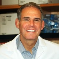
Hear Dr. Eric Topol (and others) speak at ENGAGE on the new landscape of patient engagement 
topol.jpg
Eric Topol, M.D.
Director; Prof. Genomics
Scripps Institute
For decades, physicians have told patients what to do and wielded all the power in the relationship. In the past five years, that equation has been turned on its head. With the advent of digital health technologies and the focus on genomics and personalized medicine, patients have squarely placed themselves at the center of the conversation.
Dr. Eric Topol, who has been championing this disruptive change in medicine for years, will address this great inversion and the future of patient engagement at MedCity ENGAGE, being held October 18-19 in San Diego.
Topol, Director, and Professor of Genomics at Scripps Institute, is the indefatigable champion of empowering patients through digital health and has published two bestseller books on the future of medicine.
We invite you to hear him speak on Medicine's Great Inversion: how technology and patient-centered care will change the future of patient engagement and healthcare.
Register Now
Can improved care be delivered at lower costs?
JoeSmith2.png
Joseph Smith, M.D.
President and CEO 
Reflexion Health
Entrepreneurs, payers and providers are recognizing that healthcare has to move out into the “real world” to be successful. Dr. Joe Smith, President and CEO, Reflexion Health will explain how home visits and video visits are improving access to care and lowering costs in the panel discussion: Straight to the Source: How Care is Moving Beyond the Doctor’s Office.
Smith is joined on the panel by Thompson Aderinkomi, Founder & CEO, RetraceHealth and Susan Stone, Senior VP and CEO, Sharp Coronado Hospital. You’ll hear how a data company is working with firefighters to reduce the burden on the ER and how the FDA approved physical therapy via motion trackers 
MedCity ENGAGE is the best place to discover the future of patient engagment and to meet and connect with ileading healthcare innovators.

Yoga: More Effective than Walking to Boost Anti-Anxiety Neurotransmitter

And you have a lot of anxiety since your doctor has no clue how to get you 100% recovered.
https://draxe.com/how-yoga-changes-your-brain/
Did you ever wonder how yoga changes your brain? As it turns out, that post-session happiness you feel isn’t just in your head. Using brain scans, scientists can now prove that yoga actually changes your brain chemistry. And that’s a good thing. Just like practicing tai chi moves, using yoga as a form of exercise and meditation can help naturally treat a range of health issues, particularly ones rooted in the brain.

How Yoga Changes Your Brain

While natural therapies, including yoga, don’t have a ton of funding for major studies compared to the pharmaceutical and biotech industries, we are starting to see some compelling science emerge. Some of the best science to date showing how yoga changes your brain involves yoga’s impact on anxiety, depression and pain tolerance.
Yoga Unleashes GABA
Did you know yoga is a natural remedy for anxiety? That’s because yoga impacts our brain’s GABA levels. GABA is short for gamma-aminobutryic acid, sometimes referred to as your body’s “chill out” neurotransmitter. GABA is crucial for suppressing neural activity. Your GABA neurotransmitters produce a calming effect similar to of drinking alcohol (without the harmful side effects). And, of course, alcohol’s calming effects are only temporary, with anxiety often rising once the buzz wears off. (12)
Yoga bumps up your brain’s natural GABA production without traditional anti-anxiety drugs designed to help your body release GABA. (Getting off of these benzodiazepine drugs can lead to serious withdrawal symptoms.) Yoga sounds much better than insomnia, seizures and, ironically, more anxiety linked with drug withdrawal. (3)
Bring on the asanas! And get this. While walking to lose weight really works, it may not be your best defense against anxiety. Practicing yoga unleashes more anxiety-quelling GABA in the brain’s thalamus than walking, according to a 2010 study published in the Journal of Alternative and Complementary Medicine. Compared to pleasure reading for an hour, a 60-minute yoga session increases GABA levels by 27 percent. (4) Because of its combination of breathing, meditation and movement, yoga could be one of the best exercises to combat anxiety.

Some good posters at the link.

Mechanosensing is critical for axon growth in the developing brain

How will your doctor use this to direct the axon pathfinding you need to recover?
http://www.nature.com/neuro/journal/vaop/ncurrent/full/nn.4394.html
Nature Neuroscience
doi:10.1038/nn.4394
Received
Accepted
Published online

Abstract

During nervous system development, neurons extend axons along well-defined pathways. The current understanding of axon pathfinding is based mainly on chemical signaling. However, growing neurons interact not only chemically but also mechanically with their environment. Here we identify mechanical signals as important regulators of axon pathfinding. In vitro, substrate stiffness determined growth patterns of Xenopus retinal ganglion cell axons. In vivo atomic force microscopy revealed a noticeable pattern of stiffness gradients in the embryonic brain. Retinal ganglion cell axons grew toward softer tissue, which was reproduced in vitro in the absence of chemical gradients. To test the importance of mechanical signals for axon growth in vivo, we altered brain stiffness, blocked mechanotransduction pharmacologically and knocked down the mechanosensitive ion channel piezo1. All treatments resulted in aberrant axonal growth and pathfinding errors, suggesting that local tissue stiffness, read out by mechanosensitive ion channels, is critically involved in instructing neuronal growth in vivo.

Some figures at the link.

Brain network analysis: a practical tutorial

Ask your doctor how your stroke disrupted your brain networks. Specifics only, not generic crap your doctor will try to pass off as knowledgeable. What protocols will fix those networks?
http://brain.oxfordjournals.org/content/early/2016/09/16/brain.aww232.extract

Restricted access
DOI: http://dx.doi.org/10.1093/brain/aww232 aww232 First published online: 19 September 2016

How are our brains wired? How are pathways between neurons organized? What patterns of connections allow us to think the way we do, or distinguish our ways of thinking from those of other animals? These and related questions are the bread and butter of an excellent new textbook.
FUNDAMENTALS OF BRAIN NETWORK ANALYSIS
By Alex Fornito, Andrew Zalesky and Edward Bullmore, 2016 Elsevier (Academic Press) ISBN 978-0-12-407908-3 Price: £60.99
Fundamentals of Brain Network Analysis by Fornito, Zalesky and Bullmore, is a thorough and didactic presentation of the tools available to research scientists wishing to engage in the emerging field of network neuroscience (Bullmore and Sporns, 2009). Blending computational tools and mathematical frameworks from physics, engineering, statistics, and computer science with the reams of data now being collected from diverse neural systems, network neuroscience is a truly interdisciplinary and ground-breaking field poised to transform our understanding of the brain. Rather than focusing solely on the function of single neurons or brain regions, these efforts expand the purview of our interests to the pattern of interactions between neural …
View Full Text

Relationship Between Walking Capacity, Biopsychosocial Factors, Self-efficacy, and Walking Activity in Persons Poststroke

What is your doctor doing with this to update your stroke protocols? Or are you once again on your own doing your doctors job? You will notice that there was no attempt to objectively identify dead and damaged areas in the brain. Research that will be impossible to replicate and compare results.
http://mobile.journals.lww.com/jnpt/_layouts/15/oaks.journals.mobile/articleviewer.aspx?year=2016&issue=10000&article=00004

Danks, Kelly A. PT, DPT; Pohlig, Ryan T. PhD; Roos, Margie PT, DPT, PhD; Wright, Tamara R. PT, DPT; Reisman, Darcy S. PT, PhD

Journal of Neurologic Physical Therapy
October 2016
Vol. 40 - Issue 4: p 232–238


Background/Purpose: Many factors appear to be related to physical activity after stroke, yet it is unclear how these factors interact and which ones might be the best predictors. Therefore, the purpose of this study was twofold: (1) to examine the relationship between walking capacity and walking activity, and (2) to investigate how biopsychosocial factors and self-efficacy relate to walking activity, above and beyond walking capacity impairment poststroke.
Methods: Individuals greater than 3 months poststroke (n = 55) completed the Yesavage Geriatric Depression Scale (GDS), Fatigue Severity Scale (FSS), Modified Cumulative Illness Rating (MCIR) Scale, Walk 12, Activities-Specific Balance Confidence (ABC) Scale, Functional Gait Assessment (FGA), and oxygen consumption testing. Walking activity data were collected via a StepWatch Activity Monitor. Predictors were grouped into 3 constructs: (1) walking capacity: oxygen consumption and FGA; (2) biopsychosocial: GDS, FSS, and MCIR; (3) self-efficacy: Walk 12 and ABC. Moderated sequential regression models were used to examine what factors best predicted walking activity.
Results: Walking capacity explained 35.9% (P < 0.001) of the variance in walking activity. Self-efficacy (ΔR2 = 0.15, P < 0.001) and the interaction between the FGA×ABC (ΔR2 = 0.047, P < 0.001) significantly increased the variability explained. The FGA (β = 0.37, P = 0.01), MCIR (β = -0.26, P = 0.01), and Walk 12 (β = −0.45, P = 0.00) were each individually significantly associated with walking activity.
Discussion and Conclusion: Although measures of walking capacity and self-efficacy significantly contributed to “real-world” walking activity, balance self-efficacy moderated the relationship between walking capacity and walking activity. Improving balance self-efficacy may augment walking capacity and translate to improved walking activity poststroke.
Video Abstract available for more insights from the authors (see Supplemental Digital Content 1, http://links.lww.com/JNPT/A139).

Reporting Standards for Preclinical Studies of Stroke Therapy

Our fucking failures of stroke associations should have noticed and corrected this problem with research decades ago.
http://stroke.ahajournals.org/content/47/10/2435.extract?etoc
Farhaan Vahidy, Wolf-Rüdiger Schäbitz, Marc Fisher and Jaroslaw Aronowski

The unmet need for development of new stroke therapies is enormous. Evidence generated from positive, null, or negative preclinical studies for various therapeutic agents is crucial to enhancing scientific progress. The scientific community shares a societal responsibility to practice and promote meticulous conduct and reporting of all experimental studies. A systematic survey conducted by the UK government–sponsored National Center for the Replacement, Refinement, and Reduction of Animals in Research (NC3Rs) reported that only 59% of biomedical animal studies stated the hypotheses and objectives, and ≤87% did not use randomization.1 This, in part, led to the development of the Animals in Research: Reporting In Vivo Experiments (ARRIVE) guidelines,2 modeled after the CONSORT (Consolidated Standards for Reporting …
View Full Text