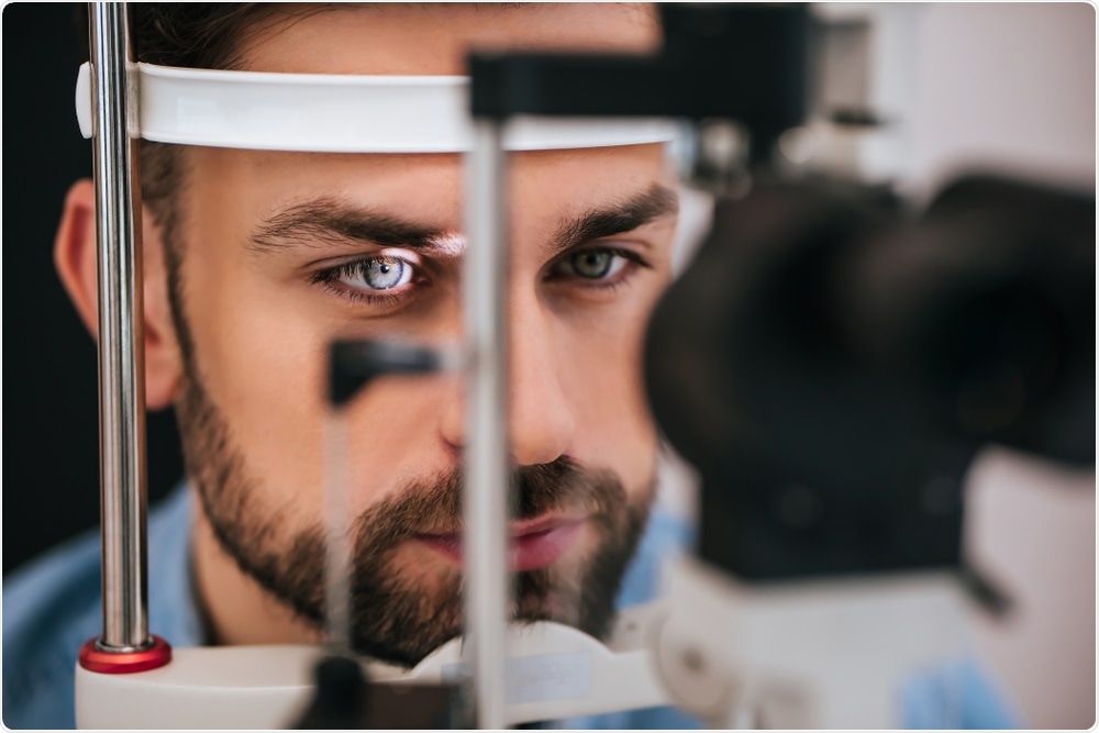
With your likely chance of dementia/Alzheimers your doctor should be using this to establish a baseline for you.
You will need this.
Your chances of getting dementia.
1. A documented 33% dementia chance post-stroke from an Australian study? May 2012.
2. Then this study came out and seems to have a range from 17-66%. December 2013.
3. A 20% chance in this research. July 2013.
2. Then this study came out and seems to have a range from 17-66%. December 2013.
3. A 20% chance in this research. July 2013.
Alzheimer’s and brain health could soon be detected using an eye exam
Soon, an eye examination may be all that is needed to confirm
a diagnosis of Alzheimer's disease, according to researchers at Duke's
Health.
 4 PM production | Shutterstock
Duke Eye Center recently recruited and studied the retinas of over
200 individuals to see if there were any differences between those with
Alzheimer's and those without. The results of the study titled, “Retinal
Microvascular and Neurodegenerative Changes in Alzheimer’s Disease and
Mild Cognitive Impairment Compared with Control Participants,” were
published in the latest issue of the journal Ophthalmology Retina.
The results showed that people who have a healthy brain function have a dense microscopic network of blood vessels in the retina, which can be observed through an eye examination. This web-like network of vessels was much less pronounced in patients with Alzheimer's disease.
The data is based on retinal images from 133 healthy participants and 39 individuals with Alzheimer's disease. Age, gender, level of education was adjusted for all cases and controls to remove the influence of bias in the results.
Duke ophthalmologist and retinal surgeon Sharon Fekrat, senior author of the study said that the differences were significant among cases and controls:
Fekrat said, “Ultimately, the goal would be to use this technology to detect Alzheimer's early, before symptoms of memory loss are evident, and be able to monitor these changes over time in participants of clinical trials studying new Alzheimer's treatments.”
The research was funded by the National Institutes of Health, 2018 Unrestricted Grant from Research to Prevent Blindness and the Karen L. Wrenn Alzheimer's Disease Award.
 4 PM production | Shutterstock
The results showed that people who have a healthy brain function have a dense microscopic network of blood vessels in the retina, which can be observed through an eye examination. This web-like network of vessels was much less pronounced in patients with Alzheimer's disease.
The data is based on retinal images from 133 healthy participants and 39 individuals with Alzheimer's disease. Age, gender, level of education was adjusted for all cases and controls to remove the influence of bias in the results.
Duke ophthalmologist and retinal surgeon Sharon Fekrat, senior author of the study said that the differences were significant among cases and controls:
We're measuring blood vessels that can't be seen during a regular eye exam and we're doing that with relatively new noninvasive technology that takes high-resolution images of very small blood vessels within the retina in just a few minutes. It's possible that these changes in blood vessel density in the retina could mirror what's going on in the tiny blood vessels in the brain, perhaps before we are able to detect any changes in cognition.”The team found that there were tell-tale signs of changes in the retinal blood vessels among those who have a mild cognitive impairment, a known precursor for Alzheimer’s disease. This is due to changes in the retinal nerve layers with mild cognitive impairment and Alzheimer’s.
Sharon Fekrat, Senior Author
We know that there are changes that occur in the brain in the small blood vessels in people with Alzheimer's disease, and because the retina is an extension of the brain, we wanted to investigate whether these changes could be detected in the retina using a new technology that is less invasive and easy to obtain.He said that they used a non-invasive technology called optical coherence tomography angiography (OCTA) to measure the blood flow in each of the layers of the retina. Some of the changes detected were in capillaries or blood vessels that measured less than the width of a human hair he explained.
Dilraj S. Grewal, Lead Author
Fekrat said, “Ultimately, the goal would be to use this technology to detect Alzheimer's early, before symptoms of memory loss are evident, and be able to monitor these changes over time in participants of clinical trials studying new Alzheimer's treatments.”
The research was funded by the National Institutes of Health, 2018 Unrestricted Grant from Research to Prevent Blindness and the Karen L. Wrenn Alzheimer's Disease Award.

No comments:
Post a Comment