So whom is putting all these mini brains together into one version so our stroke researchers can all use the same starting point to solve all the problems in stroke?
- brain in a dish (8 posts)
- brain in a lab (2 posts)
- Brain on a chip (7 posts)
Scientists grow 'mini-brain on the move' that can contract muscle
Cambridge researchers grew ‘organoid’ that spontaneously connected to spinal cord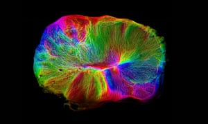
Scientists have grown a miniature brain in a dish with a spinal cord
and muscles attached, an advance that promises to accelerate the study
of conditions such as motor neurone disease.
The lentil-sized grey blob of human brain cells were seen to spontaneously send out tendril-like connections to link up with the spinal cord and muscle tissue, which was taken from a mouse. The muscles were then seen to visibly contract under the control of the so-called brain organoid.
The research is is the latest in a series of increasingly sophisticated approximations of the human brain grown in the laboratory – this time with something approaching a central nervous system attached.
Madeline Lancaster, who led the work at the Medical Research Council’s Laboratory of Molecular Biology in Cambridge, said: “We like to think of them as mini-brains on the move.”
The scientists used a new method to grow the miniature brain from human stem cells, which allowed the organoid to reach a more sophisticated stage of development than previous experiments. The latest blob shows similarities, in terms of the variety of neurons and their organisation, to the human foetal brain at 12-16 weeks of pregnancy.
However, the scientists said the structure was still too small and
primitive to have anything approaching thoughts, feelings or
consciousness.The lentil-sized grey blob of human brain cells were seen to spontaneously send out tendril-like connections to link up with the spinal cord and muscle tissue, which was taken from a mouse. The muscles were then seen to visibly contract under the control of the so-called brain organoid.
The research is is the latest in a series of increasingly sophisticated approximations of the human brain grown in the laboratory – this time with something approaching a central nervous system attached.
Madeline Lancaster, who led the work at the Medical Research Council’s Laboratory of Molecular Biology in Cambridge, said: “We like to think of them as mini-brains on the move.”
The scientists used a new method to grow the miniature brain from human stem cells, which allowed the organoid to reach a more sophisticated stage of development than previous experiments. The latest blob shows similarities, in terms of the variety of neurons and their organisation, to the human foetal brain at 12-16 weeks of pregnancy.
“It’s still a good idea to have that discussion every time we take it a step further,” said Lancaster. “But we agree generally that we’re still very far away from that.”
While a fully developed human brain has 80-90bn neurons, the organoid has a couple of million, placing it somewhere between a cockroach and a zebrafish in terms of volume of grey matter.
Previously, the sophistication of the organoids scientists had been able to achieve had been limited by the lack of a nutrient supply to the centre of the blob. Once it reached a certain size, the neurons in the centre would become cut off from their nutrient supply and start to die off, and the structure would stop developing.
In the latest research, the scientists grew the organoid and then used a tiny vibrating blade to cut it into half millimetre-thick slices which were placed on a membrane, floating on a nutrient-rich liquid. This meant the entire slice had access to energy and oxygen and it continued developing and forming new connections when it was kept in culture for a year.
Alongside the organoid, the scientists added in a 1mm-long spinal cord, taken from a mouse embryo, and the surrounding back muscle. The brain cells automatically began to send out neuronal connections, linked up with the spinal cord and began sending electrical impulses, which caused the muscles to twitch.
The ambition is to use systems like this to study how the human brain and nervous system develop and why things go wrong in illnesses such as motor neurone disease, epilepsy and schizophrenia.
“Obviously we’re not just trying to create something for the fun of it,” said Lancaster. “We want to use this to model diseases and to understand how these networks are set up in the first place.”
Gray Camp, a geneticist at the Institute of Molecular and Clinical Ophthalmology in Basel, Switzerland, who was not involved in the latest work, described the advance as “a big step for the field”. “It’s extremely exciting to see evidence of functional nerve tracts growing out of developing human brain tissue and innervating other tissues,” he said.
The findings are published in the journal Nature Neuroscience.
No comments:
Post a Comment