Use the labels in the right column to find what you want. Or you can go thru them one by one, there are only 33,532 posts. Searching is done in the search box in upper left corner. I blog on anything to do with stroke. DO NOT DO ANYTHING SUGGESTED HERE AS I AM NOT MEDICALLY TRAINED, YOUR DOCTOR IS, LISTEN TO THEM. BUT I BET THEY DON'T KNOW HOW TO GET YOU 100% RECOVERED. I DON'T EITHER BUT HAVE PLENTY OF QUESTIONS FOR YOUR DOCTOR TO ANSWER.
Changing stroke rehab and research worldwide now.Time is Brain!trillions and trillions of neuronsthatDIEeach day because there areNOeffective hyperacute therapies besides tPA(only 12% effective). I have 523 posts on hyperacute therapy, enough for researchers to spend decades proving them out. These are my personal ideas and blog on stroke rehabilitation and stroke research. Do not attempt any of these without checking with your medical provider. Unless you join me in agitating, when you need these therapies they won't be there.
What this blog is for:
My blog is not to help survivors recover, it is to have the 10 million yearly stroke survivors light fires underneath their doctors, stroke hospitals and stroke researchers to get stroke solved. 100% recovery. The stroke medical world is completely failing at that goal, they don't even have it as a goal. Shortly after getting out of the hospital and getting NO information on the process or protocols of stroke rehabilitation and recovery I started searching on the internet and found that no other survivor received useful information. This is an attempt to cover all stroke rehabilitation information that should be readily available to survivors so they can talk with informed knowledge to their medical staff. It lays out what needs to be done to get stroke survivors closer to 100% recovery. It's quite disgusting that this information is not available from every stroke association and doctors group.
Monday, March 23, 2020
New brain implant device could record activity in thousands of neurons
If we are ever going to make neuroplasticity completely repeatable we
will need to understand the signals sent between neurons. This might be
one way, Your researcher can tell you which of these other ideas would
be the best to answer that question. Nothing will ever come of this
because we have NO stroke leadership to go to to get a stroke strategy
updated.
Right now neuoplasticity is
considered the holy grail of stroke rehab but without knowing how to
make it repeatable is practically useless.
A team of Stanford University researchers has created a device that,
once implanted in the brain, could help record movies of electrical
neural activity in thousands of individual neurons.
The device, described in a paper published March 20 in Science Advances, could
be used for research or with prosthetics, and is capable of recording
more data while being less intrusive than other options.
"The design of this device is completely different from any existing
high-density recording devices, and the shape, size and density of the
array can be simply varied during fabrication. This means that we can
simultaneously record different brain regions at different depths with
virtually any 3D arrangement," Jun Ding, PhD, assistant professor of neurosurgery and neurology and co-author of the paper said in a Stanford News story. "If applied broadly, this technology will greatly excel our understanding of brain function in health and disease states."
At the heart of this invention is a bundle of microwires, each of
which is less than half the width of the thinnest human hair. These
wires, which are directed into the brain to obtain electrical signals
that pass by, are small enough to cause minimal damage but sturdy enough
to resist degrading over time.
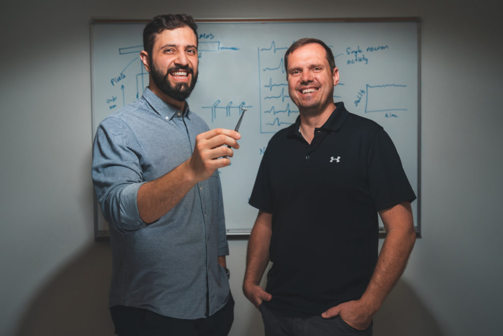
Abdulmalik
Obaid, left, and Nick Melosh with a device they invented that contains a
bundle of microwires capable of recording the activity of thousands of
neurons in the brain in real time.
The trick was figuring out how to design an orderly array of these
super thin wires that is adaptable in terms of size -- some applications
of the array may only warrant a few microwires but others would require
thousands.
The researchers spent years designing and redesigning the device and
the process for making it. Eventually, they found success by encasing
each wire in a biologically-safe polymer, then bundling them in a metal
collar. Below the collar, the polymer is removed from the wires so they
can be inserted into the brain. Topped off with a silicon chip -- like
those used in a camera -- the device can begin recording neural
activity.
Once the researchers settled on their design, they were able to run
tests in living tissues. They began with retinal cells from rats and a
138-wire array.
"We had to take kilometers of microwires and produce large-scale arrays, then directly connect them to silicon chips," Abdulmalik Obaid,
a graduate student in materials science and engineering and lead author
of the paper told Stanford News. "After years of working on that
design, we tested it on the retina for the first time and it worked
right away. It was extremely reassuring."
The team has also successfully tested the device in the brains of
living mice, using arrays that ranged from 135 to 251 wires, and are
continuing these studies so they can better understand the longevity of
their invention and the kinds of signals it is able to obtain.
"Electrical activity is one of the highest-resolution ways of looking at brain activity," said Nick Melosh,
professor of materials science and engineering and co-senior author of
the paper. "With this microwire array, we can see what's happening on
the single-neuron level."
No comments:
Post a Comment