WHOM is going to put this in a usable format so recovery can be applied to stroke survivors? We need to know the specific name of the leader and researcher so we can monitor progress. And you really think your stroke hospital subscribes to Physics World and requires the doctors to read it?
Multimodal MRI reveals brain areas that can still ‘see’ after a stroke

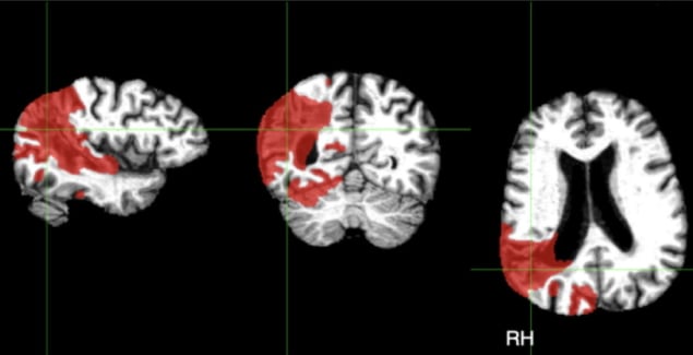
Scientists in the UK have used magnetic resonance imaging (MRI) to map the brain’s responses to visual stimuli after a stroke. In stroke survivors with vision loss, brain imaging revealed responsive areas that were inaccessible to current vision tests. The researchers’ findings could help clinicians better understand vision loss in stroke, and design tailored rehabilitation programmes for survivors. The researchers, from the University of Nottingham, describe their work in Frontiers in Neuroscience.
Every year, an estimated 100,000 people in the UK have a stroke. Globally, the number of strokes each year has risen by 70% since 1990, and stroke remains one of the leading causes of death and disability worldwide.
About a third of all stroke survivors experience visual field loss, in which a quarter or one half of their total visual field is impacted. Currently, there are no universally accepted rehabilitation strategies for vision loss in stroke, and the success of existing programmes varies significantly across survivors.
Visual field loss after stroke is usually diagnosed using perimetry, a technique that uses bright lights of differing size and brightness to test a patient’s visual response. However, perimetry only provides a coarse map of residual visual function and cannot pinpoint pathways in the brain that no longer process visual information.
To reveal more detail, the researchers used different MRI techniques – functional, anatomical and diffusion-weighted MRI – to chart affected visual pathways in the brain in four stroke survivors.

By measuring responses in the brain to visual stimuli, they were able to map the visual field in stroke survivors in much finer detail compared with perimetry. When they overlaid their visual field maps with perimetry maps, they were able to see spots in the visual field that still elicited a response from the brain.

“By examining different types of brain scans we can actually see areas of ‘residual vision’ – places where the eyes and brain can still process images, even if this doesn’t reach awareness,” explains Denis Schluppeck, senior author of the study. “Using MRI to pinpoint these areas of functional vision, clinicians could work with the stroke survivor and train them to recover some function in that particular spot.”
Rehabilitation strategies for vision loss in stroke include strengthening existing or alternative visual pathways in the brain. Combining brain imaging and optometric tests could provide a cost-effective way to understand which brain areas to focus on. The researchers note that their MRI protocol takes just one hour to perform.
“I think what was most challenging about the study was recruiting stroke survivors,” says first author Anthony Beh. “As with any clinical group, there are extra considerations to make when working closely with them, such as mobility challenges, past medical history and other stroke-related impairments. It was also important to get in touch with local stroke communities to visit and speak more about the project.”

Going forward, the researchers plan to use what they have learned to understand other forms of visual loss. “We hope to use a similar approach in children and younger individuals who have cortical visual impairment – visual loss that is not caused by damage to eyes or early parts of the visual pathway – to understand that set of disorders,” Schluppeck explains.
No comments:
Post a Comment