Changing stroke rehab and research worldwide now.Time is Brain! trillions and trillions of neurons that DIE each day because there are NO effective hyperacute therapies besides tPA(only 12% effective). I have 523 posts on hyperacute therapy, enough for researchers to spend decades proving them out. These are my personal ideas and blog on stroke rehabilitation and stroke research. Do not attempt any of these without checking with your medical provider. Unless you join me in agitating, when you need these therapies they won't be there. Did your doctor somehow miss the recovery protocol that Pedro Bach-y-Rita used to recover fully back in 1958 with only a partial brain?

What this blog is for:

My blog is not to help survivors recover, it is to have the 10 million yearly stroke survivors light fires underneath their doctors, stroke hospitals and stroke researchers to get stroke solved. 100% recovery. The stroke medical world is completely failing at that goal, they don't even have it as a goal. Shortly after getting out of the hospital and getting NO information on the process or protocols of stroke rehabilitation and recovery I started searching on the internet and found that no other survivor received useful information. This is an attempt to cover all stroke rehabilitation information that should be readily available to survivors so they can talk with informed knowledge to their medical staff. It lays out what needs to be done to get stroke survivors closer to 100% recovery. It's quite disgusting that this information is not available from every stroke association and doctors group.

Showing posts with label stimulation. Show all posts

Tuesday, June 21, 2016

Your Work Environment And The Brain: Lack Of Stimulation, Dirtiness Worsens Cognitive Health

Just like you need a mentally challenging job to prevent dementia. Burger flipping and greeter at Wal-Mart do not qualify.
http://www.medicaldaily.com/work-environment-human-brain-mental-stimulation-dirty-job-cognitive-health-390035

In a recent study published in the Journal of Occupational and Environmental Medicine, researchers from Florida State University analyzed how a dirty workplace and one that lacks stimulation can impact the long-term cognitive function of its employees.
"Psychologists say that the brain is a muscle, while industrial hygienists point to chemicals in the work environment that may cause decline," said Joseph Grzywacz, lead researcher from the Norejane Hendrickson Professor of Family and Child Sciences, in a statement. "There are real things in the workplace that can shape cognitive function: some that you can see or touch, and others you can't. We showed that both matter to cognitive health in adulthood."
Grzywacz and his colleagues collected data taken from the Midlife in the United States study, including 4,963 working adults between the ages of 32 and 84 from 48 states (47 percent male and 53 percent female).
The researchers gained insight into each participant’s cognitive function and the relationship to their workplace by examining their ability to maintain and later use learned information. Researchers also took into account executive functioning skills, including the ability to complete tasks, manage time, and pay attention. All participants were asked about any problems with memory they were experiencing.
The study produced two important takeaways: One was that greater occupational complexity, the ability to learn new skills and take on new challenges, leads to stronger cognitive performance, especially among women as they aged. The second was men and women exposed to a dirty working environment suffered cognitive decline.
"Both of these issues are important when we think about the long-term health of men and women,"  Grzywacz added. "The practical issue here is cognitive decline associated with aging and the thought of, 'if you don't use it, you lose it.’ Designing jobs to ensure that all workers have some decision-making ability may protect cognitive function later in life, but it's also about cleaning up the workplace."
Whether it’s relationships between employees or work hours, workplace standards are crucial to the health of employees. Researchers from the University of Minnesota recently compared workplaces that encourage healthy lifestyle practices such as lunchtime yoga classes and eating healthy food to those that do not. Unsurprisingly, employees from unhealthy workplaces were far more likely to be obese.
Source: Lachman M, Segel-Karpas D, Grzywacz J. Workplace Exposures and Cognitive Function During Adulthood: Evidence From National Survey of Midlife Development and the O*NET. Journal of Occupational and Environmental Medicine . 2016.

Tuesday, May 19, 2015

Rethinking Stimulation of the Brain in Stroke Rehabilitation Why Higher Motor Areas Might Be Better Alternatives for Patients with Greater Impairments

Going after the pre-motor cortex wouldn't help me at all, most of mine is dead. I wish someone would tackle the extremely difficult problem of moving functions that were in dead areas to new locations. Now that would be worthy of a Nobel prize. You will need to send your doctor after exactly how they are stimulating the PMAs.
http://nro.sagepub.com/content/21/3/225?etoc
  1. Ela B. Plow1,2
  2. David A. Cunningham1,3
  3. Nicole Varnerin1
  4. Andre Machado4
  1. 1Department of Biomedical Engineering, Lerner Research Institute, Cleveland Clinic, Cleveland, OH, USA
  2. 2Department of Physical Medicine & Rehabilitation, Neurological Institute, Cleveland Clinic, Cleveland, OH, USA
  3. 3School of Biomedical Sciences, Kent State University, Kent, OH, USA
  4. 4Center for Neurological Restoration, Neurological Institute, Cleveland Clinic, Cleveland, OH, USA
  1. Ela B. Plow, Department of Biomedical Engineering, Lerner Research Institute, Cleveland Clinic, 9500 Euclid Avenue, ND20, Cleveland, OH 44195, USA. Email: plowe2@ccf.org

Abstract

Stimulating the brain to drive its adaptive plastic potential is promising to accelerate rehabilitative outcomes in stroke. The ipsilesional primary motor cortex (M1) is invariably facilitated. However, evidence supporting its efficacy is divided, indicating that we may have overgeneralized its potential. Since the M1 and its corticospinal output are frequently damaged in patients with serious lesions and impairments, ipsilesional premotor areas (PMAs) could be useful alternates instead. We base our premise on their higher probability of survival, greater descending projections, and adaptive potential, which is causal for recovery across the seriously impaired. Using a conceptual model, we describe how chronically stimulating PMAs would strongly affect key mechanisms of stroke motor recovery, such as facilitating the plasticity of alternate descending output, restoring interhemispheric balance, and establishing widespread connectivity. Although at this time it is difficult to predict whether PMAs would be “better,” it is important to at least investigate whether they are reasonable substitutes for the M1. Even if the stimulation of the M1 may benefit those with maximum recovery potential, while that of PMAs may only help the more disadvantaged, it may still be reasonable to achieve some recovery across the majority rather than stimulate a single locus fated to be inconsistently effective across all.


Friday, March 14, 2014

Needed intellectual stimulation to prevent dementia - Italy version

We visited the Mostra di Leonardo Da Vinci museum in Rome where working models of his drawings were displayed. It takes a lot of thought to figure out how he knew so much that far ahead of others.

The official photo gallery is here:

http://www.mostradileonardo.com/index.php?option=com_content&view=article&id=8&Itemid=139&lang=en

Think long and hard how he managed to do all this.
My photos are below;


Monday, December 30, 2013

Exploring staff experience of an “enriched environment” within stroke rehabilitation: a qualitative sub-study

This was written about by Dale Corbett back in Feb. 2011

What the hell is it going to take to get this into a stroke protocol rolled out to all hospitals? Are we going to need a scenario like had in the movie

John Q in 2002 before anything gets done?

Exploring staff experience of an “enriched environment” within stroke rehabilitation: a qualitative sub-study


1Hunter Stroke Service, John Hunter Campus,
New Lambton, New South Wales
, Australia,
2Department of Medical Sciences, The University of Newcastle,
Callaghan, New South Wales
, Australia, and
3Rankin Park Unit,
New Lambton, New South Wales
, Australia
Address for correspondence:
Jennifer H. White, MMSc (Occupational Therapy), Hunter Stroke Service, Level 2, The Lodge
, John Hunter Campus,
Lookout Rd, New Lambton, New South Wales 2301
, Australia. E-mail:

Abstract

Background: Environmental Enrichment (EE) is shown to facilitate recovery of motor and cognitive function in animal models of stroke. The efficacy of EE in the clinical setting with stroke survivors remains unknown. Successful implementation of EE in a busy rehabilitation unit requires identification of barriers and enablers which are best informed by staff feedback. Aim: To qualitatively explore the experiences of nursing staff involved in a pilot study investigating the feasibility of EE in a rehabilitation ward. Methods: This was a qualitative study consisting of analysis of semi-structured interviews with nine nursing staff who were asked to reflect on “routine care” and their own “experience of the EE study”. An inductive thematic approach was used to collect and analyse data using a process of constant comparison. Results: Male and female staff with varying years of experience working in stroke rehabilitation participated in focus group and individual interviews. Three key themes were identified concerning the implementation of EE including: (i) “Nurses are so busy” – perceptions on routine work practice; (ii) “A better outlook” – perceptions of the benefit of EE; (iii) “They’re just not going to participate” – perceptions of barriers to EE. Indeed, the challenges identified in this study align with practice change literature, which indicates that staff workload, routine and attitudes can influence the implementation of a new practice. Discussion: Staff perceived the use of an EE in their rehabilitation unit promoted activity/participation and increased patient moral. The barriers and enablers experienced by staff in this study may be used to inform the design and conduct of future studies investigating the efficacy of EE during inpatient stroke rehabilitation after stroke.

Friday, May 10, 2013

A Rat’s Whiskers Point the Way toward a Novel Stimulus-Dependent, Protective Stroke Therapy

This was talked way back here in 2011,Society for Neuroscience 2011 press release. 
Why repeat the same research report?
Someone should make a hypothesis of what sensory stimulation on humans would be equivalent(face, hands,genitals?) to a whisker on a rat. This would be so easy to implement and run research on.
http://nro.sagepub.com/content/19/3/313.abstract?etoc

Abstract

Stroke is the fourth leading cause of death in the United States and the leading cause of long-term disability. Ischemic stroke, due to an interruption in blood supply, is particularly prevalent; 87% of all strokes are ischemic. Unfortunately, current options for acute treatment are extremely limited and there is a great need for new treatment strategies. This review will discuss evidence that mild sensory stimulation can completely protect the jeopardized brain from an impending stroke in a rodent model. When delivered within the first 2 hours following ischemic onset, this stimulation results in complete protection, including a full reestablishment of cortical function, sensorimotor capabilities, and blood flow. Identical stimulation, however, initiated 3 hours following ischemic onset, results in an increase in damage compared with untreated animals. The protective effect is not specific to a single sensory modality, anesthesia, or age, and increasing evoked cortical activity by increasing stimulation accelerates recovery. Taken together, these findings demonstrate that cortical activity is a critical factor for protection and suggest a new, exciting potential avenue for the development of acute stroke treatment strategies that may produce a noninvasive, drug-free, equipment-free, and side effect–free means of protecting from ischemic stroke.

Saturday, July 21, 2012

Nerve stimulation plus standard therapy may accelerate stroke recovery

This is already being used to treat depression and epilepsy. Definition here: Get our researchers on it for stroke for acute vs. chronic.

Nerve stimulation plus standard therapy may accelerate stroke recovery


UT Dallas researchers recently demonstrated how nerve stimulation paired with specific experiences, such as movements or sounds, can reorganize the brain and improve its function.
This technology could lead to new treatments for stroke, tinnitus, autism and other disorders.
A team led by Dr. Robert Rennaker and Dr. Michael Kilgard looked at whether repeatedly pairing vagus nerve stimulation with a specific movement would change neural activity within the laboratory rats’ primary motor cortex. To test the hypothesis, they paired the vagus nerve stimulation with movements of the forelimb in two groups of rats.
After five days of stimulation and movement pairing, the researchers examined the brain activity in response to the stimulation. The rats who received the training along with the stimulation displayed large changes in the organization of the brain’s movement control system. The animals receiving identical motor training without stimulation pairing did not exhibit any brain changes, or plasticity.
People who suffer strokes or brain trauma often undergo rehabilitation that includes repeated movement of the affected limb in an effort to regain motor skills. It is believed that repeated use of the affected limb causes reorganization of the brain essential to recovery.
The recent study suggested that pairing vagus nerve stimulation with standard therapy may result in more rapid and extensive reorganization of the brain, offering the potential for speeding and improving recovery following stroke, said Rennaker, associate professor in The University of Texas at Dallas’ School of Behavioral and Brain Sciences
“Our goal is to use the brain’s natural neuromodulatory systems to enhance the effectiveness of standard therapies,” Rennaker said.
“Our studies in sensory and motor cortex suggest that the technique has the potential to enhance treatments for neurological conditions ranging from chronic pain to motor disorders. Future studies will investigate its effectiveness in treating cognitive impairments,” he stated.
Since vagus nerve stimulation has an excellent safety record in human patients with epilepsy, the technique provides a new method to treat brain conditions in which the timing of brain responses is abnormal, including dyslexia and schizophrenia.
If subsequent studies confirm the UT Dallas findings, human patients may have access to more efficient therapies that are minimally invasive and avoid long-term use of drugs.
The results were published in a recent issue of Cerebral Cortex.

Tuesday, March 20, 2012

Lab Notes: Stroke Recovery Loves Company

I know this is not what Jill Bolte-Taylor would say.
http://www.medpagetoday.com/LabNotes/LabNotes/31238

Loneliness Not Good for Post-Stroke Progress

Companionship might be the key for better healing following a stroke, according to a preclinical study presented at the recent International Stroke Conference in New Orleans.

Mice that were socially isolated tended to have greater ischemic infarct volumes relative to mice paired with a stroke partner or a healthy partner. In addition, the solitary mice had significantly decreased levels of brain-derived neurotrophic factor (BDNF), a protein that helps support the survival of existing neurons.

Interestingly, the mice paired with healthy partners had significantly more BDNF than mice paired with a stroke partner. BDNF could be a marker for functional recovery, lead author Venugopal Reddy Venna, PhD, from the University of Connecticut in Farmington, Conn., told MedPage Today.

Venna said that post-stroke housing is critically important and should be considered in all patients, but especially in clinical studies evaluating various rehabilitative strategies.

Friday, December 23, 2011

Activating brain protein shown to aid in stroke recovery

I know I posted this earlier but didn't know the drug involved. Cutamesine.
A pathetic 15 years to get from basic research to a clinical trial and then another 15-20 to get into widespread use. What will make this go faster? We can't wait until the baby boomers start asking about recovery. You explain why to a Type-A personality that stroke recovery help is non-existant.
http://www.gizmag.com/activating-brain-protein-shown-to-aid-in-stroke-recovery/17953/

It's certainly not a news flash to say that being in a stimulating environment, where there's plenty to perceive and think about, is good for the brain – new neural pathways are formed, and existing ones are kept from atrophying. Now, however, researchers have discovered a way of replicating and reinforcing those good effects in any environment. It is hoped that the new technology will allow strokes to be treatable up to two days after they have occurred. Most current treatments must be administered within a matter of hours after the event.

The research was carried out by scientists at Sweden's Lund University, in collaboration with American colleagues.

Lab rats were subjected to strokes, then placed in one of two environments – some went into a regular cage, while others were placed in an "enriched" cage that contained interesting things like several levels of tubes, beams and ladders. It was found that being in the enriched cage activated a gene coded for the protein sigma-1 receptor, which plays a key role in helping the brain to recover from a stroke.

The researchers then injected those rats with cutamesine, a substance that activates the sigma-1 receptor, and found that they regained their brain function more quickly than the untreated rats. Essentially, the treatment simply recreates and enhances the brain's natural response to being in an enriched environment.

Although the basic research began over 15 years ago, the treatment is only now finally being used in a clinical trial on stroke patients by a Japanese pharmaceutical company.

"This is an excellent example of how basic research can be translated into a healthcare setting and possibly lead to new and better therapies," said Lund's Prof. Tadeusz Wieloch. "It also exemplifies the fact that, within medical research, it is a long journey from experimental studies to results that benefit the patient."

Wednesday, December 21, 2011

Training and Stimulation in Post Stroke Recovery Brain Reorganization

Another article from 2005, I wonder how many stroke units have started incorporating enriched environments for their stroke patients? Is it a part of the Joint Commission protocols on Stroke Rehabilitation? Its probably a secret(national defense and all) You wouldn't want stroke survivors to know what the latest therapies are, they might start getting vocal about them.
http://thomasland.metapress.com/content/e893m0prnjej1gxm/
Come on Joint Commission, I dare you to contact me.
Abstract

In both animal and clinical studies, training or rehabilitation increases cortical representation with subsequent functional recovery, whereas a lack of rehabilitation or training decreases cortical representation and delays recovery. Animals exposed to enriched environments post stroke have improved functional outcomes compared with animals exposed to nonenriched environments. In humans, stroke units may be the closest approximation there is to an enriched environment. However, studies indicate that patients spend the majority of time being inactive and alone while on a stroke unit. Given the animal evidence (which emphasizes increased stimulation and increased activity), there is clearly an opportunity for improving the stroke rehabilitation experience to maximize post stroke recovery.

Keywords
animal models, cerebral vascular accident, hospital units, humans, recovery of function, rehabilitation, stroke units

Monday, December 19, 2011

Transcranial magnetic stimulation in mild to severe hemiparesis early after stroke: a proof of principle and novel approach to improve motor function

The scientific details to TMS on the good side(contralesional). If I were 5-45 days out I would demand to have this regardless of the risk. But ask your doctor nicely about this therapy.Linkhttp://www.springerlink.com/content/427412375113202h/

Abstract

Low-frequency repetitive transcranial magnetic stimulation (rTMS) of the unaffected hemisphere can enhance function of the paretic hand in patients with mild motor impairment. Effects of low-frequency rTMS to the contralesional motor cortex at an early stage of mild to severe hemiparesis after stroke are unknown. In this pilot, randomized, double-blind clinical trial we compared the effects of low-frequency rTMS or sham rTMS as add-on therapies to outpatient customary rehabilitation, in 30 patients within 5–45 days after ischemic stroke, and mild to severe hand paresis. The primary feasibility outcome was compliance with the interventions. The primary safety outcome was the proportion of intervention-related adverse events. Performance of the paretic hand in the Jebsen–Taylor test and pinch strength were secondary outcomes. Outcomes were assessed at baseline, after ten sessions of treatment administered over 2 weeks and at 1 month after end of treatment. Baseline clinical features were comparable across groups. For the primary feasibility outcome, compliance with treatment was 100% in the active group and 94% in the sham group. There were no serious intervention-related adverse events. There were significant improvements in performance in the Jebsen–Taylor test (mean, 12.3% 1 month after treatment) and pinch force (mean, 0.5 Newtons) in the active group, but not in the sham group. Low-frequency rTMS to the contralesional motor cortex early after stroke is feasible, safe and potentially effective to improve function of the paretic hand, in patients with mild to severe hemiparesis. These promising results will be valuable to design larger randomized clinical trials.

Monday, October 31, 2011

Random Noise Stimulation Improves Neuroplasticity in Perceptual Learning

Even after reading this 3 times I don't understand, someone needs to tell researchers they need to put their writings into a useful understandable relation to a therapy. Noise here does not refer to sound.
http://www.jneurosci.org/content/31/43/15416.abstract


Abstract
Perceptual learning is considered a manifestation of neural plasticity in the human brain. We investigated brain plasticity mechanisms in a learning task using noninvasive transcranial electrical stimulation (tES). We hypothesized that different types of tES would have varying actions on the nervous system, which would result in different efficacies of neural plasticity modulation. Thus, the principal goal of the present study was to verify the possibility of inducing differential plasticity effects using two tES approaches [i.e., direct current stimulation (tDCS) and random noise stimulation (tRNS)] during the execution of a visual perceptual learning task.

Friday, April 15, 2011

Can brain stimulation boost learning?

I actually toured the research place with the Elderhostel Braintraining class when this research was taking place. The class is no longer being offered.
http://www.nature.com/news/2011/110413/full/472156a.html
Title: Brain Stimulation to Accelerate Learning of Threat Detection, Phase II

Purpose of Study: The research is to investigate how the brain responds to learning tasks to recognize visual targets in synthetic radar imagery. Also, we will be using Transcranial Direct Current Stimulation (TDCS) in attempt to accelerate learning.
Last year a succession of volunteers sat down in a research lab in Albuquerque, New Mexico to play DARWARS Ambush!, a video game designed to train US soldiers bound for Iraq. Each person surveyed virtual landscapes strewn with dilapidated buildings and abandoned cars for signs of trouble — a shadow cast by a rooftop sniper, or an improvised explosive device behind a rubbish bin. With just seconds to react before a blast or shots rang out, most forgot about the wet sponge affixed to their right temple that was delivering a faint electric tickle. The volunteers received a few milliamps of current at most, and the simple gadget used to deliver it was powered by a 9-volt battery.
It might sound like some wacky garage experiment, but Vincent Clark, a neuroscientist at the University of New Mexico, says that the technique, called transcranial direct-current stimulation (tDCS), could improve learning. The US Defense Advanced Research Projects Agency funded the research in the hope that it could be used to sharpen soldiers' minds on the battlefield. Yet for all its simplicity, it seems to work.
Volunteers receiving 2 milliamps to the scalp (about one-five-hundredth the amount drawn by a 100-watt light bulb) showed twice as much improvement in the game after a short amount of training as those receiving one-twentieth the amount of current1. "They learn more quickly but they don't have a good intuitive or introspective sense about why," says Clark.
The technique, which has roots in research done more than two centuries ago, is experiencing something of a revival. Clark and others see tDCS as a way to tease apart the mechanisms of learning and cognition. As the technique is refined, researchers could, with the flick of a switch, amplify or mute activity in many areas of the brain and watch what happens behaviourally. The field is "going to explode very soon and give us all sorts of new information and new questions", says Clark. And as with some other interventions for stimulating brain activity, such as high-powered magnets or surgically implanted electrodes, researchers are attempting to use tDCS to treat neurological conditions, including depression and stroke. But given the simplicity of building tDCS devices, one of the most important questions will be whether it is ethical to tinker with healthy minds — to improve learning and cognition, for example. The effects seen in experimental settings "are big enough that they would definitely have real-world consequences", says Martha Farah, a neuroethicist at the University of Pennsylvania in Philadelphia.
Getting to this point, however, was hardly straightforward. Direct-current brain stimulation has emerged from a long, touch-and-go history that ranges from the simply bizarre to the simply irreproducible. And for some, it still has much to prove.
The Italian scientist Jean Aldini first tried direct-current stimulation around 1800 — initially to induce movement in the corpses of recently executed felons. Later, he claimed in a paper to cure two acquaintances of the mood disorder then known as 'melancholy'. By the 1940s, many patients with depression were being given electric shocks to the temples that were strong enough to induce seizures — so-called electroconvulsive therapy. But for decades people toyed with the idea of treating mental illness with electric shocks that were much milder — 1,000 times less intense than electroconvulsive therapy.

Weak origins

In 1964, Joe Redfearn, a psychiatrist at Graylingwell Hospital in Chichester, UK, applied some promising results in rats directly to humans, delivering weak currents — of 50–250 microamps — to the scalps of volunteers. He reports that the volunteers became talkative, even giggly, when current was run in one direction, but withdrawn when it ran the other way2. He gave the 'giggly' treatment to 29 patients with depression and claims that half of them improved3. But no one could replicate his results, and the technique was abandoned.
In retrospect, several factors seem to have undermined his work. Among them, Redfearn used currents ten times lower than in modern tDCS — perhaps because he had no way to measure how much electricity was actually reaching his patients' brains. Within a few decades, however, the necessary methods would become available, notably as researchers began to study brain activity induced by transcranial magnetic stimulation (TMS). In TMS, a magnetic coil running at thousands of volts is positioned just outside the head, leading to electrical surges inside the brain that can be precisely measured with external electrodes. Alberto Priori, a neuroscientist now at the University of Milan in Italy, showed in the 1990s that tDCS increased the effectiveness of TMS. He stimulated the motor cortex of volunteers for seven seconds with a direct current of 0.5 milliamps, then started hitting the area with short bursts of TMS.
The assumption was that if tDCS made neurons more responsive, then more of those neurons would respond when TMS was subsequently applied. It turned out to be true — volunteers who got a short pulse of direct current had a larger response to TMS. But when Priori presented his results in 1993, colleagues doubted that the electricity was penetrating the skull. It took him until 1998 to convince reviewers that his results were bona fide4.
Michael Nitsche, a clinical neurologist at the University of Göttingen in Germany, was intrigued by the published findings. He had been experimenting with TMS to treat epilepsy at the time — but the equipment is unwieldy and expensive, and its effects on brain activity were too brief to help patients. Nitsche, a recent graduate at the time, and his supervisor, Walter Paulus, spent a year fiddling with tDCS. Their interest alarmed their colleagues. "It's fucking dangerous," Nitsche recalls being told. "You should stop this immediately." Nitsche managed to get his studies approved by university ethics boards, but a shortage of volunteers willing to have their brains zapped often forced him to experiment on his father, his sister and himself.
In 2000, Nitsche and Paulus published a paper5 showing that up to five minutes of weak current — around 1 milliamp — on the human scalp renders the motor cortex more responsive to signals for several minutes after the electricity is shut off. Like Priori, he used TMS to measure the effects.
Nitsche and others have begun to clarify how tDCS works. Physiological studies indicate that direct current creates an electric field in brain tissue that changes the voltage across the neuronal membranes. 'Anodal' stimulation, in which electrons flow into the electrode on the head, pulls neurons a few millivolts towards 'depolarization', making them more likely to fire when signals arrive from other cells. 'Cathodal' stimulation, in which electrons flow out of the electrode on the head, has the opposite effect, 'hyperpolarizing' neurons and making them less responsive to signals from other cells.
Effects seen after the electricity is shut off can last for an hour or so and seem to arise from a second mechanism. Pharmacological evidence suggests that the current increases the expression of proteins called NMDA receptors at the synapses, the connections between neurons. This heightens the plasticity of brain tissue — leaving it in a temporary state somewhat like wet clay, in which it is more apt to reshape its synaptic connections in response to stimuli, such as when learning a video game.
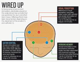
Click for larger version.
Researchers are exploring the ways in which this wet-clay state can be exploited. In a 2009 study6, Leonardo Cohen at the National Institute of Neurological Disorders and Stroke in Bethesda, Maryland, showed that tDCS improved people's ability to learn a simple coordination exercise — and that the improvement was still apparent three months after the experiment ended. Such results have led to an interest in stroke rehabilitation strategies. Small trials by Cohen, Nitsche, and others have shown improved recovery of hand function when tDCS is used this way (see 'Wired up').
Another group of researchers, led by Felipe Fregni of the Berenson-Allen Center for Noninvasive Brain Stimulation in Boston, Massachusetts, and Paulo Boggio of Mackenzie Presbyterian University in São Paulo, Brazil, is experimenting with tDCS as a way to treat depression. Several small trials done by this group and others suggest that a few sessions of tDCS to a part of the brain called the dorsolateral prefrontal cortex can improve mood for several weeks.

Risky approach

In 2007, Boggio and Fregni reported that applying tDCS to the same region can make people less likely to take risks7. They asked healthy university students to play a game in which they press a computer key to pump air into a cartoon balloon. The more they pump, the more virtual money they earn — but if the balloon bursts, they lose all their winnings. People treated with tDCS were less willing to push their luck. The results may be generalizable to addictions, in which people lack "inhibitory control", says Boggio. In 2008, he and Fregni published three studies8–10 showing that stimulation of the dorsolateral prefrontal cortex blunted cravings for alcohol, cigarettes and sweets when people later watched videos in which these were being consumed. They hope, eventually, to test the same technique in a clinical trial for smoking cessation.
“There has been a lot of hokey stuff, and it affects the credibility of the entire field.”

For a method that has seen its ups and downs, these results are encouraging. "There has been a lot of hokey stuff, frankly, and it affects the credibility of the entire field," says Marom Bikson, a biomedical engineer at the City College of New York. But in contemporary mechanistic studies to optimize tDCS, he says, "people have been much more careful".
Not everyone is convinced that the disappointments are over, though. Helen Mayberg, a clinical psychiatrist at Emory University in Atlanta, Georgia, has been experimenting with the use of deep-brain stimulation (DBS), in which electrodes are placed deep in the brain, to treat depression. She is excited about the non-invasiveness of tDCS but points out that the trials conducted to date have been short-term. The real questions, she says, will be: "How do you use it chronically, and what kinds of rebounds and relapses are there?" Boggio and others have begun a clinical trial to answer this question, in which patients being treated with tDCS for depression will be observed for up to six months.

Target practice

Still, the stimulation from tDCS is less focused than that from TMS or DBS. Its effects on neurons also drop off rapidly a few centimetres below the scalp, putting some important medical targets out of reach.
Bikson has designed a more refined version of tDCS that he hopes will address these shortcomings. Instead of one electrode, he places five on the head in an X configuration. The one in the centre pushes current in the desired direction and the four around it siphon off excess current that would otherwise spread and activate wider brain areas. The configuration could, he says, allow for slightly higher currents that would penetrate deeper into the brain in more focused areas. Such innovations might even help to persuade companies to invest in clinical trials. Currently, Cohen says, no one stands to gain enough return from therapies that can be administered using just US$1,000 worth of off-the-shelf equipment.
Aside from treatment, tDCS is also receiving attention for its potential to enhance the minds of healthy people. In addition to Clark's work showing enhanced ability to see concealed threats, other studies with tDCS have shown improvements in working memory11, word association12 and complex problem-solving13. Most of these studies address scientific questions — but one neuroscientist unabashedly aims to boost the brains of healthy people.
Allan Snyder, director of the Centre for the Mind at the University of Sydney in Australia, hopes to develop "a thinking cap", a tDCS device that corporate executives or advertising copywriters might use to bump up their creativity before walking into a brainstorming meeting. Snyder is cagey about how far he is in product development — but his latest demonstration, published this February14, garnered plenty of attention. Snyder claims to have boosted people's flair for sudden insight by stimulating their anterior temporal lobes. People who received tDCS were two to three times more likely than those receiving sham stimulation to solve a creativity problem in which they raced against the clock to spell out maths equations with matchsticks.
The jury is still out on whether these results will translate into real-world benefits. Nitsche says that it will be harder to improve cognition in young, healthy people — whose minds are theoretically already optimized — than in elderly people or those with addictions, for instance. "I wouldn't say it wouldn't be possible," says Nitsche. "But things might be a little more complicated."
“With wires and batteries and home hobbyists trying to run electricity through their heads, somebody could get hurt.”

That's not stopping some people from trying it at home. Discussions are already appearing on the Internet: buy a 9-volt battery, some wire and a resistor, and you're theoretically there. One person, hoping to improve his concentration, was alarmed by the flashing lights he experienced — a commonly reported side effect, along with burning or itching at the site of the electrode. "I probably won't be doing this again," he said in a message posted online. Another wrote in an online patients' forum that the tDCS treatments he was giving to his wife were alleviating her chronic pain. Safety is an important issue. "With wires and batteries and home hobbyists trying to run electricity through their heads, somebody could get hurt," says Farah.

And wider adoption raises ethical concerns similar to those that surround mind-enhancing drugs such as Adderall and Modafinil, which some students take as study aids. Students might secretly 'electrodope' with tDCS before a university entrance exam to inflate their scores. Ethicists worry that this will give some an unfair advantage or create a culture in which people feel pressured to use such devices. None of the studies published so far have shown a type of mind-sharpening that would help in such exams, says Farah, but that might simply be a matter of targeting the right brain areas. "It would not surprise me" if such effects were possible, she says.
Overall, though, the optimism among tDCS's believers remains high. Although it has generated some disappointments, many are convinced that the present buzz is warranted. "Sometimes in the history of medicine you have to try again after one century or so," says Priori. "You use a novel technical device, and you succeed where somebody else failed."


I wonder if this has any applicability to stroke recovery?

Friday, March 11, 2011

music listening and stroke rehab

There has been a number of studies suggesting music during acute phase is good for you. I'm not sure I would have been able to stay awake regardless of the music played. This falls into the enhanced stimulation being good for you
http://brain.oxfordjournals.org/cgi/content/full/131/3/866
Music listening enhances cognitive recovery and mood after middle cerebral artery stroke
http://www.therapytimes.com/content=0402J84C48968A84406040441
Music Therapy Speeds Post-Stroke Recovery
music therapy
http://www.msnbc.msn.com/id/35502970/ns/technology_and_science-science/
http://www.epsychology.us/rhythm-of-life-music-shows-potential-in-stroke-rehabilitation/

including Kenny Rogers, I couldn't have handled this. Ask your doctors if you can listen to music to make sure it doesn't have negative side effects.

Wednesday, February 16, 2011

Stimulate senses and stroke rehab

I liked this line in the article; researchers discovered that tickling rats’ whiskers helps them recover from ischemic stroke.

Senses Stoke Stroke Recovery

I can just see nurses going around with feathers.


In this presentation by Dale Corbett he talks about enriched environment similar to baby stimulation. I started visualizing mobiles hanging above each patients bed.

This is 45 minutes long but worth listening to.

Neuroplasticity and Stroke Recovery: Past, Present and Future


I know if you read Jill Bolte-Taylors' book or saw her on video, she highly recommended not having much sensory stimulation at all because it caused her to shut down. To each his or her own, I could have used the extra stimulation to keep me awake.

Saturday, November 13, 2010

What therapy-exercise worked best for your stroke rehab?

Therapists ask me what therapies have worked in my rehab. I can understand why because they want to add that therapy to their roster of abilities.

Survivors ask me what exercises worked in my rehab. They are hoping that if they can just find the right exercise to do they will recover.

Both of these questions are invalid because the first thing to understand is how recovery occurs and where you are in the process. Until you know that can you select a therapy or exercise to work on. If you are working on penumbra recovery in the first 6-12 months then you take the little pieces of movement you do have and keep extending them longer and farther. If you are trying to get back functions that were in the dead brain area then you need to work on neuroplastic therapies that move those functions; try passive movement, mental imagery, thermal stimulation, action observation. All of these are discussed in other posts on my blog.

Until we get the whole concept of what needs to be done to recover changed from this specific therapy or exercise will we finally come up with a therapy model for stroke rehab. So don't enable the doctors and therapists by accepting a therapy or exercise without them specifying how it meets the protocol of recovery. See my blog on restructure stroke rehab model and theoretical basis of stroke rehab for my ideas. I actually think they are pretty good.

Monday, October 4, 2010

dead brain recovery options

Since I have huge amounts of dead brain these are the therapies I am using to try to get them moved to another location. This is not actually recovering the dead brain, it is trying to move the functions that area of brain controlled to another place. This is probably the hardest thing to do, especially with no research guidance or medical support on how to do this. So this is just my opinion only, try your doctors to see if they have anything better and then reply here.

mental imagery:
Andrea Zimmermann-Schlatter*1,2, Corina Schuster2,3, Milo A Puhan4,
Ewa Siekierka5 and Johann Steurer4
http://www.jneuroengrehab.com/content/pdf/1743-0003-5-8.pdf
Using Motor Imagery in the Rehabilitation of Hemiparesis ,
Mental imagery for promoting relearning for people after stroke: A randomized controlled trial1 , *1 .
Archives of Physical Medicine and Rehabilitation , Volume 85 , Issue 9 , Pages 1403 - 1408

Mirror-box therapy: Rehabilitation of hemiparesis after stroke with a mirror
Altschuler EL, Wisdom SB, Stone L, Foster C, Galasko D, Llewellyn DME, Ramachandran V
The Lancet - Vol. 353, Issue 9169, 12 June 1999, Pages 2035-2036

Passive movement: http://www.ncbi.nlm.nih.gov/pubmed/15003755
The effects of repetitive proprioceptive stimulation on corticomotor representation in intact and hemiplegic individuals.

Thermal therapy: http://stroke.ahajournals.org/cgi/content/full/strokeaha;36/12/2665
researchers found that cycles of heat and cold significantly enhanced the
sensory and motor function in the arms and hands of stroke survivors after a few weeks of therapy.
Music therapy: I am way too late for this to help but this should be part of every survivors' therapy in the hospital.
http://www.msnbc.msn.com/id/35502970/ns/technology_and_science-science/
http://www.epsychology.us/rhythm-of-life-music-shows-potential-in-stroke-rehabilitation/
https://web.archive.org/web/20090726073749/http://hubpages.com/hub/Music-Therapy-Healing including Kenny Rogers
I don't think I could have handled Kenny in the hospital.
Lucid dreaming: This one is just my completely off-the-wall idea. Why waste the time spent sleeping. Of course I have no proof/research that supports this. I try to dream using my pre-stroke abilities.
Do not take any of this as medical advice

Friday, September 24, 2010

Stroke rehab for sensory input

My sensory cortex supposedly was not affected but is less than before so I am assuming that it was routed thru the motor cortex across the central sulcus to get to the correct nerve endings. And since the motor cortex is mostly dead I have to find a new pathway to those nerves. This is rather depressing that I have to do all this self-diagnosis myself. I take a fingernail brush and run it across my left hand. In the shower I hold my left hand under the shower spray. I haven't yet gotten to the thermal stimulation yet, that is dunking your hand in ice water alternating with warm/hot water.

A hot/cold session consisted of two alternating cycles performed five days a week for six weeks. The sessions lasted between 20 minutes and 30 minutes. Thermal packs were wrapped in towels, and applied to the patient's hand and wrist. The hot pack was 167 degrees Fahrenheit, while the cold pack was just below 32 degrees Fahrenheit.

A lot of these ideas came from Sensory re-education of the hand after stroke by Yekeutiel, Margaret.