Changing stroke rehab and research worldwide now.Time is Brain! trillions and trillions of neurons that DIE each day because there are NO effective hyperacute therapies besides tPA(only 12% effective). I have 523 posts on hyperacute therapy, enough for researchers to spend decades proving them out. These are my personal ideas and blog on stroke rehabilitation and stroke research. Do not attempt any of these without checking with your medical provider. Unless you join me in agitating, when you need these therapies they won't be there.

What this blog is for:

My blog is not to help survivors recover, it is to have the 10 million yearly stroke survivors light fires underneath their doctors, stroke hospitals and stroke researchers to get stroke solved. 100% recovery. The stroke medical world is completely failing at that goal, they don't even have it as a goal. Shortly after getting out of the hospital and getting NO information on the process or protocols of stroke rehabilitation and recovery I started searching on the internet and found that no other survivor received useful information. This is an attempt to cover all stroke rehabilitation information that should be readily available to survivors so they can talk with informed knowledge to their medical staff. It lays out what needs to be done to get stroke survivors closer to 100% recovery. It's quite disgusting that this information is not available from every stroke association and doctors group.

Thursday, September 30, 2021

Development of an Aesthetic for a Stroke Rehabilitation System

 You'll have to go to the link to see the 2:47 video.

Development of an Aesthetic for a Stroke Rehabilitation System

Published:21 September 2021

ABSTRACT

Works around stroke rehabilitation devices have largely focused on improving their performance to aid in physical training. By providing better physical training for patients, it allows them to quickly achieve adequate autonomy in life. However, this leaves little development in their aesthetics, potentially failing to address the impact on patients’ self-integrity. This paper proposes the aesthetic development of an immersive multi-sensorial stroke rehabilitation system, entitled MIDAS (Multisensorial Immersive Dynamic Autonomous System), based on the improvement of self-affirmation of aesthetic products. MIDAS consists of three subsystems, a hand exoskeleton, a Virtual Reality (VR), and an Olfactory subsystem. The functional requirement of the system and the design language of the VR subsystem were used as the basis for the aesthetic framework for the rest of the subsystems. The outcomes were a hand exoskeleton with a minimalist linkage system paired with an organic casing, and an olfactory device with a rounded form attached to a frameless face-shield.


New remote PT and AI wearable helps stroke patients to walk more easily

You can tell the competency of your hospital by how long it takes to bring this in. Anything longer than 2 months is total incompetence, your board of directors needs to be fired.  

New remote PT and AI wearable helps stroke patients to walk more easily

y


People who have experienced neurological conditions such as strokes, multiple sclerosis, or Parkinson’s disease, tend to drag their affected foot. These are termed functional gait disorders.

To help address this, a new wearable device, called the EvoWalk, which is used in conjunction with a remote physical therapy platform, has been developed by Evolution Devices. The company was founded by Pier Mantovani.

The objective of the new technology is to make an improvement with a person’s mobility. The device uses machine learning to bypass the leg nerve. The foot can be lifted at just the right time to help users avoid falls and walk more freely.

Inspiration for the device came about when the founder’s father was diagnosed with multiple sclerosis and he struggled to walk. Physical therapy was critical for addressing the issue. However, getting to the health clinic in person proved not to be easy.

Looking at the experience and the associated issues, Mantovani devised the wearable and founded Evolution Devices, a company that combines the new, virtual physical therapy platform paired with the wearable.

This holistic approach of combining remote physical therapy with an artificial intelligence powered stimulation device and patient app to personalize care is not currently available with any other fall-prevention or rehabilitation therapy.

With the EvoWalk platform, users are able to work with a neuro-certified physical therapists without having to go to therapy in person. Built-in sensors feed real-time motion data to artificial intelligence algorithms and provide actionable metrics through the connected mobile apps.

The EvoWalk device gathers detailed walking data and provides electrotherapy to stimulate muscles at the right time to rehab walking and prevent falls.

A video that shows the technology in action is available on YouTube.

Through this, dedicated physical therapists are able to work remotely with the patient every step of the way, adjusting the wearable as patients make progress.

Based on trials conducted ahead of the launch of the technology, pilot users have seen an up to tenfold increase in walking activity.

The launch of the new device was on September 21st, 2021. The launch was supported by grants from the U.S. National Science Foundation, the National Institutes of Health, and the Toyota Mobility Foundation among others.


Hypoxic postconditioning promotes neurogenesis by modulating the metabolism of neural stem cells after cerebral ischemia

Nothing in here tells me if ANY RECOVERY WAS ACHIEVED. Useless.

Hypoxic postconditioning promotes neurogenesis by modulating the metabolism of neural stem cells after cerebral ischemia


https://doi.org/10.1016/j.expneurol.2021.113871Get rights and content

Highlights

Cerebral ischemia modulated the FAO and glycolysis of neural stem cells.

HPC promoted the migration and proliferation of neural stem cells after MCAO.

HPC modulated the FAO and glycolysis of neural stem cells in vivo and in vitro.

Abstract

Ischemic stroke is one of the most lethal and severely disabling diseases that seriously affects human health and quality of life. The maintenance of self-renewal and differentiation of neural stem cells are closely related to metabolism. This study aimed to investigate whether hypoxic postconditioning (HPC) could promote neurogenesis after ischemic stroke, and to investigate the role of neuronal stem cell metabolism in HPC-induced neuroprotection. Male C57BL/6 mice were subjected to transient middle cerebral artery occlusion (MCAO), and HPC was performed for 3 h per day. Immunofluorescence staining was used to assess neurogenesis. The cell line NE-4C was used to elucidate the proliferation of neuronal stem cells in 21% O2 or 8% O2. HPC promoted the recovery of neurological function in mice on day 14. HPC promoted neuronal precursor proliferation in the subventricular zone (SVZ) on day 7 and enhanced neuronal precursor migration in the basal ganglia and cortex on day 14. Fatty acid oxidation (FAO) and glycolysis of neural stem cells in the SVZ changed after MCAO with or without HPC. HPC promoted the proliferation of NE-4C stem cells, decreased FAO and increased glycolysis. All these beneficial effects of HPC were ablated by the application of an FAO activator or a glycolysis inhibitor. In conclusion, cerebral ischemia modulated the FAO and glycolysis of neural stem cells. HPC promoted the proliferation and migration of neural stem cells after MCAO, and these effects may be related to the regulation of metabolism, including FAO and glycolysis.

Rehabilitation of patients with cognitive impairment after stroke during the late recovery period

Hope your doctor or your research analyst knows Russian.

Rehabilitation of patients with cognitive impairment after stroke during the late recovery period

[Article in Russian]
Affiliations

Abstract

Some aspects of late and delayed post-stroke rehabilitation of patients with cognitive impairment and various manifestations of brain neuroplasticity are considered. Particular attention is paid to comprehensive rehabilitation programs for post-stroke patients with cognitive impairment, based on the choice of the optimal modes of physiotherapeutic, drug effects and the duration of the course of therapeutic treatment. Similar programs that can be implemented both in a sanatorium and on an outpatient basis, are effective for patients of working age, including in the late recovery period after ischemic stroke. These programs will lead to the restoration of cognitive and motor functions, the normalization of the psychoemotional state of patients.

Keywords: cognitive impairment; neuroplasticity; physiotherapy; rehabilitation; spa rehabilitation; stroke.

MeSH terms

LinkOut - more resources


Patients who develop cognitive impairment had changes in the brain before their stroke

This changes nothing; it is still your doctor's responsibility to bring your cognition back to normal.

My doctor told me I had a bunch of white matter hyperintensities but never showed me them on any scan, so I don't know the size, location or any intervention needed, because my doctor knew nothing and did nothing. I have zero cognitive impairment.

Patients who develop cognitive impairment had changes in the brain before their stroke

Figure 1. Examples of stroke lesion segmentation. Examples of voxel-based segmentation of stroke lesions in MNI space, as shown in the coronal, sagittal, and horizontal plane. Color map depicts a larger number of voxels and thus also a larger stroke increasing from red to yellow. Credit: DOI: 10.3389/fnagi.2021.705889

Stroke is caused by disturbances in the blood circulation of the brain due to blood clots or cerebral hemorrhage. This causes damage to parts of the brain, and the patient experiences a sudden loss of particular body functions. Individuals affected by stroke need urgent, life-saving treatment followed by rehabilitation. In Norway, stroke affects approximately 12,000 people each year.

"It appears that about 50 percent of get some form of cognitive impairment after the stroke," says Till Schellhorn, who has written his doctoral thesis at the Institute of Clinical Medicine based on the Nor-COAST study.

Cognitive impairment means that an individual's ability to think and to perform intellectual and mental processes has been reduced.

Identifying at-risk patients using MRI images

The researchers examined the brains of patients who developed cognitive impairment after stroke using MRI images. MRI stands for magnetic resonance imaging, and is a method used to take pictures of the body's internal organs.

"Using MRI images of the , we can identify risk patients for the development of cognitive impairment after stroke with good certainty," says the researcher.

The MRI images showed that patients who develop cognitive impairment after stroke already had brain changes prior to the stroke that may be associated with the development of cognitive impairment.

"It was mainly the case for patients with small vessel disease, who have a reduced circulation in the small blood vessels in the brain. This can lead to many small cerebral infarctions, and was associated with the development of cognitive impairment after stroke," Schellhorn explains.

Furthermore, the results showed that the size of the stroke was associated with increased risk of developing cognitive impairment. The larger the stroke, the higher the risk of developing cognitive impairment afterwards.

There was also an association between loss of substance in the temporal lobe, which is a part of the brain associated with memory, and the development of cognitive impairment.

"But this association was less pronounced than we had expected," he points out.

Has put cognitive impairment after stroke on the agenda—The Nor-COAST study has put cognitive impairment after stroke on the agenda. In the past, the focus has mostly been on paralysis and other physical ailments, and therefore cognitive impairment has received little attention, Schellhorn explains, and adds:

"But cognitive function means a great deal for the quality of life, both of the patient and their families."

The researchers have set themselves the goals of contributing to more knowledge about the causes of cognitive impairment and developing methods for identifying at-.

"You have to know whom, and why the individual patient develops cognitive impairment in order to be able to start a treatment," he points out.

Aim for better preventive measures and treatment: "We hope that our research can lead to better treatment of cognitive impairment after stroke in the future. We hope that we can prevent and alleviate cognitive impairment," Schellhorn says.

The researchers are now testing potential preventive measures and novel forms of treatment.

"This could entail preventive treatment among stroke patients with a high risk of developing cognitive impairment, or treatment of the causes, such as cardiovascular disease," the researcher says.

Schellhorn further states that increased physical or social activity as part of rehabilitation after a stroke might slow down the development of cognitive impairment.

A model for clinical use

Eight hundred and fifteen stroke patients were included in the study. The researchers took MRI images of the brain of 410 of them.

The researchers examined these images thoroughly. On the MRI images, which were taken after the stroke, they could see which patients had changes in the brain that had been present already prior to the stroke. To do this, they used different types of MRI images, some that show that brain changes are acute and some that show brain changes of older date.

"It is a combination of specific MRI technique and interpretation that helps us say something about the age of the brain changes," Schellhorn points out.

The researchers believe that some of these changes in the brain were related to the development of cognitive impairment after stroke.

"We investigated the association between the image findings and cognitive impairment. We created a model for predicting the risk of based on the findings on the images," Schellhorn explains.

So far, the model has only be

en tested within the scope of the Nor-COAST study, but the researchers already have plans to test it on more patients. However, there are also a number of practical challenges that must be solved before the model can be used in a clinical context, i.e. for use in connection with the treatment of patients.

"Before using new computer programs at the hospital, the models must go through a routine security check. Such things typically take some time. But I hope that we can use our models in the clinic at the hospital over the next two to three years."


Explore further

Cognitive impairment reported after COVID-19 infection

More information: Eva Birgitte Aamodt et al, Predicting the Emergence of Major Neurocognitive Disorder Within Three Months After a Stroke, Frontiers in Aging Neuroscience (2021). DOI: 10.3389/fnagi.2021.705889
Provided by University of Oslo
 

Surf therapy: a growing movement to address mental health

I'll guess you'll have to have your stroke and rehab in the Netherlands. This will never get approved in the US. Eventually I'll get around to stand up paddleboarding. The only surfing around here is in winter in Lake Michigan and Superior and that would require a 5mm wetsuit, totally impossible to get on one-handed.

Surf therapy: a growing movement to address mental health


September 29, 2021 | Surfing
Surf therapy: an established form of therapeutic support for mental and physical health worldwide | Photo: The Wave Project

Surf therapy is a method of health intervention that combines surfing and surf instructing alongside structured group or individual activities that promote psychological, physical, and psychosocial well-being.

Over the past ten years, this type of therapy has become an established form of therapeutic support for mental and physical health worldwide.

In the United Kingdom, it is recognized by the National Health Service (NHS) as an effective form of therapy for children and young people at risk of mental illness.

Surf therapy has also been used to support veterans in the United States, stroke victims in the Netherlands, deprived communities in South Africa, and adults with psychological conditions in Australia and South America.

In 2021, it's safe to say that surf therapy has become a truly global movement.

Surf therapy: it promotes positive attitudes, better communication skills, a healthier lifestyle, and progress in education | Photo: The Wave Project

Mounting Research Shows Evidence-Based Health Benefits

While it's easy to find people who are able to talk about the transformational impact surf therapy has had on their lives, these positive effects are increasingly backed up by research.

Several studies now show the beneficial, tangible impact of surf therapy on mental and emotional health.

For example, independent research commissioned by The Wave Project - the surf therapy charity I founded for children and young people based in the UK - revealed a statistically significant change to the well-being of participants after our intervention.

A peer-review by the journal Community Practitioner, published in January 2015, found that The Wave Project intervention resulted in a significant and sustained increase in health and happiness levels and behavior change.

Seventy-nine percent of parents reported a more positive attitude, and sixty-two percent revealed better communication skills in their children.

Fifty-six percent thought they showed a healthier lifestyle after participation, and 46 percent knew of progress in education since the course.

The conclusions from this analysis were clear.

When delivered in a supportive environment, with opportunities for continuation and volunteering, surfing provides profound and long-term benefits to the well-being of children facing social and emotional isolation.

The Wave Project: surfing provides profound and long-term benefits to the well-being of children facing social and emotional isolation | Photo: The Wave Project

Surfing Against Post-Conflict Mental Health Disorders

There is interesting evidence to show the profound impact of surf therapy on young people in post-conflict settings.

Young people in post-conflict and post-epidemic contexts such as Sierra Leone face many mental health challenges as part of their daily lives.

A recent study involving surf therapy pilots run by five youth-focused and community development organizations around Freetown found that participation in the organization's intervention generated large positive effects on mental health.

A separate study alongside Waves for Change in neighboring Liberia suggests that such positive effects are down to safe spaces, positive social connections, and respite from negative emotions inherent within surf therapy delivery.

These elements were identified as often lacking in participants' wider lives within a challenging post-conflict environment.

Increasingly, surf therapy interventions are used to provide support to those operating in "blue light" services - namely, the police, fire services, and emergency responders.

In 2017, two police sergeants, Sam Davies and James Mallows set up Surfwell, a health promotion project that uses a recreational surfing adventure experience to promote workplace mental health within the police force, after an officer they supervised was left traumatized by an attack.

The program is specifically designed to address the escalating mental health crisis in the UK emergency services linked to workplace stress.

A 2016 online poll conducted by the charity MIND found that one in four emergency service workers in the UK had contemplated suicide, and two-thirds had considered leaving their job.

In many instances, traditional clinical interventions had failed to help, showing a clear need for other forms of support for police officers and other emergency services workers.

Surf therapy: five days of intensive surfing during the chronic phase of stroke or trauma has an impressive, positive effect on functional outcomes, participation levels, and mental well-being | Photo: The Wave Project

Surf Therapy for Neurological Disorders

There is also increasing interest in the beneficial impact of surf therapy interventions on those who have experienced strokes and other neurological disorders.

In The Netherlands, surf therapist Tijs van Bezeij founded the surftherapie.nl foundation after an experience working with a 20-year-old boy who had been hospitalized for rehabilitation after a major stroke.

The center is predicated on the understanding that challenging therapy in a rigorous, high-intensity environment generates the best results for stroke rehabilitation.

A pilot study by the foundation shows that five days of intensive surfing during the chronic phase of stroke or trauma has an impressive, positive effect on functional outcomes, participation levels, and mental well-being.

The study represents the participants' experience, demonstrates the continued recovery of the brain and the acceleration of recovery through challenging activities such as surfing.

What Next for Surf Therapy?

Increasingly, independent research is confirming what many of us engaged in surf therapy witness all the time: surf therapy works.

As a surf therapist, the challenge now is to build on this research surf therapy reaches more people who really need it.

At the 2021 International Surf Therapy Organization conference, surf therapy practitioners - including speakers from the organizations mentioned above - will join academics and campaigners in the UK's surfing hotspot Cornwall to discuss what lies ahead for surf therapy as a mental health intervention.

Hosted by The Wave Project, the theme of the very first International Surf Therapy Organization conference to be held in Europe is "Building Trust."

For many people who participate in surf therapy, this is a crucial element of their recovery.

The conference aims to bring together leading surf therapy organization heads to build on best practices, learn from one another and discuss how to broaden accessibility and improve our services.

For tickets to the 2021 International Surf Therapy Organization Conference on October 6-8, please visit The Wave Project website at www.waveproject.co.uk.


Words by Joe Taylor | CEO and Founder of The Wave Project

Designing an app for home-based enriched Music-supported Therapy in the rehabilitation of patients with chronic stroke: a pilot feasibility study

 You doctor should have created a music protocol years ago. All you would need is a smartphone and a library of music to load onto it. Cheap and easy yet your doctor incompetently still has not done it. That failure requires the board of directors to be fired.

Designing an app for home-based enriched Music-supported Therapy in the rehabilitation of patients with chronic stroke: a pilot feasibility study

Received 28 Apr 2021, Accepted 25 Aug 2021, Published online: 23 Sep 2021


Objective

After completing formal stroke rehabilitation programs, most patients do not achieve full upper limb motor function recovery. Music-supported Therapy (MST) can improve motor functionality post stroke through musical training. We designed a home-based enriched Music-supported Therapy (eMST) program to provide patients with chronic stroke the opportunity of continuing rehabilitation by themselves. We developed an app to conduct the eMST sessions at home with a MIDI-piano and percussion instruments. Here, we tested the feasibility of the eMST intervention using the novel app.

Method

This is a pilot study where five patients with chronic stroke underwent a 10-week intervention of 3 sessions per week. Patients answered feasibility questionnaires throughout the intervention to modify aspects of the rehabilitation program and the app according to their feedback. Upper limb motor functions were evaluated pre- and post-intervention as well as speed and force tapping during daily piano performance.

Results

Patients clinically improved in upper limb motor function achieving the Minimal Detectable Change (MDC) or Minimal Clinically Important Difference (MCID) in most of motor tests. The app received high usability ratings post-intervention.

Conclusion

The eMST program is a feasible intervention for patients with chronic stroke and its efficacy should be assessed in a clinical trial.


A Serving Of Spinach Every Day Could Make Your Brain 11 Years Younger

 Does your doctor even know she is responsible for correcting your

5 lost years of brain cognition due to your stroke?

Is this enough to get the dietician to create a protocol on this? You can't do this on your own if you are on warfarin.

Why Vitamin K Can Be Dangerous If You Take Warfarin

A Serving Of Spinach Every Day Could Make Your Brain 11 Years Younger


While Popeye ate spinach everyday to maintain strong muscles, he was also bettering his memory, maybe without even knowing it.

Consuming just a daily serving of spinach — or any type of leafy green, for that matter — may help slow the process of age-associated cognitive decline, according to a new study. The reason? Vitamin K.

A group of researchers at Rush University in Chicago who analyzed the diets and mental functions of 954 elderly people over a five year period found that those who incorporated more green in their diets were more likely to be mentally sharp.

The researchers tracked the diets of participants, whose age averaged 81, for an average of five years. They found that people who ate one or two daily portions of green leafy vegetables had the same cognitive abilities as someone 11 years younger who never consumed leafy greens.

Beyond spinach, vegetables like kale, collards and mustard greens could also be effective in slowing down the brain’s aging process. Previous studies have found that both folate and beta-carotene are brain boosters, but this study is the first to evaluate the benefits vitamin K has on the brain.

“No other studies have looked at vitamin K in relation to change in cognitive abilities over time, and only a limited number of studies have found some association with lutein,” said Martha Clare Morris, Sc.D., assistant provost for community research at Rush University Medical Center and the leader of the study’s research team. The team believes that other foods high in these nutrients, like asparagus, brussels sprouts and carrots could provide the same benefits as the darker greens, and they intend to expand their research to explore this possibility.

The research holds promise for a brain booster that is accessible and affordable. “Since declining cognitive ability is central to Alzheimer’s disease and dementias, increasing consumption of green leafy vegetables could offer a very simple, affordable and non-invasive way of potentially protecting your brain from Alzheimer’s disease and dementia,” Morris said.

The study was presented at the Experimental Biology meeting on March 30.


By  for huffingtonpost.com

Medical researchers on the cusp of a breakthrough treatment using common bush tick spit

All these others I'm sure your doctor is extremely familiar with.


Medical researchers on the cusp of a breakthrough treatment using common bush tick spit

 Aussie researchers are close to developing a medication that could revolutionise treatment for one of the country’s biggest killers.

Bush ticks understandably have a bad reputation but Australian researchers have discovered their saliva can be used to make potentially lifesaving drugs to treat a leading cause of death.

In a world-first, scientists at the Heart Research Institute and the University of Sydney found tick spit contained proteins that could be used to develop anti blood clotting drugs to treat strokes.

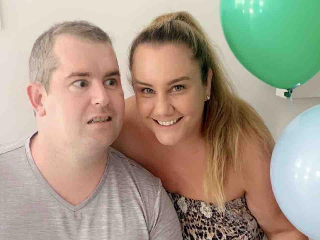
Their research could prove life changing for people like Shaun Bickley, a Brisbane father who is lucky to be alive and was left without his sight after suffering a stroke last year, aged 38.

He was one of about 50,000 Australians – and 16 million people worldwide – who suffer strokes annually, with about 85 per cent of strokes caused by a clot restricting blood flow to the brain.

Mr Bickley was admitted to Princess Alexandra Hospital on October 5 after fighting a raging headache the prior week.

“We had no idea the headache was being caused by numerous clots throughout his brain, neck and lung,” his wife Lauren said.

“The following day, after an MRI, we learnt that Shaun had suffered an embolic shower, a pulmonary embolism and that his two major arteries in his neck supplying blood to his brain were 100 per cent and 75 per cent blocked with clots.”

Shaun Bickley and his wife Lauren are hopeful that new medication could help future stroke victims. Picture: Supplied

Within two weeks Mr Bickley had lost his sight and suffered full right side paralysis and aphasia, a language disorder caused by brain damage.

He suffered several more strokes that same day and his family was told to prepare for the worst.

Mr Bickley went on to do rehab but suffered another stroke at Christmas that led to a five-month hospital stay.

Doctors finally came to the conclusion Mr Bickley’s strokes were due to an auto-immune condition that triggered a clotting storm in his body.

He survived and regained the ability to walk and talk, but not to see.

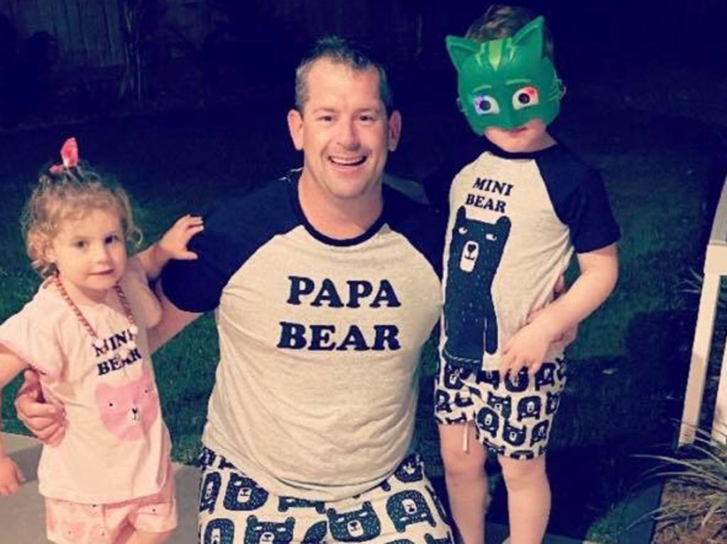
Shaun Bickley suffered multiple debilitating strokes, with his family now hopeful that new medication could help future victims. Picture: Supplied

His wife set up a fundraiser to help cover his therapy and rehabilitation costs, paying tribute to “the old Shaun”.

“I miss our old life, a life where you chased the kids and played until they would collapse,” she wrote on the GoFundMe page in May.

“A life where you would just stop and chat to strangers. You had the time of day for anyone … I’m grieving the old you. But you are still here.”

He and his family hope that research into anticoagulant medications could one day mean better outcomes for stroke victims, who are currently limited to just one drug therapy, which has limited success.

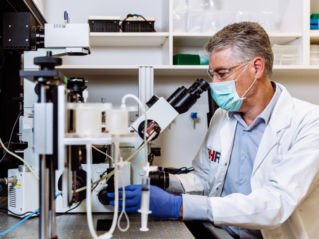
A team of leading Australian scientists at the Heart Research Institute and Sydney University, including Shaun Jackson (pictured), believe they’re on the cusp of developing a drug to save people from deadly strokes. Picture: Supplied.

University of Sydney’s Richard Payne said ticks made important bioactive molecules that could potentially be used for the breakthrough medical treatment.

“We’ve been modifying the proteins found in the saliva of these ticks and we really wanted to know what these modifications were doing to the activity of the protein – that’s never been done before,” the professor said.

The research team has received a $750,000 federal government grant to continue their work.

If this phase of the project proves successful, the team will seek further funding to move to phase one trials in humans.

Critical time window for rehabilitation after a stroke

 I totally disagree;

For each day a patient is in hospital lying in bed with minimal activity approximately 13% of muscular strength is lost (Ellis, Jackson, Liu, Molloy, & Paterson, 2013). (Correcting this is your doctors' responsibility. Don't let her weasel out of that responsibility.) 

Maybe this?

'Exercise-in-a-pill' boosts athletic endurance by 70 percent May 2017

The latest here:

Critical time window for rehabilitation after a stroke

 
  • Researchers found that intensive therapy, added to standard rehabilitation, produces the greatest improvement when administered 2-3 months after a stroke.
  • The results could lead to improved rehabilitation programs for stroke patients.

Every 40 seconds, an American has a stroke. About 750,000 new strokes occur in the United States each year.

Restoring brain function after a stroke remains a challenge. Functional recovery from brain damage requires networks of nerves to adapt and reorganize. This “neuroplasticity” naturally occurs during early development. But studies in rodents suggest that there is a brief period of similarly high neuroplasticity after a stroke. Intensive motor training provided to rodents during this window can lead to nearly full recovery. But no evidence for a similar recovery window in humans has previously been found.

To find out if such a window exists in people, a team led by Dr. Alexander Dromerick of Georgetown University Medical Center and MedStar National Rehabilitation Hospital conducted a randomized phase II clinical trial. NIH’s National Institute of Neurological Disorders and Stroke (NINDS), Eunice Kennedy Shriver National Institute of Child Health and Human Development (NICHD), and National Institute on Deafness and Other Communication Disorders (NIDCD) supported the study. Results appeared in the Proceedings of the National Academy of Sciences on September 20, 2021.

The researchers recruited 72 patients from a rehabilitation hospital in Washington, DC. They randomly assigned participants to one of four groups. All participants received standard stroke rehabilitation therapy. Those in three of the four groups received an extra 20 hours of intensive motor skills therapy. In the first group, the extra therapy began within 30 days of stroke onset. In the second group, extra therapy began 2-3 months after stroke onset. In the third, it began 6-7 months after stroke onset. Participants in the fourth (control) group received no extra therapy.

The researchers assessed arm and hand function at various points before and after treatment, up to 12 months after stroke onset. People in the 2–3 month therapy group showed the greatest improvement one year after their strokes. Participants in the 30-day group showed smaller but still significant improvement. By contrast, participants in the 6-7 month group showed no significant improvement over controls.

“Our results suggest that more intensive motor rehabilitation should be provided to stroke patients at 60 to 90 days after stroke onset,” co-author Dr. Elissa Newport says.

“Previous clinical trials have found few or very small improvements in motor function post-stroke, so our research could be an important breakthrough in finding ways we can make substantial improvements in arm and hand recovery,” Dromerick says.

The results strongly suggest that there is a critical time window for rehabilitation following a stroke. For this study, that window was 2-3 months after stroke onset. Larger clinical trials are needed to better pin down the timing and duration of this critical window. A larger trial could also determine what dose of therapy would achieve the best results during this window.

This article was written by Brian Doctrow, Ph.D. 

As with anything you read on the internet, this article should not be construed as medical advice; please talk to your doctor or primary care provider before making any changes to your wellness routine.

Content may be edited for style and length.

Task Complexity and Image Clarity Facilitate Motor and Visuo-Motor Activities in Mirror Therapy in Post-stroke Patients

So write this up as a specific stroke rehab protocol and deliver it to all 10 million yearly stroke survivors  now and into the future.

Your responsibility since we have fucking failures of stroke associations that can't accomplish that simple task for all stroke researchers.

Task Complexity and Image Clarity Facilitate Motor and Visuo-Motor Activities in Mirror Therapy in Post-stroke Patients

  • 1Department of Rehabilitation Sciences, The Hong Kong Polytechnic University, Kowloon, Hong Kong, SAR China
  • 2Department of Physiotherapy, Yobe State University Teaching Hospital, Damaturu, Nigeria
  • 3Department of Psychology, The Education University of Hong Kong, Tai Po, Hong Kong, SAR China

Introduction: Mirror therapy is effective in the recovery of upper-limb function among post-stroke patients. An important component of mirror therapy is imagining finger movements. This study aimed to determine the influence of finger movement complexity and mirror image clarity on facilitating motor and visuo-motor activities in post-stroke patients.

Methods: Fifteen post-stroke patients and 18 right-handed healthy participants performed simple or complex finger tapping while viewing mirror images of these movements at varying levels of clarity. The physical setup was identical to typical mirror therapy. Functional near infrared spectroscopy (fNIRS) was used to capture the brain activities elicited in the bilateral primary motor cortices (M1) and the precuneus using a block experimental design.

Results: In both study groups, the “complex finger-tapping task with blurred mirror image” condition resulted in lower intensity (p < 0.01) and authenticity (p < 0.01) of the kinesthetic mirror illusion, and higher levels of perceived effort in generating the illusion (p < 0.01), relative to the “simple finger-tapping with clear mirror image” condition. Greater changes in the oxygenated hemoglobin (HbO) concentration were recorded at the ipsilesional and ipsilateral M1 in the “complex finger-tapping task with blurred mirror image” condition relative to that recorded in the “simple finger-tapping task with clear mirror image” condition (p = 0.03). These HbO concentration changes were not significant in the precuneus. Post-stroke patients showed greater changes than their healthy counterparts at the ipsilesional M1 (F = 5.08; p = 0.03; partial eta squared = 0.14) and the precuneus (F = 7.71; p < 0.01; partial eta squared = 0.20).

Conclusion: The complexity and image clarity of the finger movements increased the neural activities in the ipsilesional motor cortex in the post-stroke patients. These findings suggest plausible roles for top-down attention and working memory in the treatment effects of mirror therapy. Future research can aim to corroborate these findings by using a longitudinal design to examine the use of mirror therapy to promote upper limb motor recovery in post-stroke patients.

Introduction

A plane mirror is a simple tool used daily to provide instant visual feedback on body appearance, thereby influencing self-recognition (1). Due to these properties, the plane mirror has been used in rehabilitation clinics as a biofeedback apparatus for balance and postural training (2). Advanced therapeutic application of a plane mirror is an essential element of Ramachandran's mirror therapy or the mirror-induced visual illusion (MVI) paradigm (3).

Mirror therapy has been widely used as a rehabilitative intervention to enhance motor performance in post-stroke patients (4, 5). Moderate evidence supports the effect of mirror therapy for the regain of motor function of upper and lower limbs in this population (57). The key feature of mirror therapy is viewing images of motor activity of the unaffected limb to generate an illusion of moving the hidden, affected limb. The generation of the mirror illusion has been associated with activations in the primary motor cortex (M1), precuneus, premotor cortex, primary somatosensory cortex, cerebellum, dorsolateral pre-frontal cortex, superior temporal gyrus and posterior cingulate cortex (811), which subserve motor, cognitive and perceptual processes (8, 12). Long-term training using mirror therapy leads to upward regulation of activity in the ipsilesional M1 in post-stroke patients (13), which contributes to post-stroke patients' upper-limb functional regain (14).

Recent studies conducted by our research team highlighted mental imagery as one potential explanation for the useful changes in upper limb function during mirror therapy due to consistent overlap of neural activity reported in the M1, precuneus, primary somatosensory cortex, and cerebellum (10, 11). Another theory explaining the mental processes of mirror therapy in the literature is the mirror neuron system theory (15, 16). However, the theory is limited in substantiating some of the basic neural mechanisms of the mirror therapy paradigm [see Bello et al. (10) and Bello et al. (11)]. For example, lack of significant number of neural substrates of the mirror neuron system, found sub-serving the mirror therapy mental procesess (8), and the activation of ipsilateral M1 in the mirror therapy paradigm, while viewing an immobile image of a hand holding a pencil (17, 18). On the other hand, “Mental imagery” which denotes the rehearsal of limb movement without actual execution (19, 20), shares overlapping neural substrates and associated mental processes with mirror therapy. It can further be classified into kinesthetic motor imagery and visual motor imagery, with the former yielding greater activity in the motor areas and inferior parietal lobule (21, 22).

Briefly, mental imagery theory postulates that the mirror therapy paradigm generates visual image of an “imagined action” of the hidden static limb, owing to the similarities between the hidden limb and the mirror inverted image (23, 24). Kinaesthetic motor imagery, as a modality of mental imagery is associated with the internal generation of sensory components of movement (for example kinesthesia, muscle stretching and joint mobility), associated with the execution of similar movement (20, 22). Existing literature indicated that kinaesthetic motor imagery evokes activation of the motor system without visible mobility of the imagined body part (22, 25, 26). This could serves as the basis for the presence of kinaesthetic mirror illusion (27, 28) in the mirror therapy paradigm (10).

More at link.