What is your doctor doing to stop brain atrophy in their stroke survivors? With all this earlier research available for years your doctor has absolutely no excuse for not having a stroke protocol to prevent brain atrophy. Do not self treat yourself, your doctor has to do this even if they know nothing about it.
From June, 2013;
Low Diastolic Pressure Linked to Brain Atrophy
And this from November, 2012:
Relationship between Physical Activity and Brain Atrophy Progression.
And from September, 2014:
Lack of sleep may shrink your brain
From June, 2013:
Preventing Alzheimer’s disease-related gray matter atrophy by B-vitamin treatment
The latest here:
Brain Atrophy Estimated from Structural Magnetic Resonance Imaging as a Marker of Large-Scale Network-Based Neurodegeneration in Aging and Stroke
Michele Veldsman 1,2
1
Nuffield Department of Clinical Neurosciences, University of Oxford, Oxford OX3 9DU, UK
2
The Florey Institute for Neuroscience and Mental Health, University of Melbourne, Melbourne VIC 3084, Australia
Received: 18 August 2017 / Revised: 30 October 2017 / Accepted: 9 November 2017 / Published: 10 November 2017
(This article belongs to the Special Issue Stroke in Ageing)
Abstract
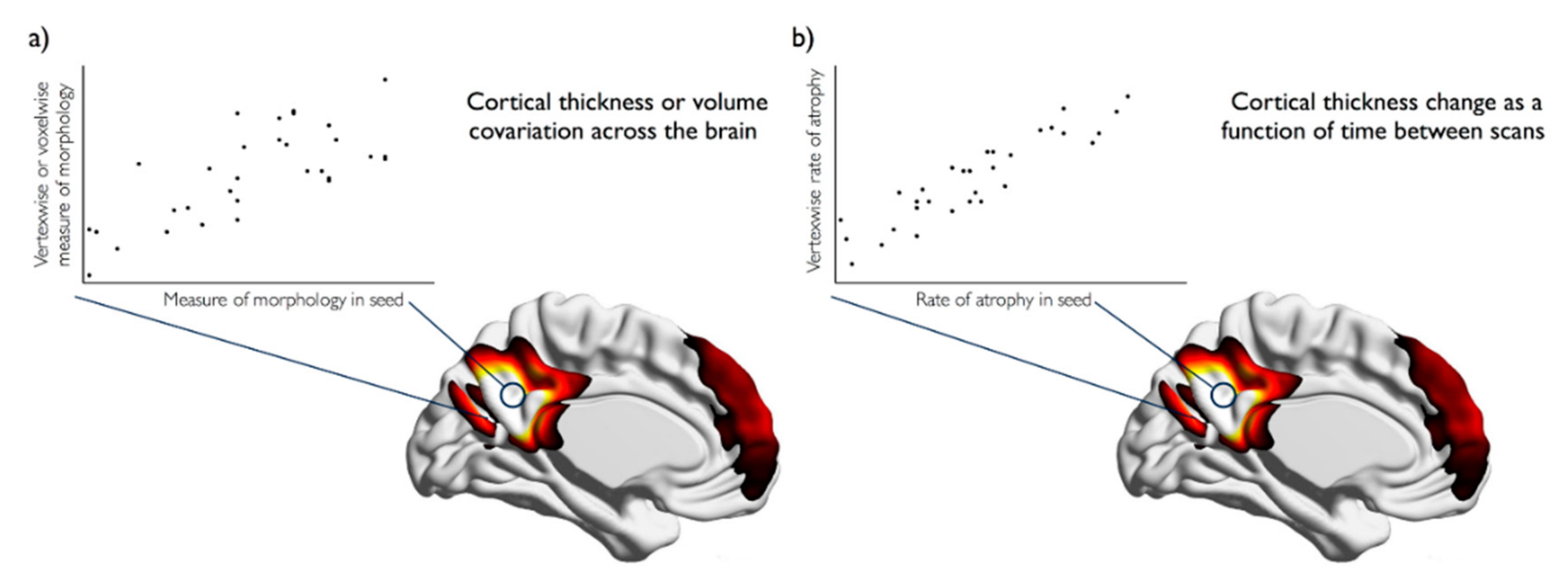
Brain atrophy is a normal part of healthy aging, and stroke appears to have neurodegenerative effects, accelerating this atrophy to pathological levels. The distributed pattern of atrophy in healthy aging suggests that large-scale brain networks may be involved. At the same time, the network wide effects of stroke are beginning to be appreciated. There is now widespread use of network methods to understand the brain in terms of coordinated brain activity or white matter connectivity. Examining brain morphology on a network level presents a powerful method of understanding brain structure and has been successfully applied to charting the course of brain development. This review will introduce recent advances in structural magnetic resonance imaging (MRI) acquisition and analyses that have allowed for reliable and reproducible estimates of atrophy in large-scale brain networks in aging and after stroke. These methods are currently underutilized despite their ease of acquisition and potential to clarify the progression of brain atrophy as a normal part of healthy aging and in the context of stroke. Understanding brain atrophy at the network level may be key to clarifying healthy aging processes and the pathway to neurodegeneration after stroke. View Full-Text Figure 1
This is an open access article distributed under the Creative Commons Attribution License
which permits unrestricted use, distribution, and reproduction in any
medium, provided the original work is properly cited. (CC BY 4.0).
No comments:
Post a Comment