Changing stroke rehab and research worldwide now.Time is Brain! trillions and trillions of neurons that DIE each day because there are NO effective hyperacute therapies besides tPA(only 12% effective). I have 523 posts on hyperacute therapy, enough for researchers to spend decades proving them out. These are my personal ideas and blog on stroke rehabilitation and stroke research. Do not attempt any of these without checking with your medical provider. Unless you join me in agitating, when you need these therapies they won't be there.

What this blog is for:

My blog is not to help survivors recover, it is to have the 10 million yearly stroke survivors light fires underneath their doctors, stroke hospitals and stroke researchers to get stroke solved. 100% recovery. The stroke medical world is completely failing at that goal, they don't even have it as a goal. Shortly after getting out of the hospital and getting NO information on the process or protocols of stroke rehabilitation and recovery I started searching on the internet and found that no other survivor received useful information. This is an attempt to cover all stroke rehabilitation information that should be readily available to survivors so they can talk with informed knowledge to their medical staff. It lays out what needs to be done to get stroke survivors closer to 100% recovery. It's quite disgusting that this information is not available from every stroke association and doctors group.

Friday, April 17, 2026

‘Dancing’ Molecules Spur Healing in Spinal Cord Tissue

 Do we have ANY INNOVATIVE thinkers in stroke that will look at this and say;'

 'Maybe this could work for stroke'! I bet we have shit for brains that can't think AND WILL DO NOTHING!

We already have these brain representatives:

‘Dancing’ Molecules Spur Healing in Spinal Cord Tissue

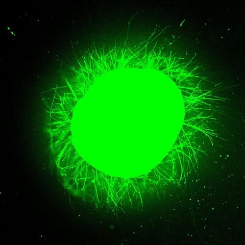
Under a fluorescence microscope at Northwestern University, a sphere of human spinal cord tissue — about 3 mm across, roughly the diameter of a mouse spinal cord — erupted with green light. It had been stained with a calcium-sensitive dye that lights up living neurons and their extending fibers. This green light provided unmistakable visual proof of neurites streaming outward from the tissue’s injured surface and into a synthetic gel, where they were growing in organized, parallel structures.

photo of Fluorescence microscopy reveals axons (green) extending from an injured spinal cord organoid
Fluorescence microscopy reveals axons (green) extending from an injured spinal cord organoid after treatment with the supramolecular therapy.

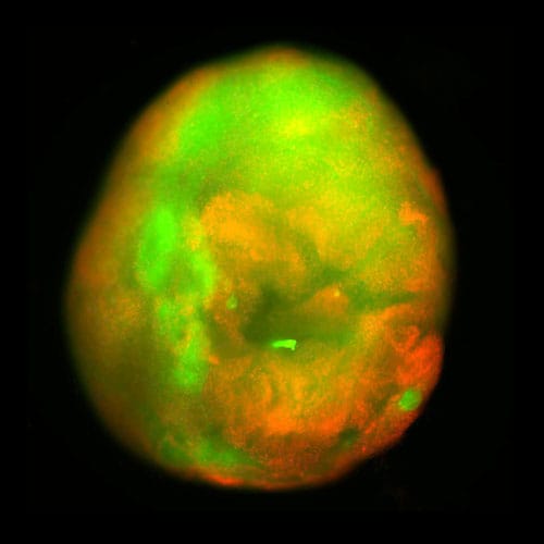
That sphere of spinal cord tissue was a spinal cord organoid — a three-dimensional, miniaturized tissue model grown in a dish from human induced pluripotent stem cells (iPSCs) to recapitulate key features of an actual organ. This one had been differentiated over 24 weeks into a range of spinal cord cell types: neurons, astrocytes, oligodendrocyte progenitor cells, and Schwann cells.

The Northwestern team also incorporated microglia — the brain and spinal cord’s resident immune cells — by coaxing iPSC-derived progenitors to infiltrate the organoid. The result was an immune-competent model capable of mounting an inflammatory response to injury.

photo of Contusion from a mechanical impactor
Contusion from a mechanical impactor produces diffuse cell death (red), simulating the blunt-force trauma behind real-world spinal cord injuries.

Then, to make the organoid useful, the team injured it — either slicing it with a scalpel to model surgical hemisection or crushing it with a mechanical impactor to deliver the kind of compressive contusion that causes most real-world spinal cord injuries.

No comments:

Post a Comment