Use the labels in the right column to find what you want. Or you can go thru them one by one, there are only 34,365 posts. Searching is done in the search box in upper left corner. I blog on anything to do with stroke. DO NOT DO ANYTHING SUGGESTED HERE AS I AM NOT MEDICALLY TRAINED, YOUR DOCTOR IS, LISTEN TO THEM. BUT I BET THEY DON'T KNOW HOW TO GET YOU 100% RECOVERED. I DON'T EITHER BUT HAVE PLENTY OF QUESTIONS FOR YOUR DOCTOR TO ANSWER.
Changing stroke rehab and research worldwide now.Time is Brain!trillions and trillions of neuronsthatDIEeach day because there areNOeffective hyperacute therapies besides tPA(only 12% effective). I have 523 posts on hyperacute therapy, enough for researchers to spend decades proving them out. These are my personal ideas and blog on stroke rehabilitation and stroke research. Do not attempt any of these without checking with your medical provider. Unless you join me in agitating, when you need these therapies they won't be there.
What this blog is for:
My blog is not to help survivors recover, it is to have the 10 million yearly stroke survivors light fires underneath their doctors, stroke hospitals and stroke researchers to get stroke solved. 100% recovery. The stroke medical world is completely failing at that goal, they don't even have it as a goal. Shortly after getting out of the hospital and getting NO information on the process or protocols of stroke rehabilitation and recovery I started searching on the internet and found that no other survivor received useful information. This is an attempt to cover all stroke rehabilitation information that should be readily available to survivors so they can talk with informed knowledge to their medical staff. It lays out what needs to be done to get stroke survivors closer to 100% recovery. It's quite disgusting that this information is not available from every stroke association and doctors group.
With ANY BRAINS AT ALL in our stroke medical 'professionals' we could do research that tells us exactly what interventions work to stop the neuronal cascade of death in the first week! Putting the brain on a chip with brain organoids and this and we could solve a lot of stroke recovery questions. But nothing will occur; OUR 'PROFESSIONALS' ARE FUCKING INCOMPETENT!
Imagine what brain surgeons could do with a three-dimensional (3D)-printed model of the brain. Not a structural model printed in plastic and uniform throughout, but one that mirrors the heterogeneity of real human brain tissue — its mechanical, thermal, and electromagnetic properties.
Christopher O’ Bryan, PhD, MS
“If somebody has a brain tumor, you could take an MRI of their brain and make a 3D model from it that includes the tumor, and then practice surgery on that model,” said Christopher O’ Bryan, PhD, MS, assistant professor of mechanical and aerospace engineering at the University of Missouri (UM) in Columbia, Missouri. “You could learn how sound, electromagnetic waves, or physical insults interact with the brain to better understand concussions and traumatic brain injury.”
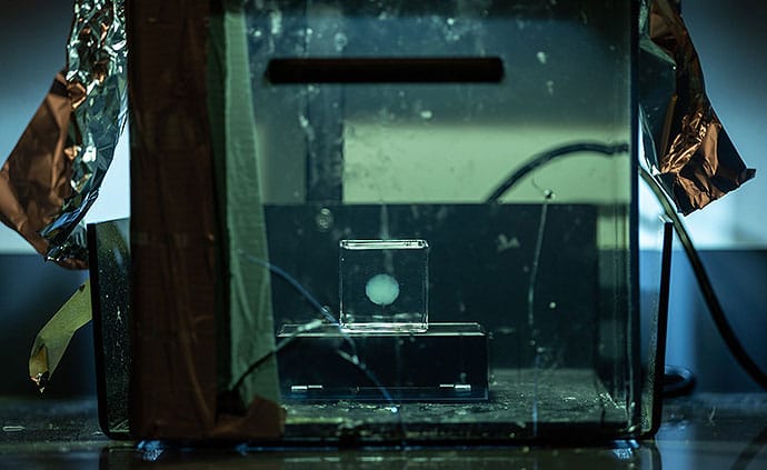
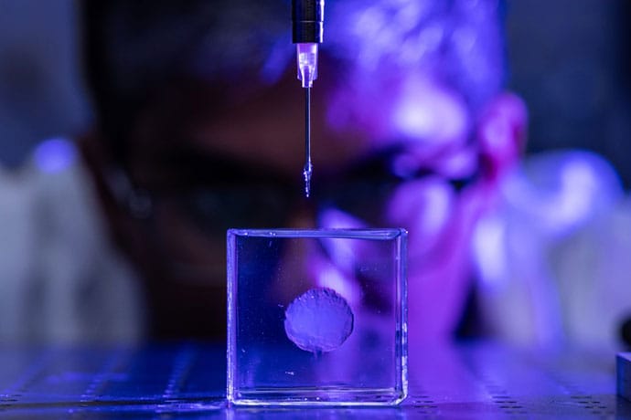
A scaled-down 3D model of the brain, about 15% of a real brain’s size.
O’Bryan and colleagues in UM’s College of Engineering recently published a study in the journal Materialia describing how the researchers designed a unique 3D printer ink made from a modified polymer — called a photo-crosslinkable poly(vinyl alcohol) methacrylate polymer — and then used it to print a scaled-down 3D model of the brain, about 15% of a real brain’s size. The scientists relied on a method called embedded 3D printing, crosslinking the polymer in a microgel bath, to achieve a model that mimicked the soft tissue and the structural complexities of a real human brain.
How They Did It
To provide a broader picture of why they conducted the study, O’Bryan described his lab’s goals. “The focus areas in our lab include the designing of soft materials, the testing of these materials, and then how to actually manufacture things out of these soft materials. And that’s especially challenging when you take into account that most soft materials are hydrogels and biopolymers which start out in a liquid phase ,” he said.
Researchers used embedded 3D printing, crosslinking the polymer with a gel bath to mimic brain tissue.
The researchers’ embedded 3D printing approach uses “sacrificial support baths,” jelly-like baths, which act as a supportive matrix during the printing phase. “What this allows us to do is to print materials as liquids and keep them as liquids until we’ve completely printed our structure, and then we crosslink the printed structure using heat or UV [ultraviolet] light to make it solid. Then we take it out of the gel bath, and it’s become a soft, deformable structure — meaning it can change shape when a force is applied,” O’Bryan said.
We don't SPECIFICALLY know why
a neuron gives up its' current job and takes on a neighbors. Thus
nothing on neuroplasticity is scientifically repeatable on demand. So, DEMAND your doctor give you EXACT PROTOCOLS to use. Don't allow your doctor to give you generalities or guidelines.
Ischemic stroke induces irreversible cerebral tissue damage, a condition exacerbated by the brain’s limited endogenous neuroplasticity and inability to regenerate neurons. While neural circuit reorganization holds therapeutic potential, its efficacy is hindered by pathological barriers such as glial scarring, chronic inflammation, and neurotrophic factor deficiency. Although pharmacological and interventional methods for stroke have been well developed, their functional recovery outcomes remain suboptimal. Emerging neural regeneration paradigms, particularly stem cell-based strategies (encompassing neural stem cell transplantation, neural progenitors grafts, and 3D brain organoid implantation), offer novel solutions to these challenges. However, critical limitations persist in conventional stem cell approaches: (1) compromised synaptic integration efficacy hinders functional neural circuit reconstruction; (2) the absence of functional vascular niches coupled with deficient astrocyte-mediated support and extracellular matrix signaling; (3) restricted regenerative capacity despite theoretical multipotent differentiation potential. Recent breakthroughs in cerebral organoid technology have revolutionized neurological disease modeling and neural repair research. Building upon this paradigm shift, our study mechanistically interrogates neuroplastic remodeling processes following ischemic stroke, while critically evaluating the therapeutic efficacy and inherent limitations of stem cell-based interventions. This affirms the critical role of 3D human cerebral organoid transplantation in the reconstruction of neural circuits. Additionally, we further summarize the utility of organoid-based disease models and address associated ethical and societal concerns. Future efforts should prioritize the clinical translation of organoid transplantation for ischemic stroke, aiming to mitigate neurological deficits and restore functional recovery.
Introduction
Stroke, a cerebrovascular disease, is the second leading cause of death globally and the third leading cause when disability and mortality are included as compounding factors [1]. Accounting for 62.4% of all strokes, ischemic stroke is the most frequent type and is the leading cause of neurological dysfunction encountered in clinical practice. Stroke imposes a significant economic and social burden and severely impacts the healthy lives of individuals worldwide [2]. Despite the high incidence of cerebrovascular diseases such as stroke, only a limited number of effective therapies are available in clinical practice. This, along with the fact that most patients do not seek timely medical attention for treatment, delays the optimal time for treatment and leads to extensive necrosis in the infarcted area, which can cause irreversible damage to the neurons in the brain. After patients progress through the acute phase of a stroke, they are left with serious sequelae, such as motor dysfunction. Post-stroke motor impairments typically affect the face, arms, and legs on one side of the body, impacting approximately 80% of patients [3]. Recovery is often prolonged and can severely impact the patient’s daily work and life. Longitudinal follow-up data reveal that approximately 50% of patients continue to experience motor deficits four years after their stroke occurred [4]. Clinics currently use pharmacological thrombolysis as the primary treatment to restore cerebral perfusion [5]. Although many drugs show neuroprotective effects in both preclinical studies and animal models, they have not demonstrated satisfactory results in clinical practice. Differences in physiology between species pose a challenge to the clinical translation of disease research [6].
Neurorestorative therapies for stroke typically extended therapeutic timeframes from days to weeks after stroke onset and aim to improve neurovascular remodelling and synaptogenesis while decreasing inflammatory and immune responses [7]. Stem cell therapy in regenerative medicine offers novel therapeutic paradigms [9]. Cell therapy enhances the recovery of neurological function of the body through sophisticated neuroimmune modulation and trophic factor secretion [8], thereby avoiding the suboptimal functional outcomes associated with conventional stroke treatments and showing substantial developmental potential [10]. Neural stem cells (NSCs) exhibit multimodal therapeutic actions that can stimulate functional neural replacement or induce multimodal therapeutic actions across multiple central nervous system (CNS) regions [11]. Effectively combining the anti-neuroinflammatory, anti-apoptotic, and pro-angiogenic capacities properties of NSCs with other interventions (e.g., recombinant tissue plasminogen activator) may mitigate apoptosis and necrosis caused by the generation of excess reactive oxygen species (ROS). This excess ROS is generated when the therapeutic time window for restoring blood flow during rt-PA therapy is missed, resulting in reperfusion injury (RI). However, transplantation of cells after 2D monolayer culture is less effective in repairing the tissue structure of the infarcted core compared with culturing cortical organoid systems in a 3D structure. More importantly, cerebral organoids (COs) not only demonstrate efficacy in repairing the ischemic core but also exhibit lesion-targeted neurogenesis into lesion-specific neurons. These neurons extend axons to cortical-basal ganglia-thalamic circuits, establishing functional synaptic connections with the host neural network to reconstruct complete neural circuits. They also demonstrates measurable efficacy in ameliorating post-stroke sensorimotor dysfunction [12]. Currently, several technologies are available to improve the reproducibility of cerebral organoid culture, such as 3D printing to help manipulate the size, shape, and organisation of organoids [13] and Notch signalling inhibition to prevent cell proliferation, induce terminal differentiation, and promote cerebral organoid maturation [14]. Therefore, based on the feasibility and therapeutic potential of generating and optimizing human cerebral organoids, the clinical translation of organoids for enhancing the neuroregenerative capacity and functional remodelling of the brain is a translational research priority.
Mechanisms of neuroplasticity after stroke onset
Brain recovery after stroke injury is dynamic and phase-dependent process. In early neurodevelopment (0–3 years), as the brain begins to develops, a large number of synapses are formed to sensory processing and multimodal integration and explore the comprehensive neural network [15]. During the initial development of the brain, the number of dendritic spines and synapses increases substantially, peaking at five years of age, after which the number decreases [15]. As brain volume expansion slows in the later stages, the energy needs of neuronal and synaptic activities cause the brain to adjust the density of these structures through selective synaptic elimination, transitioning from high plasticity to functional [16]. Stroke survivors show a sustained recovery of motor abilities over many years following brain injury. This recovery process is somewhat similar to how infants learn motor function [17]. The Stroke Recovery and Rehabilitation Roundtable has established a time frame for recovery of stroke patients, which is differentiated into the following four phases: the hyper-acute phase (0–24 h), the early subacute phase (7 days–3 months), the late subacute phase (3–6 months), and the chronic phase (>6 months) [18]. After a stroke occurs, the brain undergoes a series of ischemic cascade changes during the acute phase. Due to the disruption of blood flow to the brain and ATP depletion, neurons are unable to maintain normal transmembrane ion concentrations, resulting in a cascade of apoptosis and necrosis [19]. Hypoxia and depletion of metabolic substrates, as well as a substantial reduction in neuronal energy failure, subsequently causes structural damage to the brain [20].Therefore, ionic imbalance and cell death are the primary mechanisms at the onset of a stroke. In the subacute phase of stroke recovery, neuroinflammation diminishes while functional brain networks exhibit activity-dependent neuroplastic changes [21]. Microglia and monocyte-derived macrophages (MDMs) release factors that facilitate adaptation to resolving inflammation by removing neutrophils, cellular debris, and apoptotic cells [22]. These spontaneous neuroplastic changes include the release of brain-derived neurotrophic factor [23], structural remodelling at synaptic, axonal, and dendritic levels [24], and the activation, migration, and differentiation of endogenous neural stem cells [25]. During the chronic phase, experience-dependent reorganization of the neural network stabilizes, and long-term adaptation begins. At this point, “auto-reactive” lymphocytes sensitive to brain antigens return to the brain, triggering chronic inflammation and cytotoxicity, which likely contribute to the chronic sequelae of stroke [26]. Figure 1 Rehabilitation during the early subacute phase, when heightened neuroplasticity is at its peak, substantially increases both the speed and extent of patient recovery [18]. Although spontaneous remodeling occurs after ischemic stroke, these changes alone are insufficient to produce significant functional recovery [27]. In the future, functional recovery through cortical neuronal replacement may be the key to curing human neurodegenerative diseases [28, 29]. Compared with the use of dissociated neural progenitor cells, cerebral organoid transplantation offers several advantages, including a diverse cellular composition, maintenance of a native microenvironment, greater surgical feasibility, and higher vascularization potential, making it a promising therapeutic approach for restoring brain function in both the subacute and chronic phases of the disease [30].
Fig. 1
The main pathophysiological mechanisms of IS in the acute and convalescent phases. In the acute phase of IS, the ischaemia and reperfusion injury lead to mitochondrial dysfunction, cellular damage, and impairment of the blood–brain barrier. As IS interrupts the normal synaptic plasticity, during the recovery period, homeostatic plasticity is up-regulated and neurogenesis and synaptic remodelling occur. In addition, new synapse formation builds new circuits and aids brain recovery. In the cellular response mode, astrocytes secrete neurotrophic and angiogenic factors to repair damaged blood vessels and nerves. IS ischaemic stroke
When will our stroke leadership do similar research? Oh, that's right, we have NO leadership on stroke, which is why stroke never gets solved. Our fucking failures of stroke associations aren't even trying for 100% recovery and obviously don't give a shit about what survivors want!
Brain Organoids Illuminate TBI’s Link to Neurodegeneration
Summary: Researchers have advanced our understanding
of how traumatic brain injuries (TBI) contribute to neurodegenerative
diseases using lab-grown brain organoids. By simulating TBI in organoids
derived from human stem cells, the team observed nerve cell death and
pathological changes similar to those in TBI patients, particularly in
proteins associated with ALS and dementia.
The discovery of the
gene KCNJ2 as a protective factor against TBI effects opens new avenues
for treatments. This study, supported by a blend of federal and private
funding, underscores the potential of organoids in medical research and
the critical role of genetics in TBI outcomes.
Key Facts:
Simulated
TBIs in brain organoids show pathological changes mirroring those in
patients, highlighting a genetic predisposition to neurodegenerative
diseases post-injury.
The gene KCNJ2, involved in potassium ion
transport, was identified as a protective agent against TBI-induced
nerve cell death in both organoids and mouse models.
The
research, combining federal and private funding, demonstrates the value
of brain organoids in exploring the mechanisms of TBI and identifying
potential therapeutic targets.
Source: USC
A
traumatic brain injury (TBI) can quadruple your risk for developing
dementia and increase your chances of developing neurodegenerative
diseases such as ALS.
In a new study published in Cell Stem Cell,
USC scientists use lab-grown human brain structures known as organoids
to offer insights into why this is the case and how to mitigate the
risk.
In the study, former postdoc Jesse Lai and PhD student Joshua Berlind
from the USC Stem Cell laboratory of Justin Ichida used human
patient-derived stem cells to grow rudimentary brain structures known as
organoids in the lab. They then injured these organoids with
high-intensity ultrasound waves.
“This
could have potential as either a post-injury treatment or as a
prophylactic for athletes and others at high risk for TBI.” Credit:
Neuroscience News
The injured organoids showed
some of the same features seen in TBI patients, including nerve cell
death and pathological changes in tau proteins, as well as in a protein
called TDP-43.
The scientists found that the pathological changes
in TDP-43 were more prevalent in organoids derived from patients with
ALS or frontotemporal dementia, making their nerve cells more
suspectable to dysfunction and death following injury.
This
suggests that TBI might increase the risk of developing these diseases
even more for patients with a genetic predisposition. The worst injuries
were sustained by nerve cells that share information—called excitatory
neurons—located in the deep layers of the organoids.
In their search for ways to protect these neurons against the effects
of TBI, the scientists identified a gene called KCNJ2, which contains
instructions for making channels that selectively allow potassium to
pass through the cell membrane, helping to enable muscle contraction and
relaxation.
Inhibiting this gene had a protective effect on
organoids derived from patients with and without ALS, as well as on
mice, following a TBI.
“Targeting KCNJ2 may reduce the death of
nerve cells after TBI,” said Ichida, who is the John Douglas French
Alzheimer’s Foundation Associate Professor of Stem Cell Biology and
Regenerative Medicine at USC, and a principal investigator at the Eli
and Edythe Broad Center for Regenerative Medicine and Stem Cell Research
at USC.
“This could have potential as either a post-injury treatment or as a prophylactic for athletes and others at high risk for TBI.”
About the authors and the study
Co-corresponding
author Ichida is also a co-founder of AcuraStem and Modulo Bio, a
Scientific Advisory Board (SAB) member at Spinogenix and Vesalius
Therapeutics, and an employee in the Research and Early Development
group at BioMarin Pharmaceutical. Co-corresponding author Lai and
co-author Violeta Yu were both employees of Amgen during the study, and
currently work at Dewpoint Therapeutics. Named companies were not
involved in this research project.
First
author Berlind is a PhD student in the Ichida Lab. Additional
co-authors are Gabriella Fricklas, Cecilia Lie, Jean-Paul Urenda, Kelsey
Lam, Naomi Sta Maria, Russell Jacobs, and Zhen Zhao from USC.
Funding: Fifty percent of the work was supported by
federal funding from the National Institute of Neurological Disorders
and Stroke (NINDS) and the National Institute on Aging (grant
F31NS117075), NINDS (grant R01 1R01NS097850-01), and the Department of
Defense (grant 12907280). The project was also privately funded by an
Amgen postdoctoral fellowship, the New York Stem Cell Foundation, the
Tau Consortium, the Harrington Discovery Institute, the Alzheimer’s Drug
Discovery Foundation, the Association for Frontotemporal Dementia, and
the John Douglas French Alzheimer’s Foundation.
About this neurology and genetics research news
Author: Laura LeBlanc Source: USC Contact: Laura LeBlanc – USC Image: The image is credited to Neuroscience News
Summary: Researchers have developed human brain
organoids that contain microglia, the immune cells of the brain. These
organoids allow researchers to study how microglia develop and function
in a more realistic setting than previous models.
The researchers
found that microglia are influenced by the environment in which they
develop and that they play a role in both development and disease. Their
findings could lead to new treatments for neurological disorders.
Key Facts:
Microglia are responsible for clearing away cellular debris and pathogens, and they also play a role in neuroprotection.
The researchers found that microglia from individuals with autism spectrum disorder were more reactive to damage or intruders.
The
researchers hope that their findings will lead to new treatments for
neurological disorders such as autism spectrum disorder and Alzheimer’s
disease.
Source: Salk Institute
Situated
at the intersection of the human immune system and the brain are
microglia, specialized brain immune cells that play a crucial role in
development and disease. Although the importance of microglia is
undisputed, modeling and studying them has remained a difficult task.
Unlike
some human cells that can be studied outside of the body or in nonhuman
models, human microglia are difficult to study when removed from the
human-brain-like environment.
To overcome this barrier, Salk
scientists developed an organoid model—a three-dimensional collection of
cells that mimics features of human tissues. This model allows
researchers to study human microglial development and function for the
first time in living human-derived tissue.
Further, the scientists
examined patient-derived microglia from children with macrocephalic
autism spectrum disorder (a condition where infant head circumference is
greater than 97 percent of other infants’) to determine whether brain
environment influences the development of more reactive microglia.
The findings, published in Cell on May 11, 2023, highlight the importance of immune cell and brain interaction, and improve the understanding of neurodegenerative and developmental diseases, such as autism spectrum disorder and Alzheimer’s disease.
“Outside
of the brain environment, microglia lose almost all function and
meaning,” says Professor Rusty Gage, senior author and holder of the Vi
and John Alder Chair for Research on Age-Related Neurodegenerative
Disease.
“We knew that if we found a way to replicate the human
brain environment in an organoid in order to study human microglia, then
we would finally have a tool for examining how the heathy and diseased
brain influence microglia and, reciprocally, how healthy and diseased
microglia influence the brain.”
Emerging roughly 10 years ago,
organoids have become a prevalent tool to bridge the gap between cell
and human studies. Organoids can mimic human development and organ
generation better than other laboratory systems, allowing researchers to
study how drugs or diseases affect human cells in a more realistic
setting.
Brain organoids are typically grown in culture dishes,
but the organoids are structurally and functionally limited by the lack
of blood vessels, short survival time, and inability to sustain diverse
cell types (like microglia).
“To create a brain organoid model
that contains mature microglia and enables us to research them, we used a
novel transplantation technique to create a human-brain-like
environment” says co-first author Abed Mansour, a former postdoctoral
researcher in Gage’s lab and now an assistant professor at the Hebrew
University of Jerusalem.
“So we could finally make a human brain
organoid that had all the features necessary to orchestrate human
microglia growth, behavior, and function.”
Unlike previous models, the researchers created a human brain organoid that had microglia and a
human-brain-like environment, which finally allowed them to look at
environmental influences on microglia throughout brain development.
They
found that a characteristic protein called SALL1 appeared as early as
eleven weeks into development and served to confirm microglial identity
and promote mature function. Additionally, they found that brain
environment-specific factors, like the proteins TMEM119 and P2RY12, were
necessary for microglia to function.
“Creating a human brain
model that can effectively replicate the human brain environment is very
exciting,” says Associate Professor Axel Nimmerjahn, another author of
the study.
“With this model, we can finally investigate how human microglia function within the human brain environment.”
As
the team learned more about microglia, the importance of the
relationship between brain environment and microglia became
clear—especially in disease scenarios.
The lab previously
examined neurons derived from people with autism spectrum disorder and
found their neurons grew faster and had more complex branches than
neurotypical counterparts.
With the new organoid model, the team
could ask whether those neuronal differences altered the brain
environment and influenced microglia development.
To do so, they
compared microglia derived from skin samples from three individuals with
macrocephalic autism spectrum disorder versus three neurotypical
individuals with macrocephaly.
The researchers found that
individuals with autism spectrum disorder exhibited the neuronal
differences the team had previously noted, and that the microglia were
influenced by those differences in their growth environment.
Because
of this neuron-dependent environmental change, the microglia became
more reactive to damage or intruders—a finding that may explain the
brain inflammation observed in some individuals with autism spectrum disorder.
Since
this was a preliminary study with a small sample size, the team plans
to examine more microglia from additional people in the future to verify
their findings. They also aim to expand their research to study other
developmental and neurodegenerative diseases to see how microglia are
contributing to disease onset.
“Rather than deconstruct the brain,
we decided to construct it ourselves,” says co-first author Simon
Schafer, a former postdoctoral researcher in Gage’s lab and now an
assistant professor at Technical University of Munich.
“By
building our own brain model we can work from the bottom up and see
solutions that may be impossible to see from the top down. We are eager
to continue improving on our model and unravelling the relationship
between the brain and immune system.”
Other authors include
Monique Pena, Saeed Ghassemzadeh, Lisa Mitchell, Amanda Mar, Daphne
Quang, Sarah Stumpf, and Clara Baek of the Salk Institute; Johannes C.
M. Schlachetzki, Addison J. Lana, and Christopher K. Glass of UC San
Diego; Irene Santisteban of the Technical University of Munich; and
Raghad Zaghal of the Hebrew University of Jerusalem.
Funding: The
work was supported by the National Institutes of Health (R01 AG056306,
R01 AG057706, R01 AG056511, R01 AG061060, R01 NS108034, U19 NS123719,
NCI CCSG: P30 014195, NCI CCSG: P30 014195), the American Heart
Association and Paul G. Allen Frontiers Group (grant 19PABHI34610000),
the Brain and Behavior Research Foundation (27685 and 30421), the German
Research Foundation (500300695), the Milky Way Research Foundation,
Annette C. Merle-Smith and the Robert and Mary Jane Engman Foundation,
the European Molecular Biology Organization (ALTF 1214-2014), the Human
Frontier Science Program (LT001074/2015), the European Research Council,
the Chapman Foundation, the JBP Foundation and the Helmsley Charitable
Trust.
About this neuroscience research news
Author: Salk Communications Source: Salk Institute Contact: Salk Communications – Salk Institute Image: The image is credited to Neuroscience News
Summary: Researchers proposed the need for a legal
framework to guide the conversation on whether or not human brain
organoids can be considered people.
Brain organoids are grown from
stem cells in a lab, mimicking the growth and structure of real brains.
However, they do not fulfill the requirements to be considered natural
persons, according to the researchers.
The study explores the
potential juridical personhood of human brain organoids, and whether
they can be considered legal entities.
Key Facts:
Human
brain organoids are laboratory-grown miniature models of human brain
development that can be used to investigate the origins and potential
treatments of neural diseases.
Researchers from Japan and Taiwan
suggest that a legal framework is needed to guide the conversation
about the potential personhood of human brain organoids, to clarify
issues such as informed consent and acceptable uses.
While human
brain organoids currently do not fulfill the basic requirements of
natural persons, their potential to become natural persons in the future
requires more thorough consideration in advance, especially as brain
organoid technology advances.
Source: University of Hiroshima
Grown
in labs, human brain organoids are cultivated from stem cells, feed on
nutrient broth, and serve as a model of human brain development in
miniature. Their growth and structure mimic portions of real brains,
allowing scientists to better investigate the origins and potential
treatments of neural diseases.
How similar are they to actual human brains, though? Are they close enough to be considered people in their own right?
The
question is complicated in myriad ethical and moral ways, but
researchers based in Japan and Taiwan propose that the legal lens may
prove critical when understanding the potential personhood of human
brain organoids.
They published their argument for a legal framework to guide the conversation on April 3 in the Journal of Law and Biosciences.
“The
moral status of human brain organoids has often been discussed, but
their legal status has rarely been discussed,” said corresponding
author Tsutomu Sawai, associate professor at the Graduate School of
Humanities and Social Sciences at Hiroshima University.
“To
clarify the legal status of human brain organoids will illuminate issues
such as what information should be informed to the cell donor, to what
extent the donor’s consent justifies the research, and what uses are
acceptable.”
Legal person refers to any entity that holds legal rights and obligations — it can be human or not, according to Sawai.
Human
legal persons are called natural persons, and they are defined by being
born of a womb and whether they can be considered legally dead once
their heart or brain stops working. Nonhuman legal persons are called
juridical persons and can include corporations and governmental
agencies.
In their paper, Sawai and his team explore how the
legal definition of what it means to be a natural or a juridical person
applies to human brain organoids. Currently, the organoids do not
fulfill basic requirements of what it means to be a natural person,
Sawai said, but research may bridge the existing gaps.
“Although
human brain organoids do not constitute natural persons at present, the
likelihood of their potential to become natural persons in the near
future requires more thorough consideration in advance of that reality
occurring,” said first author Masanori Kataoka, a researcher in
Hiroshima University’s Graduate School of Humanities and Social
Sciences.
“Research on linking human brain organoids with bodies
is expected to advance rapidly in the coming years, whereas the
conditions of natural personhood, especially viability and birth are
becoming increasingly flexible and contentious.”
The researchers
noted that previous discussions on this topic have focused almost
exclusively on natural personhood, overshadowing questions about the
potential juridical personhood of human brain organoids. Corporations,
for example, are considered juridical persons so that they can
participate in legal matters, such as entering contracts.
According
to Kataoka, whether this applies to human brain organoids would depend
on what legal purposes such consideration may have, and the matter
should be considered separately from whether they are natural persons.
“Current
brain organoid technology is in many ways quite limited, and it has not
yet reached a stage where human brain organoids could become natural or
juridical persons,” Sawai said.
“However, as we have
emphasized, this issue will soon become urgent once brain organoid
technology has been further developed. In preparation for that time, it
is essential to examine the accompanying questions thoroughly and in
advance; we have taken the first step in that direction.”
Sawai is
also affiliated with Kyoto University’s Institute for the Advanced
Study of Human Biology. Tsung-Ling Lee with Taipei Medical University’s
Graduate Institute of Health and Biotechnology Law also co-authored this
paper.
Funding: The Japan Agency for Medical
Research and Development, the Japan Society for the Promotion of
Science, the Uehiro Foundation, the Mitsubishi Foundation, the Japan
Science and Technology’s Research Institute of Science and Technology
for Society funded this work.
About this neuroethics research news
Author:Lika Araki Source: Hiroshima University Contact: Lika Araki – Hiroshima University Image: The image is credited to Neuroscience News
Which built brain should our researchers be using? I expect our researchers to be using the best one. Shades of Christine O’Donnell famously insisting that scientists were putting human brains into mice.
Fri 3 Feb 2023 04.57 ESTLast modified on Fri 3 Feb 2023 05.14 EST
Blobs
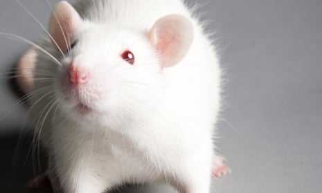
of human brain tissue have been transplanted into the brains of rats in
work that could pave the way for new treatments for devastating brain
injuries.
The groundbreaking study showed that
the “human brain organoids” – sesame seed-sized balls of neurons – were
able to integrate into the rat brain, linking up with their blood
supplies and communicating with the rat neurons.
The
team behind the work suggest that eventually doctors might be able to
grow blobs of brain tissue from a patient’s own cells in the lab and use
them to repair brain injuries caused by stroke or trauma.
“This
is incredibly exciting to me as a physician,” said Isaac Chen, a
physician and assistant professor of neurosurgery at the University of
Pennsylvania.
The study is the latest in the
rapidly growing and ethically complex field of brain organoids.
Scientists have shown that when cultivated in the right conditions,
neurons begin to form tiny brain-like structures, allowing scientists to
investigate developmental conditions such as autism and a wide range of
basic neuroscience questions.
The new work is
the first demonstration that the lab-grown brain tissue can be
successfully implanted into an injury site to repair an adult brain,
suggesting there could be future clinical applications.
Chen
and colleagues grew human brain organoids in a dish until they were
about 1.5mm in diameter. The balls of tissue were then transplanted into
the brains of adult rats that had sustained injuries to their visual
cortex. Within three months, the grafted organoids had integrated with
their host’s brain, hooking up with the blood supply, expanding to
several times the initial volume and sending out projections that linked
up with the rat’s neurons, according to the study published in Cell Stem Cell.
“We
were not expecting to see this degree of functional integration so
early,” says Chen. “[This] suggests that neural tissue transplantation
in the adult mammalian brain, especially one that has been disrupted
with some sort of injury, really is a viable path forward for neural
repair.”
The scientists did not assess whether
the implants improved how well the rats were able to function, but tests
showed that the human neurons fired off electrical signals when the
rats were exposed to flashing lights. Chen said that this supported the
idea that organoids could act as “blank processing units” that the brain
could absorb and use to rebuild itself after injury.
“By
rationally introducing these engineered processing units to specific
areas of the injured brain, we think that the increased computational
capacity of those areas would result in sufficient restoration of brain
networks to restore neurological function,” said Chen.
In
theory, personalised brain organoids could be created in the lab from a
patient’s own cells, although Chen predicted that clinical applications
would be at least five to 10 years away. “We are at the very beginning
of this journey,” he said.
Dr
Serena Barral, a lecturer in developmental neuroscience at UCL, who was
not involved in the work, described it as an “incredible” demonstration
of the sheer adaptability of neurons. “There’s a lot of information
that’s in the DNA itself and that allows the neurons to work wherever
they are – whether they’re in a plastic box in the lab or in the brain,”
she said.
She added that in future clinical
applications, the degree to which the brain could be repaired was likely
to depend on which functions had been lost.
“The
visual cortex is more simple, but if you think about replacing the
areas that are important for speech, mathematical calculations, thinking
– that could be a little bit more tricky because there are a lot of
abilities of the brain that are grown with experience,” she said.