Use the labels in the right column to find what you want. Or you can go thru them one by one, there are only 33,864 posts. Searching is done in the search box in upper left corner. I blog on anything to do with stroke. DO NOT DO ANYTHING SUGGESTED HERE AS I AM NOT MEDICALLY TRAINED, YOUR DOCTOR IS, LISTEN TO THEM. BUT I BET THEY DON'T KNOW HOW TO GET YOU 100% RECOVERED. I DON'T EITHER BUT HAVE PLENTY OF QUESTIONS FOR YOUR DOCTOR TO ANSWER.
Changing stroke rehab and research worldwide now.Time is Brain!trillions and trillions of neuronsthatDIEeach day because there areNOeffective hyperacute therapies besides tPA(only 12% effective). I have 523 posts on hyperacute therapy, enough for researchers to spend decades proving them out. These are my personal ideas and blog on stroke rehabilitation and stroke research. Do not attempt any of these without checking with your medical provider. Unless you join me in agitating, when you need these therapies they won't be there.
What this blog is for:
My blog is not to help survivors recover, it is to have the 10 million yearly stroke survivors light fires underneath their doctors, stroke hospitals and stroke researchers to get stroke solved. 100% recovery. The stroke medical world is completely failing at that goal, they don't even have it as a goal. Shortly after getting out of the hospital and getting NO information on the process or protocols of stroke rehabilitation and recovery I started searching on the internet and found that no other survivor received useful information. This is an attempt to cover all stroke rehabilitation information that should be readily available to survivors so they can talk with informed knowledge to their medical staff. It lays out what needs to be done to get stroke survivors closer to 100% recovery. It's quite disgusting that this information is not available from every stroke association and doctors group.
Showing posts with label Brain on a chip. Show all posts
Showing posts with label Brain on a chip. Show all posts
Isn't your competent? doctor already working with these other researchers on other brain initiatives? That's right, you have an incompetent? doctor, don't you? As proven by not even having or attempting 100% recovery protocols!
Artificial Brain(1 post to February 2015) A decade for your doctor to become competent
Brain on a chip(8 posts to October 2012) Even longer incompetence!
One of the things that we're trying to do with Li-Huei Tsai and Alice Stanton, who's a postdoc with me, is make models of the human brain. So, you might have a brain on a chip someday. That might, to the extent that we're successful, lead to new ways of finding molecules that could interfere with different brain diseases.
What Alice has done is make a personalized brain on a chip, because it's outside the body. If you try to study humans or animals, it's very hard to get much data. But if it's outside the body on a microscope slide or a chip, you can do many experiments and get a lot of data, and hopefully understand things better.
Ultimately, you’d of course have to go back to animals or humans. But with the idea that we could really make something like that, I'm hopeful it will lead to new ways of studying brain disease and maybe lead to new treatments someday.
Summary: Researchers developed the world’s first
3D-printed brain tissue that grows and behaves similarly to natural
brain tissue, marking a significant leap forward for neurological and
neurodevelopmental disorder research.
This novel 3D-printing
technique uses a horizontal layering approach and a softer bio-ink,
allowing neurons to interconnect and form networks akin to human brain
structures.
The ability to precisely control cell types and
arrangements provides unparalleled opportunities to study brain
functions and disorders in a controlled environment, offering new
avenues for drug testing and understanding brain development and
diseases like Alzheimer’s and Parkinson’s.
Key Facts:
The 3D-printed brain tissue can form networks and communicate through neurotransmitters, similar to human brain interactions.
This
new printing method allows for precise control over cell types and
arrangements, surpassing the capabilities of traditional brain
organoids.
The technique is accessible to many labs, not
requiring special equipment or culture methods, and can significantly
impact the study of various neurological conditions and treatments.
Source: University of Wisconsin
A
team of University of Wisconsin–Madison scientists has developed the
first 3D-printed brain tissue that can grow and function like typical
brain tissue.
It’s an achievement with important
implications for scientists studying the brain and working on treatments
for a broad range of neurological and neurodevelopmental disorders,
such as Alzheimer’s and Parkinson’s disease.
“This could be a
hugely powerful model to help us understand how brain cells and parts of
the brain communicate in humans,” says Su-Chun Zhang, professor of
neuroscience and neurology at UW–Madison’s Waisman Center.
“Our
tissue stays relatively thin and this makes it easy for the neurons to
get enough oxygen and enough nutrients from the growth media,” Yan says.
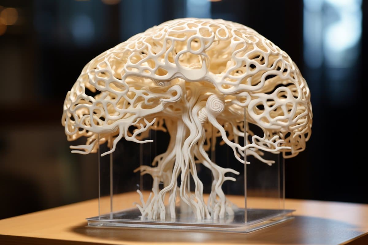
Credit: Neuroscience News
“It could change the way we look at stem cell biology, neuroscience,
and the pathogenesis of many neurological and psychiatric disorders.”
Printing
methods have limited the success of previous attempts to print brain
tissue, according to Zhang and Yuanwei Yan, a scientist in Zhang’s lab.
The group behind the new 3D-printing process described their method
today in the journal Cell Stem Cell.
Instead of using the
traditional 3D-printing approach, stacking layers vertically, the
researchers went horizontally. They situated brain cells, neurons grown
from induced pluripotent stem cells, in a softer “bio-ink” gel than
previous attempts had employed.
“The tissue still has enough
structure to hold together but it is soft enough to allow the neurons to
grow into each other and start talking to each other,” Zhang says.
The cells are laid next to each other like pencils laid next to each other on a tabletop.
“Our
tissue stays relatively thin and this makes it easy for the neurons to
get enough oxygen and enough nutrients from the growth media,” Yan says.
The
results speak for themselves — which is to say, the cells can speak to
each other. The printed cells reach through the medium to form
connections inside each printed layer as well as across layers, forming
networks comparable to human brains.
The neurons communicate, send signals, interact with each other
through neurotransmitters, and even form proper networks with support
cells that were added to the printed tissue.
“We printed the
cerebral cortex and the striatum and what we found was quite striking,”
Zhang says. “Even when we printed different cells belonging to different
parts of the brain, they were still able to talk to each other in a
very special and specific way.”
The printing technique offers
precision — control over the types and arrangement of cells — not found
in brain organoids, miniature organs used to study brains. The organoids
grow with less organization and control.
“Our lab is very special
in that we are able to produce pretty much any type of neurons at any
time. Then we can piece them together at almost any time and in whatever
way we like,” Zhang says.
“Because we can print the tissue by
design, we can have a defined system to look at how our human brain
network operates. We can look very specifically at how the nerve cells
talk to each other under certain conditions because we can print exactly
what we want.”
That specificity provides flexibility. The printed
brain tissue could be used to study signaling between cells in Down
syndrome, interactions between healthy tissue and neighboring tissue
affected by Alzheimer’s, testing new drug candidates, or even watching
the brain grow.
“In the past, we have often looked at one thing at
a time, which means we often miss some critical components. Our brain
operates in networks. We want to print brain tissue this way because
cells do not operate by themselves. They talk to each other. This is how
our brain works and it has to be studied all together like this to
truly understand it,” Zhang says.
“Our
brain tissue could be used to study almost every major aspect of what
many people at the Waisman Center are working on. It can be used to look
at the molecular mechanisms underlying brain development, human
development, developmental disabilities, neurodegenerative disorders,
and more.”
The new printing technique should also be accessible to many labs. It
does not require special bio-printing equipment or culturing methods to
keep the tissue healthy, and can be studied in depth with microscopes,
standard imaging techniques and electrodes already common in the field.
The
researchers would like to explore the potential of specialization,
though, further improving their bio-ink and refining their equipment to
allow for specific orientations of cells within their printed tissue..
“Right
now, our printer is a benchtop commercialized one,” Yan says. “We can
make some specialized improvements to help us print specific types of
brain tissue on-demand.”
Funding: This study was
supported in part by NIH-NINDS (NS096282, NS076352, NS086604), NICHD
(HD106197, HD090256), the National Medical Research Council of Singapore
(MOH-000212, MOH-000207), Ministry of Education of Singapore
(MOE2018-T2-2-103), Aligning Science Across Parkinson’s (ASAP-000301),
the Bleser Family Foundation, and the Busta Foundation.
About this neurotech research news
Author: Emily Leclerc Source: University of Wisconsin Contact: Emily Leclerc – University of Wisconsin Image: The image is credited to Neuroscience News
3D bioprinting of human neural tissues with functional connectivity
Highlights
Functional human neural tissues assembled by 3D bioprinting
Neural circuits formed between defined neural subtypes
Functional connections established between cortical-striatal tissues
Printed tissues for modeling neural network impairment
Summary
Probing
how human neural networks operate is hindered by the lack of reliable
human neural tissues amenable to the dynamic functional assessment of
neural circuits. We developed a 3D bioprinting platform to assemble
tissues with defined human neural cell types in a desired dimension
using a commercial bioprinter.
The printed neuronal progenitors
differentiate into neurons and form functional neural circuits within
and between tissue layers with specificity within weeks, evidenced by
the cortical-to-striatal projection, spontaneous synaptic currents, and
synaptic response to neuronal excitation.
Printed astrocyte
progenitors develop into mature astrocytes with elaborated processes and
form functional neuron-astrocyte networks, indicated by calcium flux
and glutamate uptake in response to neuronal excitation under
physiological and pathological conditions.
These designed human
neural tissues will likely be useful for understanding the wiring of
human neural networks, modeling pathological processes, and serving as
platforms for drug testing.
With the various brains on a chip our researchers can figure out what intervention needs to pass thru the blood brain barrier to accelerate neuroplasticity and neurogenesis. Five steps to solve this. This is so fucking simple, getting it right might take a while but the concept is simple.
1. Describe the problem exactly. 2. Write an RFP to researchers to solve that problem.
3. Fund them with foundation grants.
4. Write stroke rehab protocols based on the research.
5. Get the Nobel prize in medicine
Wake Forest Institute for Regenerative Medicine (WFIRM) scientists have developed a 3-D brain organoid that could have potential applications in drug discovery and disease modeling. This is the first engineered tissue equivalent to closely resemble normal human brain anatomy, containing all six major cell types found in normal organs including, neurons and immune cells.
In a study published this month in Scientific Reports, the researchers report that their advanced 3-D organoids promote the formation of a fully cell-based, natural and functional barrier - the blood brain barrier - that mimics normal human anatomy.
The blood brain barrier is a semipermeable membrane that separates the circulating blood from the brain, protecting it from foreign substances that could cause injury. This development is important because the model can help to further understanding of disease mechanisms at the blood brain barrier, the passage of drugs through the barrier, and the effects of drugs once they cross the barrier.
"The shortage of effective therapies and low success rate of investigational drugs are due in part because we do not have a human-like tissue models for testing," said senior author Anthony Atala, M.D., director of WFIRM. "The development of tissue engineered 3D brain tissue equivalents such as these can help advance the science toward better treatments and improve patients' lives."
The development of the model opens the door to speedier drug discovery and screening, both for neurological conditions and for diseases like HIV where pathogens hide in the brain and avoid current treatments that cannot cross the blood brain barrier. It may also allow for disease modeling of neurological conditions such as Alzheimer's disease, multiple sclerosis and Parkinson's disease so that researchers can better understand their pathways and progression.
Thus far the researchers have used the brain organoids to mimic strokes in order to measure impairment of the blood brain barrier and have successfully tested the model's permeability with large and small molecules.
"Using an engineered tissue model provides a platform that can be used to understand the fundamental principles at play with the blood brain barrier and its function, as well as the effects of chemical substances that cross it," said Goodwell Nzou, a Ph.D. candidate at WFIRM who co-authored the paper.
The Houston Methodist Research Institute is making mini brains from human stem cells that put researchers on a fast track to repair the nervous system after injury or disease of the brain and spinal cord.
Houston Methodist neuroscientist Robert Krencik, Ph.D., and his team have developed a new system to reduce the time it takes to grow these brain models, which will give them the ability to screen drugs and study what's behind disease-causing mutations more quickly. Their findings are described in an article titled "Systematic three-dimensional coculture rapidly recapitulates interactions between human neurons and astrocytes," in the Dec. 12 issue of Stem Cell Reports.
"We always felt like what we were doing in the lab was not precisely modeling how the cells act within the human brain," Krencik said. "So, for the first time, when we put these cells together systematically, they dramatically changed their morphological complexity, size and shape. They look like cells as you would see them within the human brain, so now we can study cells in the lab in a more natural environment."
And why is this important? Krencik says cells grown in traditional lab cultures are put on a flat petri dish, broken up and otherwise manipulated, disturbing their interactions. This results in not being able to reproduce the form, structure and developmental growth of the brain's cells in the lab, leading to very simplistic-looking and immature cells. In the human brain, however, these cells are very complex-looking and interact in intricate ways with each other and the environment. New technologies are now focused on 3-D culture systems, but the exhaustive time for these studies is not feasible for accelerating discoveries.
"Normally, growing these 3-D mini brains takes months and years to develop," Krencik said. "We have new techniques to pre-mature the cells separately and then combine them, and we found that within a few weeks they're able to form mature interactions with each other. So, the length of time to get to that endpoint for studies is dramatically reduced with our system."
Krencik's lab focused on a star-shaped cell type called astrocytes, because they are a key factor in getting the brain's neurons to connect and talk to each other by helping to increase the number and strength of neuronal connections in the brain and spinal cord. They are involved in most neural diseases and also are responsible for maintaining a healthy nervous system. With the model Krencik's team bioengineered, the incorporation of astrocytes accelerated the connections of the surrounding neurons.
Krencik's group is the first to specifically engineer astrocytes into these 3-D mini brains. By doing so, this led to the accelerated maturation of both the astrocytes and the surrounding neurons. Introducing them for the first time in this paper, he coined these bioengineered mini brains "asteroids" to distinguish them from other types of 3-D sphere cultures, known as organoids. Krencik's "asteroids" contain specific populations of astrocytes, whereas organoids have undefined numbers and types of cells.
"Using our system, we can generate mature astrocytes and have them interact intimately with neurons to a greater extent than has been done before," Krencik said. "Unlike other cells in the brain and in the rest of the body, astrocytes have unique properties in humans. It's thought they are partly responsible for the unique cognitive functions of humans and also may underlie aspects of human diseases, such as Alzheimer's and autism spectrum disorders."
Ultimately, Krencik is using these "asteroids" to form functional neural circuits that researchers can experimentally manipulate for developing treatments and deciphering what makes diseases tick. Krencik says they can make induced pluripotent stem cells, commonly termed iPS cells, from any disease or patient and then form these mini brains to study the disease process, as well as screen therapeutic compounds on them to aid in the development of drugs. Within about five years, his goal is to use this system to develop clinical trials to improve or regenerate a person's impaired nervous system.
With all this earlier research our researchers should be able to put this all together and create real human brains that can be damaged by stroke and show how to stop and reverse such damage. But that is way too pie in the sky for our stroke medical professionals to understand and implement.
Scientists in Australia have used a 3D printer to create nerve cells
found in the brain using a special bio-ink made from stem cells.
Key points:
Stem cells from adult cells used to make "bio-ink"
Bio-ink printed into 3D scaffold and then stem cells turned into nerve cells found in the brain
Process could be used in the future to make replacement brain tissue from patient's own skin cells
The research takes us a step closer to making replacement
brain tissue derived from a patient's own skin or blood cells to help
treat conditions such as brain injury, Parkinson's disease, epilepsy and
schizophrenia.
The bio-ink is made of human induced pluripotent
stem cells (iPSC), which have the same power as embryonic stem cells to
turn into any cell in the body, and possibly form replacement body
tissues and even whole organs.
Jeremy Crook, who led the research,
said the ability to customise brain tissue from a person's own body
tissue was better for transplantation.
"That circumvents issues of
immune rejection, which is common in organ transplantation," said Dr
Crook, from the University of Wollongong and ARC Centre of Excellence
for Electromaterials Science.
Correcting chemical imbalances
Dr Crook said many
neuropsychiatric disorders result from an imbalance of key chemicals
called neurotransmitters, which are produced by specific nerve cells in
the brain.
For example, he said, defective serotonin and
GABA-producing nerve cells are implicated in schizophrenia and epilepsy
while defective dopamine-producing cells are implicated in Parkinson's
disease.
The team used 3D printing to make neurones involved in
producing GABA and serotonin, as well as support cells called neuroglia,
they reported in the journal Advanced Healthcare Material.
In the future, they plan to print neurones that produce dopamine.
"That's absolutely achievable."
To make the neurones,
Dr Crook and colleagues used their bio-ink to print layers of a hatched
pattern to create a 5 millimetre-sized cube.
They then "crosslinked" the cube into a firm jelly-like substance.
Growth
factors and nutrients were then fed into the holes of this spongey
"scaffold", encouraging the stem cells to grow and turn into neurons and
support cells, linking up to form tissue.
Waste was also removed via the holes in the scaffold.
Dr
Crook said once scaled up, blood vessels would be needed, but small
transplants could be theoretically possible using the tissue developed
so far.
Impressive but risky too
Tissue engineer Makoto Nakamura from Toyama University in Japan said the study was "very impressive".
"This
article indicates the good feasibility of 3D bioprinting with human iPS
cells to engineer neural tissues," said Professor Nakamura, who
recently wrote an overview on the use of 3D bioprinting in the journal
Tissue Engineering.
But he said there were also risks with the technology.
(Supplied: Gu et al/Advanced Healthcare Materials)
One of the challenges of using iPSCs is that, like embryonic
stem cells, they have the potential to develop into teratomas —
disturbing looking tumours that contain more than one type of tissue
type (think toenails growing in brain tissue, or teeth growing in ovary
tissue).
According to Professor Nakamura, it would be important to
ensure all the stem cells had turned into nerve cells in the final
transplanted material.
"Undesired tissue may grow if even only one immature [stem] cell contaminates [the tissue to be transplanted]," he said.
Dr
Crook said the team was currently carrying out animal experiments to
test if teratomas developed from the 3D printed nerve cells.
3D brains?
While
this is a first step towards 3D printing of whole organs, Dr Crook said
a whole functioning brain would be a much more complex task.
"That's
a whole different scale. The tissue we print is uniform, and not made
up of different regions like a brain," said Dr Crook.
Still, it is a goal the researchers are heading towards.
Apart from providing customised transplants, 3D printed tissue could be useful for medical research.
For
example, tissue from a patient with epilepsy or schizophrenia could be
created, specifically to study their particular version of the
condition.
"You can compare how neuronal networks form differently compared to healthy patient," said Dr Crook.
And the tissue could also be used to screen for effective drugs or electrical stimulation treatments.
Your doctor, IF ANY GOOD AT ALL, should be contacting these researchers to determine how this could be used to determine the connectivity problems you have post-stroke and what can be done to reconnect them properly. But that will never occur so you will need to contact the researchers yourselves. 10 million yearly stroke survivors contacting them might show how important this is to stroke recovery. Or we could have a great stroke association do it once and distribute the information worldwide. ROFLMAO.
Harvard University researchers have developed a
multiregional brain-on-a-chip that models the connectivity between three
distinct regions of the brain. The in vitro model was used to
extensively characterize the differences between neurons from different
regions of the brain and to mimic the system’s connectivity.
The research was published in the Journal of Neurophysiology.
“The brain is so much more than individual neurons,” said Ben Maoz,
co-first author of the paper and postdoctoral fellow in the Disease
Biophysics Group in the Harvard John A. Paulson School of Engineering
and Applied Sciences (SEAS). “It’s about the different types of cells
and the connectivity between different regions of the brain. When
modeling the brain, you need to be able to recapitulate that
connectivity because there are many different diseases that attack those
connections.”
“Roughly twenty-six percent of the US healthcare budget is spent on
neurological and psychiatric disorders,” said Kit Parker, the Tarr
Family Professor of Bioengineering and Applied Physics Building at SEAS
and Core Faculty Member of the Wyss Institute for Biologically Inspired
Engineering at Harvard University. “Tools to support the development of
therapeutics to alleviate the suffering of these patients is not only
the human thing to do, it is the best means of reducing this cost."
Researchers from the Disease Biophysics Group at SEAS and the Wyss
Institute modeled three regions of the brain most affected by
schizophrenia — the amygdala, hippocampus and prefrontal cortex.
They began by characterizing the cell composition, protein
expression, metabolism, and electrical activity of neurons from each
region in vitro.
“It’s no surprise that neurons in distinct regions of the brain are
different but it is surprising just how different they are,” said
Stephanie Dauth, co-first author of the paper and former postdoctoral
fellow in the Disease Biophysics Group. “We found that the cell-type
ratio, the metabolism, the protein expression and the electrical
activity all differ between regions in vitro. This shows that it does
make a difference which brain region’s neurons you’re working with.”
Next, the team looked at how these neurons change when they’re
communicating with one another. To do that, they cultured cells from
each region independently and then let the cells establish connections
via guided pathways embedded in the chip.
The researchers then measured cell composition and electrical
activity again and found that the cells dramatically changed when they
were in contact with neurons from different regions.
“When the cells are communicating with other regions, the cellular
composition of the culture changes, the electrophysiology changes, all
these inherent properties of the neurons change,” said Maoz. “This shows
how important it is to implement different brain regions into in vitro
models, especially when studying how neurological diseases impact
connected regions of the brain.”
To demonstrate the chip’s efficacy in modeling disease, the team
doped different regions of the brain with the drug Phencyclidine
hydrochloride — commonly known as PCP — which simulates schizophrenia.
The brain-on-a-chip allowed the researchers for the first time to look
at both the drug’s impact on the individual regions as well as its
downstream effect on the interconnected regions in vitro. The brain-on-a-chip could be useful for studying any number of
neurological and psychiatric diseases, including drug addiction, post
traumatic stress disorder, and traumatic brain injury.(maybe stroke?)
"To date, the Connectome project has not recognized all of the
networks in the brain,” said Parker. “In our studies, we are showing
that the extracellular matrix network is an important part of
distinguishing different brain regions and that, subsequently,
physiological and pathophysiological processes in these brain regions
are unique. This advance will not only enable the development of
therapeutics, but fundamental insights as to how we think, feel, and
survive."
This research was coauthored by Sean P. Sheehy, Matthew A. Hemphill,
Tara Murty, Mary Kate Macedonia, Angie M. Greer and Bogdan Budnik. It
was supported by the Wyss Institute for Biologically Inspired
Engineering at Harvard University and the Defense Advanced Research
Projects Agency. http://jn.physiology.org/content/early/2016/12/28/jn.00575.2016
Attached files
New model mimics the connectivity of the brain by connecting
three distinct brain regions on a chip (Image courtesy of the Disease
Biophysics Group/Harvard University)
Full bibliographic informationNeurons derived from different brain regions are inherently different in vitro: A novel multiregional brain-on-a-chip
Stephanie
Dauth, Ben M Maoz, Sean P Sheehy, Matthew A Hemphill, Tara Murty, Mary
Kate Macedonia, Angie M Greer, Bogdan Budnik, Kevin Kit Parker Journal of Neurophysiology Published 28 December 2016 Vol. no. , DOI: 10.1152/jn.00575.2016
And since we already can grow brains on a chip this style of testing might solve rodent model in inflammation is not the same as humans. At least that could be the case if we had a great stroke association directing where research should be going. http://news.ubc.ca/2013/12/30/organs-on-a-chip-2/
Imagine
if medical research and clinical drug tests could be done on
artificially grown organs on microchips to save time, costs, and ease
ethical concerns?
That’s the dream of James Feng, a professor in biological and chemical engineering at UBC.
“The potential is tremendous,” says Feng. “The main impact of organs
grown this way will be on the design of drugs; the understanding of the
pathological processes.”
Dr. Feng’s group carries out research in three broad areas: mechanics
of biological cells and tissues, interfacial fluid dynamics,
and mechanics and rheology of complex fluids.
The group has an inter-disciplinary flavour–crosscutting applied
mathematics, cell biology, soft-matter physics and chemical and
biomedical engineering—that is well-suited for exploring this burgeoning
technology. Implications for the pharmaceutical industry
Feng cites a Harvard study
using a small silicon device that holds a thin layer of real cell
membranes capable of producing motion similar to the heaving and
breathing of a lung.
Organ models designed this way have the potential to be more accurate
in drug and treatment trials, says Feng, as they can better mimic the
functions of human organs, as opposed to animal models which are the
current research standard.
“It’s more controlled and you can simplify the process much faster,” said Feng.
“Harvard researchers also injected drugs into their chip model to see
how it changed its behaviour and to see the tissue’s reaction to
mechanical or chemical disturbance,” he added.
“It’s very important for drug design and discovery and the pharmaceutical industry would be tremendously interested in that.”
In addition, organs on a chip present a less controversial option for
organ model testing compared to stem cell research. According to Feng,
this is because their ultimate goals are very different from each other.
“The research that tried to grow organs directly from stem cells is
aiming for eventually implantable organs,” he said. “The idea of making
the chip is to work toward replacing animal models, so as to be more
accurate and realistic like human organs. While the ability to replicate
a complex human organ function remains far off, the direction appeals
to anyone who is hoping to reduce the use of animals in research.” Simulating organ functions on a chip
Feng says this kind of organ testing offers the possibility of greatly reducing cost and time required for clinical trials.
“By using computer simulations we can generate results and insights,
and run virtual tests much more easily and quickly,” he says.
“We can test maybe hundreds or thousands of designs of organ chips to
be able to tell you whether you should try those ten designs instead of
the hundreds one by one.”
Feng, who has a background in aerospace engineering, says this new
bio-technology has the potential to transform the development of
artificial organs and drugs the way computer simulations have replaced
the use of wind tunnels for designing aircrafts.
“That used to be the dominant mode of designing crafts,” he said,
“but that’s being replaced by online computer simulations because we
understand the principles of aerodynamics so well.”
While UBC’s efforts in the field are in the early stages, Feng is
reaching out to researchers from other backgrounds. He will be inviting
leading scientists to UBC in July 2014 for a workshop that will centre
on the growth of artificial organs and computer simulations. He is also
exploring ideas of his own.
“I have a collaboration with an engineering colleague on how to use
the microfluidic chip, the technology used to emulate the lung in the
Harvard study, as a way of measuring malaria-infected red cells,” he
said, suggesting that this is just one of the countless ways this new
technology could be used to fuel future innovation.
The actual research abstract here: http://pubs.rsc.org/en/content/articlelanding/2012/lc/c2lc41033h
In this work, we describe the fabrication and working of a modular
microsystem that recapitulates the functions of the “Neurovascular
Unit”. The microdevice comprised of a vertical stack of poly
(dimethylsiloxane) (PDMS) neural parenchymal chamber separated by a
vascular channel via a microporous polycarbonate (PC) membrane. The
neural chamber housed a mixture of neurons (~4%), astrocytes (~95%), and
microglia (~1%). The vascular channel was lined with a layer of rat
brain microvascular endothelial cell line (RBE4). Cellular components in
neural chamber and vascular channel showed viability (>90%). The
neural cells fired inhibitory as well as excitatory potentials following
10 days of culture. The endothelial cells showed diluted-acetylated low
density lipoprotein (dil-a-LDL) uptake, expressed von Willebrand factor
(vWF) and zonula occludens (ZO-1) tight junctions, and showed decreased
Alexafluor™-conjugated dextran leakage across their barrier
significantly compared with controls (p < 0.05). When the vascular
layer was stimulated with TNF-α for 6h, about 75% of resident microglia
and astrocytes on the neural side were activated significantly (p <
0.05 compared to controls) recapitulating tissue-mimetic responses
resembling neuroinflammation. The impact of this microsystem lies in the
fact that this biomimetic neurovascular platform might not only be
harnessed for obtaining mechanistic insights for neurodegenerative
disorders, but could also serve as a potential screening tool for
central nervous system (CNS) therapeutics in toxicology and
neuroinfectious diseases.