Use the labels in the right column to find what you want. Or you can go thru them one by one, there are only 33,793 posts. Searching is done in the search box in upper left corner. I blog on anything to do with stroke. DO NOT DO ANYTHING SUGGESTED HERE AS I AM NOT MEDICALLY TRAINED, YOUR DOCTOR IS, LISTEN TO THEM. BUT I BET THEY DON'T KNOW HOW TO GET YOU 100% RECOVERED. I DON'T EITHER BUT HAVE PLENTY OF QUESTIONS FOR YOUR DOCTOR TO ANSWER.
Changing stroke rehab and research worldwide now.Time is Brain!trillions and trillions of neuronsthatDIEeach day because there areNOeffective hyperacute therapies besides tPA(only 12% effective). I have 523 posts on hyperacute therapy, enough for researchers to spend decades proving them out. These are my personal ideas and blog on stroke rehabilitation and stroke research. Do not attempt any of these without checking with your medical provider. Unless you join me in agitating, when you need these therapies they won't be there.
What this blog is for:
My blog is not to help survivors recover, it is to have the 10 million yearly stroke survivors light fires underneath their doctors, stroke hospitals and stroke researchers to get stroke solved. 100% recovery. The stroke medical world is completely failing at that goal, they don't even have it as a goal. Shortly after getting out of the hospital and getting NO information on the process or protocols of stroke rehabilitation and recovery I started searching on the internet and found that no other survivor received useful information. This is an attempt to cover all stroke rehabilitation information that should be readily available to survivors so they can talk with informed knowledge to their medical staff. It lays out what needs to be done to get stroke survivors closer to 100% recovery. It's quite disgusting that this information is not available from every stroke association and doctors group.
All this information out there on backward walking and no one in stroke is smart enough to put this all together in a protocol? WHAT STROKE 'LEADER' DO I REAM OUT FOR COMPLETE INCOMPETENCY? It took me 1 minute to find this.
Been done already eight years before this came out.
If your doctor/hospital didn't do anything with this from
14 years ago or any of the following then s/he needs to be fired. Why
is your board of directors being so incompetent in not demanding new
interventions from any stroke research?
2014, International Journal of Stroke Stella Maris Michaelsen 1*,
Angélica Cristiane Ovando 2,
Fernanda Romaguera 1,
and Louise Ada 3 Rationale Residual walking deficits are common in people after stroke. Treadmill training can increase walking speed andwalking distance. A new way to increase the challenge of walking is to walk backwards. Backward treadmill walking may provide advantages by promoting improvement in balance, walking spatiotemporal parameters and quality that may reflect in improving walking distance. Aim This study will test the hypothesis that backward treadmill walking is superior to forward treadmill walking in improving walking capacity, walking parameters, quality and balance in people with stroke. Design A prospective, single-blinded, randomized trial will randomly allocate 88 community-dwelling people after stroke into either an experimental or control group. The experimental group will undertake 30-min sessions of backward treadmill walking, three-days/week for six-weeks, while the control group will undertake the same dose of forward treadmill walking. Training will begin at the baseline overground walking speed and will increase each week by 10% of baseline speed. Study outcomes The primary outcome will be distance walked in the 6-min Walk Test. Secondary outcomes will be walking speed, step length, cadence, and one-leg stance time. Outcomes will be collected by a researcher blinded to group allocation at baseline (Week 0), at the end of training period(Week 6), and three-months after the cessation of intervention(Week 18). Discussion If backward treadmill walking can improve walking capacity more than forward treadmill training in stroke, it may have broader implications because walking capacity has been shown to predict physical activity level and community participation. Key words: ambulation, clinical trial, intervention, rehabilitation, stroke Introduction and rationale Walking is one of the most important activities to enable community participation. After stroke, although the majority of patients recover independent walking, many have residual walking disabilities. Many individuals after stroke cannot walk fast or far enough, which reduces their ability to walk outside the house. Even for those undergoing inpatient rehabilitation,walking quality can be surprisingly poor at the time of discharge from hospital,with one study finding that only 7% of people after stroke are able to have independent community ambulation at discharge(That is an appalling failure rate. WHOM IS BEING FIRED FOR THAT?) of rehabilitation (1) and mobility problems persist in the chronic phase after stroke (2). The walking speed of community-dwelling people after stroke has been reported to be40% less, and six-minute walking distance about half the value expected in age- and gender-matched people (3). This reduced capacity to walk long distances can restrict the access for community exercise programs as well as result in major limitations in community participation.The treadmill is frequently used for walking training in people after stroke. It allows a controlled speed and an intensive amount of practice (4), working as a ‘forced use’ (5). A recent review on treadmill training (6) suggests that treadmill training can increase walking speed by 0·12 m/s and improve walking distance by 40 m.A relatively new way to increase the challenge of walking is to walk backwards. At a given speed, when compared with forward walking, backward walking elicits more electromyographic activity which in turn results in higher physiological cost and greater perceived exertion (7–9). Hip extension is more active in backward walking due to the concentric contraction of knee flexors during early swing phase (10,11) which may be beneficial at improving lower limb coordination after stroke. Also, backward walking is more difficult and demanding than forward walking due to its postural instability and may therefore provide a training challenge to balance (12). There have been three clinical trials of backward walking. One trial (13) in chronic stroke found that backward overground walking increased walking speed while another trial (14) found that backward treadmill walking also increased speed compared with overground walking. The only trial to compare backward treadmill walking with forward treadmill walking (15) was carried out in acute just-ambulatory patients and was therefore performed with body weight support, but it found that backward walking produced more independent walking.The main objectives of this trial are: to determine whether six weeks of backward treadmill walking is more effective than six weeks of forward treadmill walking in improving walking capacity, walking parameters, walking quality, and balance in Correspondence: Stella Maris Michaelsen * , Department of PhysicalTherapy – Centro de Ciências da Saúde e do Esporte, Universidade doEstado de Santa Catarina, Rua Pascoal Simone, 358 – Coqueiros,88080-350, Florianópolis, SC, Brazil.E-mail: michaelsenstella@hotmail.com 1 Departmentof PhysicalTherapy,PhysicalTherapyMasterProgram,Uni-versidade do Estado de Santa Catarina, Florianópolis, Santa Catarina,Brazil 2 Department of Physical Therapy, Universidade do Estado de Santa Cata-rina, Florianópolis, Santa Catarina, Brazil 3 Discipline of Physiotherapy, The University of Sydney, Lidcombe, New South Wales, AustraliaReceived: 30 August 2013; Accepted: 16 December 2013; Published online15 April 2014Conflict of interest: The authors declare that there is no conflict of interest.Funding: The trial is funded by the Brazilian National funding agency (Conselho Nacional de Pesquisa). DOI: 10.1111/ijs.12255
Academic Editor:Michelle PloughmanReceived: 22 February 2021Accepted: 10 March 2021Published: 13 March 2021Publisher’s Note:MDPI stays neutral with regard to jurisdictional claims in published maps and institutional affiliations.
Post-stroke rehabilitation often aims to increase walking speeds, as faster walking is associated with improved functional status and quality of life. However, for successful community ambulation, ability to modulate (increase and decrease) walking speeds is more important than walking continuously at constant speeds. Increasing paretic propulsive forces to increase walking speed has been extensively examined; however, little is known about the mechanics of slow walking post-stroke. The primary purpose of this study was to identify the effects of increased and decreased walking speeds on post-stroke kinetics and ankle kinematics. Fifteen individuals with chronic post-stroke hemiparesis and 15 non-neurologically impaired controls walked over an instrumented treadmill under: slow, self-selected, and fast walking speeds. We examined the peak propulsive forces, propulsive impulse, peak braking forces, braking impulse, and ankle kinematics under each condition. When walking at slow walking speeds, paretic limbs were unable to reduce braking impulse and peak propulsive force or modulate ankle kinematics. Impaired modulation of paretic gait kinetics during slow walking places people post-stroke at high risks for slip-related falls. These findings suggest the need for developing gait retraining paradigms for slow walking in individuals chronically post-stroke that target the ability of the paretic limb to modulate braking forces.Keywords:post-stroke hemiparesis; walking speed; gait; slow walking; ground reaction forces1.
Introduction
Stroke is the leading cause of adult long-term disabilities [1]. Individuals with hemiparesis resulting from a stroke possess significant impairments in locomotor function,resulting in slow walking speeds, asymmetrical gait patterns and fall risks, which negatively impacts functional or mobility independence and safety [2–4]. Faster walking speed is associated with enhanced quality of life [5], thus improving walking speed has been an important goal in stroke rehabilitation.Despite the significance of aiming to increase walking speed in people post-stroke,in the context of functional community ambulation, long durations of continuous steady-state comfortable speed walking behavior has been found to be less important and also occurs less frequently [6]. Rather, gait speed modulation (i.e., increasing and decreasing walking speed) and gait initiation/termination has been identified as important and more frequent functional tasks for successful community mobility. For example, walking is often combined with talking, or other attention demanding activities, resulting in slower walking speeds [7], or in the attempt to change directions during walking, when negotiating a turn,walking speeds would be decreased compared to walking in a straight line [8]. Additionally,when an individual walks past a stationary object, or when a moving object approaches the individual, walking speed has been observed to be decreased [9]. In non-neurologically impaired individuals, during steady state walking (constant speed conditions), gait is characterized by symmetric anterior-posterior ground reaction forces generated by the two legs. Walking speed is regulated by anterior-posterior ground reaction force impulses (i.e., propulsion and braking), where, with increases in walking speed, propulsive and braking impulses are observed to increase [10]. With decreases in walking speed, magnitude of anterior-posterior ground reaction forces, and joint angles decreased [11]. In individuals post-stroke, during steady state walking, asymmetry in anterior-posterior ground reaction forces generated have been reported, with less propulsive forces generated by the paretic limbs. Furthermore, the more severe the hemiparesis,the greater the asymmetry in the propulsive and braking impulses observed [12]. With respect to the ability of foot-force control, during a posturally supported locomotor task,the stroke-impaired system has been reported to be capable of generating foot forces that are appropriately controlled in magnitude and direction [13]. The propulsive forces generated by the paretic leg have been reported to be predictive of walking speed [12], and,in fast treadmill walking, increased paretic propulsive forces have been observed [14],suggesting the limited but potential capabilities of the stroke-impaired system to modulate foot forces under controlled locomotor conditions. However, the kinetics during slower walking speeds have yet to be examined in people with chronic post-stroke hemiparesis. Reduction in walking speed to below comfortable walking speeds has been considered to be a relatively more complex task, where different locomotor and postural control strategies are adopted, compared to comfortable walking speeds [15–17], and thus individuals post stroke may not exhibit similar speed mediated changes compared to fast walking over the treadmill. In an intact nervous system, when walking speeds are reduced from comfortable to very slow walking speeds, the system switches from locomotor towards greater postural muscular control [15]. Faster locomotor speeds are potentially associated with greater suppression of vestibular drive, thus a reduction in walking speed would be associated with reduced suppression of this destabilizing vestibular drive. In conjunction with reduced proprioceptive input when speeds are reduced, increased dynamic instability makes slow walking more complex [18–20]. The primary goal of this study was to identify the effects of increased (120% of self-selected) and decreased (80% of self-selected) walking speeds on kinetics and ankle kinematics changes in people with chronic post-stroke hemiparesis. We hypothesized that in individuals with post-stroke hemiparesis, during fast walking (120% of self selected speed), paretic legs would exhibit increased speed-related changes in kinetics (i.e., increased peak propulsive forces, propulsive impulse, peak braking forces and braking impulses)and ankle kinematics, compared to self-selected walking speed, but these variables would remain unchanged during slow walking (80% of self-selected speed).
Investigations
of real-time brain activations during walking have become increasingly
important to aid in recovery of walking after a stroke. Individual brain
activation patterns can be a valuable biomarker of neuroplasticity
during the rehabilitation process and can result in improved
personalized medicine for rehabilitation. The purpose of this systematic
review is to explore the brain activation characteristics during
walking post-stroke by determining: (1) if different components of gait
(i.e., initiation/acceleration, steady-state, complex) result in
different brain activations, (2) whether brain activations differ from
healthy individuals. Six databases were searched resulting in 22
studies. Initiation/acceleration showed bilateral activation in frontal
areas; steady-state and complex walking showed broad activations with
the majority exploring and finding increases in frontal regions and some
studies also showing increases in parietal activation. Asymmetrical
activations were often related to performance asymmetry and were more
common in studies with slower gait speed. Hyperactivations and
asymmetrical activations commonly decreased with walking interventions
and as walking performance improved. Hyperactivations often persisted in
individuals who had experienced severe strokes. Only a third of the
studies included comparisons to a healthy group: individuals post-stroke
employed greater brain activation compared to young adults, while
comparisons to older adults were less clear and limited. Current
literature suggests some indicators of walking recovery however future
studies investigating more brain regions and comparisons with healthy
age-matched adults are needed to further understand the effect of stroke
on walking-related brain activation.
Background
Stroke
is a leading cause of adult long-term disability worldwide. The
restoration of gait is rated as a high priority for stroke survivors [1, 2]. Yet, more than 50% of individuals living post-stroke do not independently walk within their community [3, 4]. Arguably, the efficacy of gait rehabilitation could be advanced with an individual’s personal brain activations [5, 6].
This notion of personalized medicine has become an important avenue of
exploration and is now stated as a research priority within national
funding agencies [7].
Determining neural correlates of walking is an important starting point
in investigating how brain activation can be a valuable biomarker or
indicator of neuroplasticity during the rehabilitation process.
Until
recently, neural correlates of human walking were informed by studies
with simulated or imagined walking tasks while under constrained brain
imaging environments. The recent advancement in technologies such as
portable electroencephalography (EEG), functional near-infrared
spectroscopy (fNIRS), and radioactive tracing with positron emission
topography (PET) or single-photon emission computerized tomography
(SPECT) have allowed for investigation of brain function during
real-time walking. Comparisons of simulated/imagined walking and
real-time walking in healthy adults show many similarities in activation
areas along the cortex, basal ganglia, brainstem, and cerebellum [8]
and differences in motor preparatory areas (e.g., bilateral
supplementary motor area (SMA)) and executive function areas (e.g.,
dorsolateral prefrontal cortex (PFC)) [9].
The
ability to obtain measurements during real-time walking allows for
investigation of brain activations associated with walking components
that are necessary for successful community ambulation, such as
acceleration/deceleration phases, steady-state walking, and complex
situations that involve avoiding obstacles or doing multiple tasks at
once (e.g., talking and walking). Previous studies show differing brain
activities during these various components. In healthy adults, walking
preparation increases PFC, premotor cortex (PMC), SMA and medial
sensorimotor cortex (SMC) activity, whereas walking execution mainly
activates SMA and medial SMC [10].
As the complexity of walking increases (e.g., walking while doing a
secondary task), further increases of bilateral PFC activation are shown
in healthy older adults [11].
When assessing brain activation during different components of walking
within neurological populations, results are quite varied [12,13,14].
Other reviews investigating brain activation during real-time walking
focus on the general neurological population category, rather than
stroke specifically [8, 15].
Two systematic reviews exclusively looking at fNIRS studies in
individuals with stroke only included three and five real-time walking
studies [16, 17].
Their narrow inclusion criteria excluded some pertinent studies and
more investigations have since been conducted in the stroke population.
To facilitate rehabilitation of community ambulation post-stroke, a
thorough understanding of how stroke affects functional brain activation
during various walking components is necessary.
Thus, the purpose
of the current systematic review is to consolidate work investigating
the spatial and temporal brain activation of real-time walking in
individuals with stroke. Specifically, studies will be described within
three components:
1.
Intention/acceleration: prior to walking onset or immediately post initiation of walking
2.
Steady-state: during walking at a steady pace without additional tasks
3.
Complex walking: walking with a secondary task or an externally cued gait
Methods
The
protocol for this systematic review was registered in the International
Prospective Register of Systematic Reviews, PROSPERO (CRD42019127401,
April 2019). This systematic review was conducted in accordance with
PRISMA guidelines. A narrative synthesis of results was completed. If
sufficient homogeneity in studies was present, quantitative pooling was
planned (none was completed due to significant heterogeneity in
methods).
Search strategy and study selection
Six
databases were used to search for studies published from inception to
July 16, 2020: Medline (Ovid), Embase (Ovid), Pubmed, Web of Science,
CINAHL (EBSCOHOST), and PsycInfo (EBSCOHOST). Search terms relating to
Population (stroke), Intervention (real-time, upright walking), and
Outcome (brain activation, fNIRS, EEG, PET or SPECT with radioactive
tracing) were created for each database with keywords and medical
subject headings (MeSH) terms as appropriate (Appendix Table 4).
Specific
brain-imaging modalities were included in the search based on the
ability to measure brain activation during real-time walking. In short,
fNIRS takes advantage of the absorption properties of hemoglobin and
utilizes near-infrared light to measure changes in regional
(de)oxyhemoglobin concentration along the cortex (i.e., limited ability
to measure subcortical structures). Similar to fMRI, fNIRS uses the
theory of neurovascular coupling to infer real-time regional brain
activity through changes in hemoglobin concentrations (i.e., more brain
activation requires more oxygen and thus more oxyhemoglobin) [18].
EEG utilizes electrodes placed along specific points on the scalp and
measures the net electrical activity across an ensemble of neurons
within the cortical and subcortical layers with high temporal
resolution. EEG is typically described through its frequency profile or
an event-related potential, with increases in higher frequencies (e.g.,
beta band: 13–30 Hz) and larger baseline deflections indicating
increased activation [19].
Finally, PET and SPECT scans utilize an injected tracer to assess
metabolic uptake during the entire task or uptake period (i.e., not in
real-time but representative of activation during the task). In most
cases for PET, a fluorodeoxyglucose (FDG) tracer is used to follow the
metabolic pathway of glucose (an excitatory neurotransmitter) and
provides an indication of regions with increased excitatory neuronal
activation [20].
Search
results were imported into Covidence (Veritas Health Innovation,
Australia) for duplicate removal and screening. Full-text reviews of the
screened articles were then assessed for inclusion based on the
criteria below. Reference lists of included full-texts and relevant
reviews were hand-searched for additional articles. Screening of titles,
abstracts, and full-text reviews were independently completed by two
authors (SBL, DRL). Inconsistencies were discussed between reviewers; if
a consensus was not reached, a third author (SP) was consulted.
Inclusion and exclusion criteria
Articles
were included if they assessed brain activity during real-time, upright
gait in adults (> 18 years of age) post-stroke. Studies were also
included if brain activity was assessed immediately prior to gait in
order to assess the preparation and initiation component of gait. All
types of study designs were considered for inclusion (i.e., case
studies, pre-post studies, cross-sectional studies, randomized
controlled trials). Published abstracts and conference abstracts were
also included if adequate information regarding brain imaging methods
and walking tasks were provided. Studies that included individuals of
various neurological conditions were only included if at least 50% of
the sample had a stroke. Due to the infancy of this field, inclusion of a
broad range of study designs and mixed groups was deliberate to ensure
that no relevant stroke findings were missed. Articles involving animal
models, pediatric strokes (< 18 years of age), and studies published
in languages other than English were not included.
Data extraction
Data
from the full-text articles were extracted independently by two authors
(SBL, SP). The data extraction form included the following article
details: title, year, author, journal, country of study, study type,
participants (number, age, time since stroke, type of stroke, severity
of stroke), technique used for measuring brain activity (type of device,
density of recording, regions of interest, rigour of measuring brain
activity), type of walking task (acceleration/initiation, steady-state,
complex), walking trial (length of trial, number of trials, speed of
walking), intervention (if applicable), comparator groups (no
comparator, older adults, young adults, other neurological groups), and
main findings. If the walking task was separated within the analysis,
results from the first portion were placed in the
acceleration/initiation category and the second portion were placed in
the steady-state category. If distinct walking tasks were not explicitly
investigated (e.g., acceleration/initiation versus steady-state) and a
study investigated a single walking period including the acceleration
phase, it was categorized as steady-state walking. Corresponding authors
were contacted for further clarification and details on the studies as
needed.
The National Institutes of Health (NIH) Study Quality Assessment Tools [21]
were used to determine the quality of each study by two authors (SBL,
DRL). This tool was designed based on quality assessment methods,
concepts, and other tools developed by numerous national and
international agencies. As indicated by the NIH Tools, separate
assessments were completed based on the study type and an overall study
rating of poor, fair, or good was provided by each assessor. According
to the NIH descriptions, “a ‘good’ study has the least risk of bias, and
result are considered to be valid. A ‘fair’ study is susceptible to
some bias deemed not sufficient to invalidate its results. The fair
quality category is likely to be broad, so studies with this rating will
vary in their strengths and weaknesses. A ‘poor’ rating indicates
significant risk of bias” [21]. Inconsistencies were discussed between assessors; if a consensus was not reached, a third author (SP) was consulted.
Results
Search yield
A
total of 6566 articles were retrieved from the six databases. Once
duplicates were removed and titles and abstracts were screened, 60
full-text articles were reviewed for inclusion. Thirty-eight articles
were excluded (see Fig. 1 for details). Twenty-two articles met the inclusion criteria for this systematic review.
Study characteristics
Articles
were published between 2000 and 2020. Studies were conducted in Canada
(n = 2), France (n = 1), Italy (n = 2), Japan (n = 6), Korea (n = 2),
the Netherlands (n = 1), Spain (n = 1), Taiwan (n = 3), United Kingdom
(n = 1), and USA (n = 3).
Eighteen studies were cross-sectional [22,23,24,25,26,27,28,29,30,31,32,33,34,35,36,37,38,39], two studies were randomized controlled trials [40, 41], and two studies were uncontrolled pre-post trials [42, 43]. One study was published as a book chapter [28] and two studies were published conference abstracts [30, 31]. Further detail on the book chapter [28] and conference abstract [31] were obtained through personal communication. Subsequent publication of the healthy older adult group [44] and a subsequent preprint currently under peer-review [45] were used to extract participant and task details.
Stroke population
A
total of 290 stroke participants (mean (SD): 59.0 (21.3) years, 24.6
(26.9) months post-stroke) were investigated in the 22 studies. All
studies included stroke-only participant groups with no studies
including participants with other neurological conditions. Studies
included a range in number of participants, from 1 to 33 individuals
with stroke. Specifically, the majority of studies (10 studies) had less
than 10 participants, five had between 10 and 20 participants, six had
between 20 and 30 participants, and only one study had greater than 30
participants. Six studies included individuals in the subacute stage of
stroke (< 6 months post), 15 studies included chronic stroke
(> 6 months), and one study did not report the post-stroke time.
Fifteen studies included individuals who could walk independently, six
studies were classified participants as having severe-moderate walking
impairments or required maximum-moderate walking assistance, and one
study did not report walking ability (Appendix Table 5).
Twelve
studies provided details on individual lesion locations, eight studies
only reported lesion side, stroke type (ischemic or hemorrhagic), or
depth of lesion (cortical or subcortical), and three did not report any
detail on the stroke. Overall, lesion locations were heterogeneous with
only three studies being more specific in inclusion criteria: Mihara et
al. [39] and Mori et al. [26]
specifically excluded participants with lesions over recording
areas—cortical lesions and PFC lesions, respectively—and Mitchell et al.
[31] only included participants with lesions around the basal ganglia or internal capsule (Appendix Table 5). Two studies specifically reported observing no brain activation over lesioned areas [24, 43]. All other studies did not report accounting for lesion location in data analysis.
Three studies [35, 36, 46] made comparisons with a younger group of adults, and seven studies made comparisons with older or age-matched adults [22, 23, 25, 26, 31, 36, 39].
Study quality
Eight studies were assessed as “good” [23, 29, 33, 36,37,38, 40], nine studies were “fair” [25, 26, 31, 34, 35, 39, 41,42,43], and five studies were “poor” [24, 27, 28, 30, 32] using the NIH Study Quality Assessment Tools (Appendix Table 6).
In general, most studies neglected to report recruitment methods,
number of eligible participants, or sample size justifications. Walking
tasks were generally described with adequate detail. Most studies assess
participants at their comfortable walking pace and walking tasks were
similar between participants within each study. Methods of recording
functional brain activation were described in good detail, though
specific details on landmarking for device set up and localization of
functional brain regions were often absent.
Brain recording details
Three
methods of measuring brain activity were used: EEG (n = 7), fNIRS
(n = 14), and [18F]-FDG-PET (n = 1). Brain recording set-ups and regions
of interest varied from investigating one brain region to whole head
measures. The majority of studies used the 10/10 or 10/20 International
system [47] to place their channels, four studies aligned their channels based on estimates from representative participants [24, 25, 39, 43], three reported a rough location of where channels were placed (e.g., high and lateral on forehead) [27, 33, 36], and two did not report how channels were placed [30, 32].
Results
within the following sections will be described in the following order:
activations in the stroke population, relationships between brain
activation and performance, and brain activations in comparison to
healthy individuals.
Brain activation during initiation and acceleration of walking
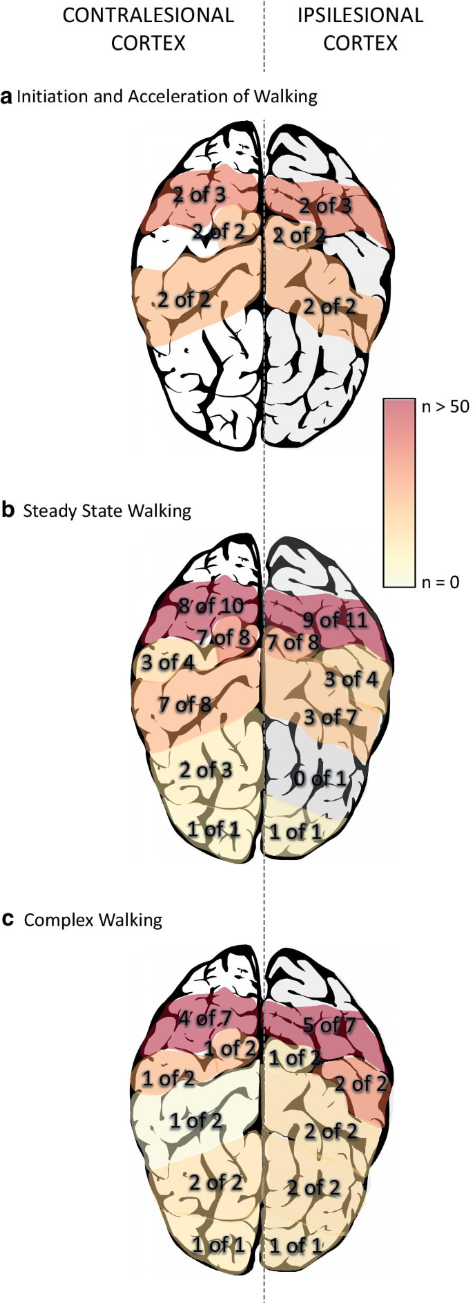
Three studies were included within this category [29, 36, 39] (Table 1).
Overall, activations were bilateral with no differences between
lesioned or non-lesioned hemispheres. For the stroke participants, two [36, 39]
of the three studies showed increased activation in bilateral PFC, and
both studies that looked at SMA and SMC showed increased activations
with walking compared to standing [29, 36] (Fig. 2a).
Table 1 Studies investigating initiation and acceleration of walking listed by increasing walking speed
Summary of regional activations for the a initiation/acceleration phase of walking, b steady-state phase of walking, and c
complex walking tasks. Numbers within the brain indicate how many
studies showed increased activation out of the total number of studies
that investigated the region. The colour gradient indicates the sum
total of subjects within the studies showing increased activation
None of these studies compared brain activation to gait performance.
When compared to young adults, PFC increases were greater in the stroke group [36]; in a follow-up study, Sburlea et al. [46]
reanalyzed their dataset and showed that brain activation increases
were similar but more widespread (i.e., larger volume of activated
areas) in the stroke group. PFC activations compared to older adults
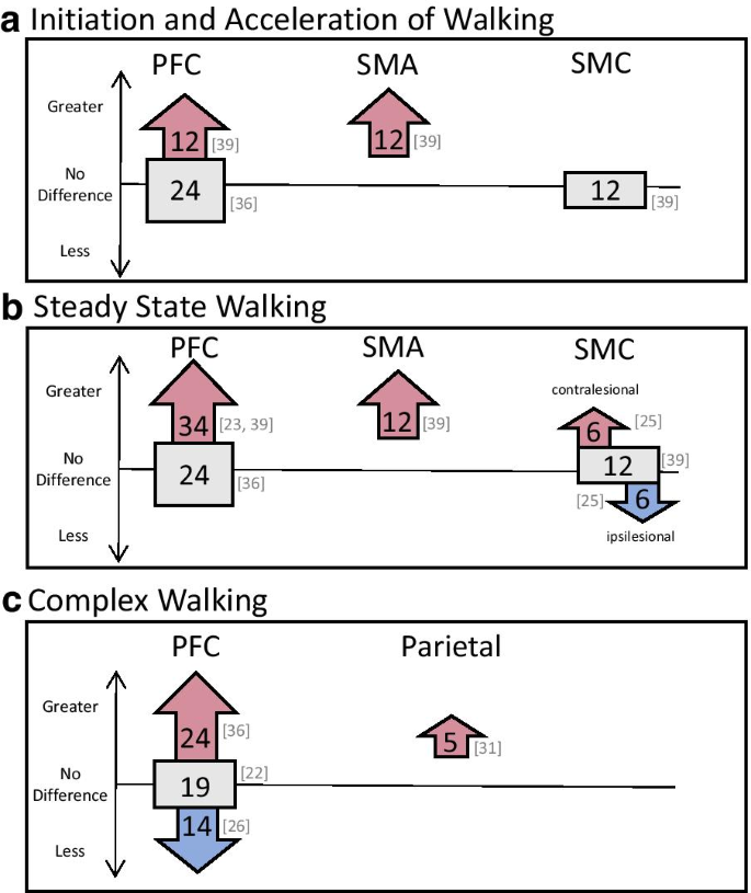
were less clear. Hawkins et al. [36] showed similar PFC increases between their chronic stroke group and older adults while Mihara et al. [39] showed greater activation over right PFC in their subacute stroke group with ataxia compared to older adults. Mihara et al. [39]
also showed greater activation over SMA for their ataxic group but no
difference in SMC activation compared to older adults (Fig. 3a).
Fig. 3
Differences
in regional activation patterns in comparison to age-matched healthy
individuals. Arrows pointing up indicate greater activation, arrows
pointing down indicate less activation, and squares indicate no
difference between stroke groups and age-matched healthy adults. Numbers
within the shapes represent total number of participants within the
studies, with the specific studies cited to the right of the shapes.
Panel a, b, and c represent studies looking at the
initiation/acceleration phase of walking, steady-state phase of walking,
and complex walking tasks, respectively
Fifteen studies investigated brain activity during steady-state walking [23,24,25, 27, 28, 30, 33,34,35,36,37, 39, 41,42,43] (Table 2).
All results will be described as activations during walking in
comparison to standing immediately prior to walking, unless otherwise
stated. Overall, stroke participants showed bilateral activations in
PFC, PMC, SMA, SMC, superior parietal and occipital lobe and greater
activations were found in the contralesional hemisphere for SMC and
parietal areas (Fig. 2b). A variety of steady-state walking tasks were compared. These included:
single-session assisted walking with sensory feedback [28], walking with body-weight support [25], and walking with robotic [23, 30, 35] or therapist [24] assistance,
multi-session gait interventions: controlled [41] and uncontrolled [42, 43].
Unassisted walking: Increased bilateral activations were observed in PFC (6 of 8 studies: [23, 28, 33, 36, 37, 39]), PMC (2 studies: [25, 28]), and SMA (3 studies: [25, 28, 39]) for the stroke groups. These studies, with the exception of Sangani et al. [28], showed no differences between hemispheres. Sangani et al. [28]
found greater overall contralesional activation; however, this was a
single-subject proof-of-concept study and specific details of precise
activations were not reported. Saitou et al. [27]
only measured from ipsilesional PFC and showed increased activation
during walking compared to standing in 15 of the 22 patients tested.
Increased PFC activations were also related to greater impairment (e.g.,
lower Fugl-Meyer scores) [33, 36] and lower balance confidence [33].
Increases in brain activity during walking were also found in SMC (4 studies: [25, 28, 34, 39]) and parietal regions (1 study: [34]).
In both these areas, more studies found greater activation in the
contralesional hemisphere with either little or no activation in the
ipsilesional hemisphere [25, 28, 34].
Compared to young healthy adults, PFC activation in stroke was greater [36]. However, compared to older adults, Hawkins et al. [36]
showed similar activations in both groups. Conversely, greater PFC
activation was reported for the stroke group in two studies [23, 39]. Mihara et al. [39]
also showed a sustained elevation in activation over SMA regions in
their ataxic stroke group and decreased PFC and SMA activation in their
healthy adult group during steady-state compared to acceleration. No
between group differences were observed for SMC [39] (Fig. 3b).
Assisted walking: Increased PFC was observed during overground exoskeleton walking compared to overground unassisted walking [23]. No difference in PMC and SMA activations were found between body weight supported walking and unassisted treadmill walking [25]. In contrast, Lee et al. [30]
found an overall decrease in PMC, SMA, and SMC activation with robotic
assistance compared to unassisted overground walking. Compared to
unassisted walking, 2 of the 3 studies showed increased brain activation
symmetry over the SMC during body weight supported walking [25] and with light finger touch on a stable surface [28] compared to unassisted walking. Increased symmetry in SMC activation was related to increased gait symmetry [25, 28] and increased SMC activity also related to increased walking cadence [25]. No correlations were found with PMC or SMA activations with gait performance.
Robotic
walking, compared to upright stationary body-weight suspension,
resulted in increased bilateral activation over SMC and contralesional
centro-parietal regions [35].
Manual assistance of the paretic leg during gait, compared to standing,
resulted in increased bilateral activation of PFC, PMC, SMA, and SMC,
with greater activation in ipsilesional (compared to contralesional) PMC
and contralesional (compared to ipsilesional) SMC [24]. Miyai et al. [24]
also compared manual assistance of the leg to facilitation at the hip
and found greater overall activations and greater symmetry of SMC
activity with facilitation. Facilitation also resulted in increased
walking cadence and symmetry [24]. No correlations were found between brain activations during robotic walking and walking performance [35].
In
healthy individuals, young adults showed differential activations based
on gait-phase whereas no phasic activations were observed after stroke [35]. In older adults, similar regions were activated compared with the stroke groups, though no asymmetries were observed [25, 28] (Fig. 3b).
Multi-session gait interventions: Interventions took place with chronic stroke groups 3 times a week for 4 weeks [41, 42] and in subacute individuals during inpatient rehabilitation for 2 months [43].
Prior to these interventions, individuals who were not walking
independently showed minimal activation over ipsilesional SMC during
body weight supported treadmill walking [43]
and individuals who were able to walk independently showed broad
activations over SMA and occipital lobe when first using robotic
assistance [42]. After turning treadmill training, Chen et al. [41]
showed increased connectivity with the middle central, contralesional
frontocentral, and ipsilesional centroparietal regions, whereas no
changes in connectivity were observed in the group that received regular
treadmill training. After overground robotic gait training,
Contreras-Vidal et al. [42]
showed greater localization of brain activation to SMA and occipital
lobe. With treadmill-based gait rehabilitation, increased activation in
ipsilesional SMC and PMC were observed [43]. Miyai et al. [43]
also found increased PFC and SMA activation that persisted throughout
rehabilitation for participants with large cortical strokes and severe
hemiparesis. Increased SMC brain activations and connectivity were
related to increased gait symmetry [41, 43] but not gait speed [41] though Contreras-Vidal et al. [42] showed a doubling in gait speed after their intervention. No correlations were found with PMC or SMA [43]. No healthy adults were included in these intervention studies.
Brain activation during complex walking
Nine studies investigated brain activation during complex walking (Table 3).
Investigation of PFC activation was the most common region of interest
amongst the studies (7 studies) whereas only one or two studies
investigated PMC, SMA, SMC, parietal and occipital regions. The majority
of these studies used an additional cognitive or motor task (i.e.
dual-task walking) to increase the complexity of walking (6 studies: [22, 26, 33, 36,37,38]).
Other studies investigated externally cued walking (i.e., real-time
direction of where and how to walk) using virtual reality [40], augmented reality [32] or objects on the ground [31]. All studies, with the exception of Calabro et al. [40], compared their complex walking task to simple, overground walking. Calabro et al. [40]
compared their complex walking paradigm to linear exoskeleton walking
on a treadmill. The following detailed results are described as
comparisons to each study’s simple walking task.
With dual-task walking, five groups solely investigated PFC activity [22, 26, 33, 36, 37] and one group investigated PFC, PMC, and SMA [38]. Four of these six studies showed increased PFC activity with dual-task walking [22, 33, 36, 38]. Two studies showed no change in PFC activation with dual-task, though other characteristics were noted: Hermand et al. [37]
showed significant decreases in walking speed and increased gait
variability with dual-task walking, and although Mori et al. [26]
also showed no group change in PFC activity, they found that more PFC
activity correlated to less change in gait acceleration magnitude (i.e.,
less walking-related detriments). On the contrary, Chatterjee et al. [33]
showed that greater PFC change was related to greater decreases in
walking speed and stride length (i.e., greater walking-related
detriments). Hawkins et al. [36]
completed a subgroup analysis and found that those with greater
impairment (i.e., lower Fugl-Meyer scores) showed greater PFC activation
compared to individuals with less impairment; there were no differences
in gait speed for these subgroups. Liu et al. [38] also showed increases in bilateral PMC and contralesional SMA with dual-task walking (Fig. 2c).
These increased activations were correlated to decreased walking speed
and cadence, and increased stride time and asymmetry (i.e., worse
walking performance).
In comparison to young adults, a larger increase in PFC activity was observed for the stroke group [36]. In contrast, comparisons with older adults were variable and showed greater [36], less [26] and similar [22] PFC activations (Fig. 3c).
Externally cued walking generally resulted in increased activations over PFC [31], PMC [40], SMA [40], SMC [32, 40], parietal areas [31, 40], and occipital areas [40]
when compared to non-cued walking. Specifically, using special glasses
to virtually cue stepping with music resulted in significant increases
in activation over SMC compared to overground, non-cued walking [32].
An 8 week, 5 sessions per week exoskeleton intervention using complex,
obstacle navigation in virtual reality led to greater ipsilesional PMC,
SMA, and SMC activations, bilateral parieto-occipital activations, and
distinct activation patterns related to the gait-phase compared to
linear exoskeleton gait training with no virtual environment [40].
Different stroke severities also resulted in different asymmetric
activations during complex walking: greater activations were observed
over ipsilesional PFC and contralesional parietal areas for more
impaired individuals; less impaired individuals showed more activation
over contralesional PFC and ipsilesional parietal areas [31]. Increased ipsilesional SMA activation correlated to increased gait and balance performance [40]. No significant correlations were found between brain activations and age, sex, stroke duration or number of comorbidities [40].
Compared to healthy age-matched adults, more asymmetrical activation
was observed over superior parietal regions in the stroke group [31].
Discussion
This
is the first review to consider patterns of spatial and temporal brain
activation during different components of real-time walking in
individuals with stroke. Overall, compared to standing, all components
of walking generally showed increased activation across all areas of the
brain that were measured: PFC, PMC, SMA, SMC, parietal, and occipital
regions. Distinct differences in symmetry of activation were observed
between walking components which depended on brain region and gait
performance. Comparisons to healthy individuals were variable and
depended on the age of the comparator group, the region of interest, and
walking category. Possible explanations for asymmetries and between
group comparisons are discussed below.
Asymmetric activations
Previous
studies and reviews have typically shown symmetrical brain activations
in healthy young and older adults for gait preparation, steady-state,
and complex gait [10, 48].
Our systematic review suggests that activation symmetry may be a
biomarker of walking recovery after stroke with activation asymmetry
correlating to asymmetrical gait biomechanics and poorer gait
performance. Further, our results suggest that rehabilitation therapies
which allow more symmetrical motor performance (e.g., body weight
support, sensory feedback using light touch, therapist facilitation or
via multiple training sessions) may improve ipsilesional SMC activation,
and consequently SMC symmetry.
In the upper extremity, greater
contralesional activation has been attributed to activation from the
uncrossed corticospinal tract [49], compensatory networks to facilitate ipsilesional movements [50] or an increase in relative interhemispheric inhibition from the contralesional to ipsilesional hemisphere [51].
Functional recovery of the paretic limb has then been associated with
either increased activation in the ipsilesional hemisphere or increased
activation in motor related areas of the ipsi- and contralesional
hemisphere [52, 53]. The current literature in the lower extremity point to some differences [54] and similarities [55]
in recovery mechanisms compared to the upper extremity. Although,
distinct mechanisms of lower limb recovery remain unclear, we can
speculate that similar models could be applied to the lower extremity.
Within
this current review, the observed asymmetries towards the
contralesional hemisphere—particularly in SMC—appeared to decrease with
gait interventions, however, the asymmetries appeared to persist for
individuals with severe walking difficulties [35], large cortical strokes [43] and slower walking speed (i.e., slower than 0.5 m/s) [24, 25, 28, 43].
It is possible that if the structures involved in motor output (e.g.,
SMC and corticospinal tract) are severely damaged, there may be limited
recovery potential in that region and greater activation in association
areas are needed to compensate [56].
Activations
within complex walking studies were highly variable with half of the
studies showing asymmetrical activations. The laterality of these
asymmetries tended to favour the ipsilesional hemisphere, though
increased contralesional activations in specific subgroups and tasks
were also observed [31, 38].
The differences between each of these tasks make it difficult to
generalize the findings and no obvious differences in study design,
stroke population, or region of interest are present between studies
that do and do not show brain activation asymmetries. Thus, it is
currently unclear under which circumstances complex walking results in
asymmetries and how these asymmetries should be interpreted.
Finally,
it is important to note that no asymmetries were reported for the
studies looking at the initiation or acceleration phases of walking.
While motor planning in the lower extremity is not well studied
post-stroke, the limited work available supports the findings within
this review. Peters et al. [57]
showed similar pre-movement EEG potentials when the paretic or
non-paretic leg was used to step onto a box. This lack of difference
between paretic and non-paretic limbs and lack of brain asymmetry may
suggest that the planning or initial brain activation associated with
walking may not be impaired post-stroke.
Brain activations compared to healthy adults
This
review found a consistent increase in brain activation in steady-state
walking compared to younger adults but mixed results in comparison to
older adults, which may suggest that the increased activation is a
function of age and not necessarily an effect of stroke. This is
consistent with previous reviews and studies showing greater brain
activity in healthy older adults [58], and individuals with Parkinson’s Disease [12, 59]
when compared to younger adults. However, more studies are needed to
explore this hypothesis. Studies on complex walking did not find
consistent results, and this may be due to differing tasks, as well as
variability in stroke chronicity. With less than half the studies making
direct comparisons between the stroke group and a group of healthy
adults, it is difficult to make any conclusions about how brain
activation may differ post-stroke.
Limitations within the literature
There
are numerous common limitations within the studies in this review.
First, the majority of studies involved less than 20 participants. This
poses a large problem as the between-subject variability (though not
commonly reported) is likely very high within these brain activation
methods [60].
This large between subject variability likely contributes to the
discrepancies in results. Additionally, less than half the studies made
comparisons with a healthy age-matched group. Direct comparisons with
healthy adults are important to fully understand if brain activation
differs after stroke, or if it is a function of the aging process.
Previous works suggest brain activations differ with aging and depending
on the specific task [12].
Without a direct comparison with a healthy older adult group, no clear
comparison to “normal” brain activations can be made. Furthermore, it is
important to note that while most participants were tasked with similar
walking goals (i.e., comfortable walking), the actual speed of walking
between studies and between groups within the same study often differed.
Previous studies have shown some scaling of brain activation with gait
speed in neurological populations [61]
though no specific investigations have been made in the stroke
population. Although not discussed in detail within this review, the
included studies used many different time windows for data analysis.
Several studies used the timeframe of the entire walking task to assess
brain activation, while others separated the acceleration or early phase
of walking from the steady-state phase. Evidence from one study that
separated walking phases showed differences in activation for each phase
[39]; so, it is possible that separating brain activation by phase of walking may result in different findings.
Within the fNIRS studies, the majority of the newer studies solely investigated the PFC region [22, 23, 27, 33, 36, 37].
Investigation of brain activation beyond the PFC, particularly the
parietal cortex, is important as several EEG and [18F]-FDG-PET studies
suggest increased activation over the parietal lobes during walking.
Finally, the method of placing channels to assess regions of interest is
overall poor or severely under reported. Individual brain morphology,
especially with aging and after stroke, is highly variable [60]
and more precise methods are required to accurately measure ROIs. Most
studies used rough estimates based on a few skull landmarks to then
align a headcap or band embedded with channels. This, on its own, is
problematic—especially for studies with multiple ROIs—as it does not
ensure that similar regions are being recorded between participants or
within participants across several sessions. Technologies that allow for
3D digitization of channels and subsequent co-registration to atlas
brains or individual structural anatomy can improve the accuracy and
consistency in channel placements. None of the studies included within
this review digitized their channels, and only four studies defined
their channels based on structural anatomy from representative subjects [24, 25, 39, 43].
Limitations of this review
Due
to the infancy of this field, we included data from all types of
studies including book chapters and conference abstracts. While the
inclusion of non-peer reviewed studies may affect the quality of the
data, we believe it was important to include all available data due to
the limited number of published studies within this field. Along with
using the NIH Study Quality Assessments (Appendix Table 6), information displayed as total number of subjects (Fig. 2)
may further inform the reader on the possible strength of a finding. In
addition, the inclusion of multiple brain recording modalities makes it
difficult to consolidate information and thus quantitative analysis of
the findings was not possible. Due to this broad inclusion,
consolidation of brain activation across sites arising from different
modalities should be taken with caution. However, the inclusion of these
modalities has shown the need for more exploration, especially with
fNIRS, in more posterior cortical regions.
Conclusion
By
separating brain activation results based on walking categories, our
findings showed distinct activation differences and apparent limitations
within the current literature. Symmetrical increases in motor planning
and execution areas (i.e., PFC, SMA, and SMC) were activated for
initiation/acceleration. Half the studies showed greater contralesional
activation in motor execution and sensory integration areas (i.e., SMC
and parietal regions) during steady-state, which was more apparent at
slower walking speeds and related to gait performance. A less distinct
tendency toward ipsilesional activations with complex walking was also
observed. Individuals post-stroke employed greater brain activation
compared to young adults, while comparisons to older adults were less
clear. With these findings we make the following recommendations for
future studies:
1.
Larger sample sizes (n > 20) of more
homogeneous stroke participants (i.e., severity, lesion side) are needed
to account for the large inter-subject variability in brain imaging
data
2.
Direct, controlled comparisons with healthy age-matched adults should be made
3.
Time frame of data analyzed should take into
account the different phases of gait (i.e., do not group together the
acceleration and steady-state phases)
4.
Stroke location should be accounted for or
detailed reports are needed in how the data is handled when measuring
over lesion locations.
Availability of data and materials
All information found within this review have been
taken from previously published data. Further detail on studies that
have not been published in a peer-review paper have been obtain from
personal communication with the authors.