Changing stroke rehab and research worldwide now.Time is Brain! trillions and trillions of neurons that DIE each day because there are NO effective hyperacute therapies besides tPA(only 12% effective). I have 523 posts on hyperacute therapy, enough for researchers to spend decades proving them out. These are my personal ideas and blog on stroke rehabilitation and stroke research. Do not attempt any of these without checking with your medical provider. Unless you join me in agitating, when you need these therapies they won't be there.

What this blog is for:

My blog is not to help survivors recover, it is to have the 10 million yearly stroke survivors light fires underneath their doctors, stroke hospitals and stroke researchers to get stroke solved. 100% recovery. The stroke medical world is completely failing at that goal, they don't even have it as a goal. Shortly after getting out of the hospital and getting NO information on the process or protocols of stroke rehabilitation and recovery I started searching on the internet and found that no other survivor received useful information. This is an attempt to cover all stroke rehabilitation information that should be readily available to survivors so they can talk with informed knowledge to their medical staff. It lays out what needs to be done to get stroke survivors closer to 100% recovery. It's quite disgusting that this information is not available from every stroke association and doctors group.

Showing posts with label wait 50 years. Show all posts

Saturday, February 21, 2026

Does Ginkgo biloba help dementia? New review examines the evidence

Not really.
But this:

There's nothing you can do with this on your own since this is an injection. You'll just have to wait 50 years before it becomes a protocol. Hope you can hold off having a stroke for that long. 

The latest here: 

 Does Ginkgo biloba help dementia? New review examines the evidence

A review of randomized controlled trials found little to no benefit of Ginkgo biloba for mild cognitive impairment or multiple sclerosis-related cognitive symptoms. Evidence suggests small symptomatic improvements in dementia, but findings are uncertain due to study variability and limited long-term data.

Study: Ginkgo biloba for cognitive impairment and dementia. Image Credit: Gondronx Studio / Shutterstock

Study: Ginkgo biloba for cognitive impairment and dementia. Image Credit: Gondronx Studio / Shutterstock

In a recent systematic review published in the Cochrane Database of Systematic Reviews, researchers evaluated the clinical efficacy and safety of Ginkgo biloba (ginkgo) for individuals with cognitive impairment and dementia. The review analyzed data from 82 randomized controlled trials (RCTs) involving 10,613 participants, with 72 studies providing extractable outcome data; however, not all outcomes were suitable for quantitative pooling. The review aimed to elucidate ginkgo’s impact on memory, cognitive function, and daily routine task performance.

Sunday, November 30, 2025

Does coffee truly boost memory and focus or is it all hype?

These pieces of research are why I do coffee, nothing about memory or focus!

Has your incompetent? doctor still not installed a 24-hour coffee station in the hospital? Massive earlier research on the benefits of coffee here!

I'm not taking chances and do a 12 cup pot of coffee a day. 

How coffee protects against Parkinson’s Aug. 2014 

Coffee May Lower Your Risk of Dementia Feb. 2013

Coffee drinkers rejoice! Drinking coffee could lower the risk of Alzheimer’s disease 

And this: Coffee's Phenylindanes Fight Alzheimer's Plaque December 2018

New research suggests drinking coffee may reduce the risk of frailty May 2025

I think I'm in this category:  I never get the jitters or flushed skin.

Genetics determine how much coffee you can drink before it goes wrong

I'm doing a 12 cup pot of coffee a day with full fat milk to lessen my chances of dementia and Parkinsons. Tell me EXACTLY how much coffee to drink for that and I'll change. Yep, that is a lot more than the 400mg. suggested limit, I don't care! Preventing dementia and Parkinsons is vastly more important than whatever problems it can cause! 

Of course, your fuckingly incompetent? doctor did nothing with this from 2 years ago!

This line is great: The findings indicate that even the Espresso Martini cocktail contains the espresso's beneficial compounds - and can contribute to staving off dementia.


The latest here:

 Does coffee truly boost memory and focus or is it all hype?

A new narrative review unpacks how coffee may sharpen thinking and protect the brain while highlighting why its true mechanisms remain elusive.

Neurocognitive and Neurological Effects of Coffee and Caffeine: A Narrative Review. Image Credit: Igor_83 / Shutterstock

Neurocognitive and Neurological Effects of Coffee and Caffeine: A Narrative Review. Image Credit: Igor_83 / Shutterstock

In a recent review published in the journal Cureus Journal of Medical Science, researchers examined the major chemical constituents of coffee and evaluated evidence from existing animal and human studies on their pain-relieving and cognitive benefits.

They concluded that coffee may offer cognitive, anti-inflammatory, and neuroprotective benefits. However, the diverse types of coffee, dosing patterns, and preparation methods make underlying mechanisms difficult to study, and the review emphasised that most evidence is associative rather than causal, and further study is needed.(I'm not waiting, Dementia and Parkinsons prevention are needed now. Not some 50 years in the future!)

Open Questions on Coffee Benefits

Epidemiological studies suggest that habitual coffee drinkers have lower risks of several neurodegenerative and cerebrovascular conditions, including Parkinson’s disease, Alzheimer’s disease, dementia, stroke, and multiple sclerosis.

Caffeine and related purine metabolites (theobromine, theophylline, and paraxanthine) are the best-known components, yet their precise roles in neuroplasticity, synaptic development, and neuronal signaling remain underexplored.

Caffeine influences multiple receptor systems, including adenosine, phosphodiesterase, and gamma-aminobutyric acid (GABA) receptors, but other caffeinated beverages do not consistently replicate coffee’s effects, suggesting coffee-specific synergies.

Given the aging global population, interest in coffee’s potential to enhance neuroprotection, memory, and cognitive performance has grown.

Experimental research in animals shows encouraging effects on memory, attention, and neurogenesis, but translating this to humans is complicated by the heterogeneity of coffee products and dosing patterns and by species differences in caffeine metabolism that limit generalisability from rodent models.

To map the current evidence, the authors conducted a narrative review. Broad searches in three medical databases retrieved 109 relevant peer-reviewed articles published in English in the past decade.

Coffee, Neuroplasticity, and Synaptic Function

Researchers found evidence of growing scientific interest in coffee’s relationship with neuroplasticity, the brain’s capacity to reorganize neural circuits through synaptic remodeling, long-term potentiation (LTP), long-term depression (LTD), and adult neurogenesis.

Aging reduces the brain’s plastic potential, making factors that maintain or enhance plasticity particularly important. Coffee constituents, especially caffeine, appear to influence several pathways involved in plasticity, including intracellular calcium regulation, receptor modulation, and neural oscillatory activity.

Evidence from animal studies indicates that caffeine can shift synaptic activity toward LTP, which supports learning and memory. However, studies also show that high or chronic caffeine exposure can attenuate LTP in the hippocampus, suggesting dose sensitivity and highlighting mechanistic uncertainty that remains unresolved in human studies.

Coffee and Human Brain Activity

Several trials reported improvements in vigilance, response time, memory accuracy, neural efficiency, and subjective alertness after consuming coffee, coffee fruit extracts, or combinations of coffee constituents with herbal supplements. These effects often appeared independent of caffeine dose, suggesting synergistic contributions from polyphenols although some trials reported neutral findings, underscoring variability across studies.

Studies also indicated benefits such as reduced fatigue, improved mood, and enhanced positive affect following regular coffee or coffee-berry extract consumption. Some interventions that combined sage or ginseng with coffee extracts produced additional benefits.

Additionally, beverages containing coffee berry extract or apple polyphenols increased cerebral blood flow and improved mood, hinting at a vascular or antioxidant contribution. Coffee can produce significant physiological, anxiety, and stress responses. 

Caffeine challenges in individuals with panic disorder induced panic symptoms in nearly half of participants, though this was not mediated by hypothalamic, pituitary, adrenal (HPA) axis activation. Conversely, coffee aroma reduced stress biomarkers and pulse rate during dental procedures. 

Regarding impacts on sleep, daily caffeine consumption in habitual coffee drinkers did not significantly alter sleep architecture, suggesting that they may adapt to its effects. Imaging studies under sleep deprivation show regional grey matter changes influenced by caffeine intake or withdrawal, highlighting coffee’s interaction with sleep-related brain plasticity. 

Population-based cohorts show that higher coffee or caffeine intake is associated with slower cognitive decline in older adults, especially women. The review notes that sex-specific hormonal interactions may contribute to these differences, though mechanisms remain unclear. 

Animal research supports caffeine’s neuroprotective roles in models of Alzheimer’s disease, metabolic disorders, stress, and seizures but results across human studies remain mixed.

Mechanisms Involving Adenosine

Caffeine’s neuroactive properties arise largely from antagonism of adenosine receptors, particularly A1 and A2A, which influence synaptic strength, neuronal excitability, inflammation, and energy balance.

Although caffeine binds all four adenosine receptors, many neuroplastic effects align most closely with A2A blockade. The review also discussed adenosine triphosphate (ATP) and adenosine as neuromodulators involved in neuroprotection, injury response, and neurodegenerative diseases.

Dysregulation of A2A and P2 receptors is implicated in Parkinson’s and Alzheimer’s disease, thus caffeine’s modulation of these pathways may underlie some epidemiological findings. 

The review also notes that caffeine’s analgesic actions, including enhanced analgesic bioavailability and modulation of nociceptive signaling, add an additional pathway through which coffee consumption may indirectly support cognitive function in people with chronic pain although this was presented as a secondary context rather than a primary mechanism of neuroprotection.

Conclusions

Current evidence suggests coffee may support cognition, neuroplasticity, and neuroprotection, but findings remain inconsistent.

Coffee’s effects are difficult to isolate because it contains many bioactive compounds, interacts with genetics and sex, and may be further modified by differences in caffeine metabolism, and is typically consumed within broader dietary patterns such as the Mediterranean diet.

Observational data show both benefits and potential risks at high intake levels, and results vary across neurodegenerative outcomes. 

However, this review’s narrative nature, reliance on heterogeneous and mainly observational studies, and limited control for factors such as bean type, preparation methods, and genetic differences in caffeine metabolism restrict firm conclusions and prevent determination of causality. 

Overall, coffee appears safe and possibly beneficial, but its mechanisms and optimal intake require more rigorous, controlled research.

Journal reference:

Saturday, June 28, 2025

Sleep Learning: How Synapses Strengthen While We Rest

Since 30% of survivors have sleep problems, has your doctor done ONE DAMN THING about it in the last 5 years? NO? So, you don't have a functioning stroke doctor, do you?

Is your doctor suggesting either of these? Never mind, way too soon for your doctor to read, understand and implement these interventions. Maybe 50 years from now. Of course your doctor needs to get this testing done on stroke survivors if they are competent at all!

Effects of saffron on sleep quality in healthy adults with self-reported poor sleep: A randomized, double-blind, placebo-controlled trial June 2020

Pink Noise Machines Improve Sleep & Fight Dementia  June 2020

 Or is your doctor so incompetent that s/he is not even trying to create recovery protocols for stroke survivors? Mine knew nothing and did nothing as proven by writing three prescriptions of E.T.(Evaluate and Treat to the therapists. I could train a chimp to do that!

In my opinion competence is immediately installing protocols upon published research. Does your incompetent? doctor think that is too high a bar? FIRE THEM! And if your doctor is still there after doing nothing your board of directors is completely incompetent!

 The latest here:

Sleep Learning: How Synapses Strengthen While We Rest

Summary: New research reveals how synaptic connections in the cerebral cortex can strengthen during sleep, offering insight into how the brain continues learning even while we rest. Using computer simulations, researchers demonstrated that synaptic activity during sleep follows known “synaptic learning rules” when neural activity reaches specific thresholds.

This means that under certain conditions, learning can occur during sleep—a concept long speculated but now theoretically supported. The findings may also shed light on sleep-related brain disorders and pave the way for new strategies in cognitive health and memory enhancement.

Key Facts:

  • Sleep-Driven Plasticity: Synaptic strength can increase during sleep if certain activity thresholds and learning rules are met.
  • Theoretical Sleep Learning: The study predicts conditions under which “sleep learning” is scientifically plausible.
  • Clinical Relevance: Insights could inform understanding and treatment of sleep-linked brain disorders like neuropsychiatric diseases.

Source: Japan Science and Technology Agency

In the cerebral cortex, numerous neurons exchange information through junctions known as synapses. The strength of each synaptic connection changes depending on the activity levels of the neurons involved, and these changes are thought to form the basis of learning and memory.

There are several established principles governing the relationship between neuronal activity patterns and changes in synaptic strength, referred to as “synaptic learning rules” (1).

Although it is well known that sleep plays a crucial role in learning and memory, how synaptic connections are altered during sleep has remained unclear.

A research group led by Professor Hiroki Ueda of the Graduate School of Medicine, The University of Tokyo, has demonstrated that the strength of synaptic connections in the cerebral cortex during sleep changes depending on synaptic learning rules and the level of neuronal activity during sleep.

They revealed that it was possible to theoretically predict the conditions under which “sleep learning (2) ” may occur.

The researchers used computational simulations to reproduce the activity of neural networks composed of various types of interconnected neurons, and they investigated changes in synaptic connections during the neural activity observed in the sleep-wake states.

These results showed that synaptic connections in the cerebral cortex are strengthened during sleep when specific levels of neural activity were accompanied by typical synaptic learning rules.

This finding clarified the conditions under which synaptic strengthening can occur even during sleep, thereby enabling theoretical predictions of when “sleep learning” is possible.

Based on these predictions, these insights are expected to lead to a deeper understanding of the relationship between sleep, learning and memory.

Moreover, they may contribute to elucidating the mechanisms of brain disorders associated with sleep disturbances, such as neuropsychiatric conditions.

These findings were published in the online version of the American scientific journal PLOS Biology on June 12, 2025.

This result was achieved in the Ueda Biological Timing Project, a research area of the Exploratory Research for Advanced Technology (ERATO) by the Japan Science and Technology Agency (JST). Under this project, JST pursues “systems biology for understanding humans” using the sleep-wake rhythm as a model system and aims to understand information on “biological time,” which transcends from molecules to individual humans livingin society.

Notes:

(*1) Synaptic learning rules

Rules that describe how the strength of synaptic connections between neurons changes depending on the timing and frequency of neural activity. Examples include Hebbian rule and spike-timing-dependent plasticity (STDP).

(*2) Sleep learning

The enhancement of memory and learning performance through the organization and integration of new information by the brain during sleep.

About this sleep and learning research news

Author: Satomi Kobayashi
Source: Japan Science and Technology Agency
Contact: Satomi Kobayashi – Japan Science and Technology Agency
Image: The image is credited to Neuroscience News

Original Research: Open access.
A unified framework to model synaptic dynamics during the sleep–wake cycle” by Hiroki Ueda et al. PLOS Biology

Friday, June 27, 2025

How a hated bacteria could protect the brain from Alzheimer’s disease

 Do you really think your competent? doctor and hospital will ensure further research gets done on this? I highly doubt it, but I could be surprised in 50 years when the research is finally done. 

How a hated bacteria could protect the brain from Alzheimer’s disease

Every three seconds, someone in the world develops dementia. Alzheimer’s disease is the most common form of dementia, accounting for between 60 per cent and 70 per cent of all cases.

Although scientists have made significant progress in understanding the disease, there’s still no cure. That’s partly because Alzheimer’s disease has multiple causes – many of which are still not fully understood. 

Two proteins which are widely believed to play central roles in Alzheimer’s disease are amyloid-beta and tau. Amyloid-beta forms sticky plaques on the outside of brain cells. This disrupts communication between neurons. Tau accumulate inside brain cells, where it twists into tangles. This ultimately leads to cell death. These plaques and tangles are the hallmark features of Alzheimer’s disease.This understanding, known as the amyloid hypothesis, has shaped research for decades and led to treatments that aim to clear amyloid from the brain. Monoclonal antibody drugs have been approved in recent years for this purpose.

But they only work in the early stages of the disease. They do not reverse existing damage and may cause serious side effects such as brain swelling and bleeding. Most importantly, they only target amyloid-beta, leaving tau untreated.But in a surprise twist, recent research published by my colleagues and me has found that a protein from Helicobacter pylori – a bacteria best known for causing stomach ulcers – can block the toxic buildup of both amyloid-beta and tau. This unexpected finding may point to a new strategy for the fight against Alzheimer’s disease.
Our discovery began with a very different question. We were initially studying how H pylori interacts with other microbes. Some bacteria form protective communities called biofilms, which rely on amyloid assemblies (similar in structure to the plaques which form in the brain) as a structural scaffold. This led us to wonder: could H pylori influence bacterial biofilms by also interfering with amyloid assemblies in humans?We turned our attention to a well-known H pylori protein called CagA. While half of the protein is known to trigger harmful effects in human cells (referred to as the C-terminal region), the other half (the protein’s N-terminal region) may have protective properties. To our surprise, this N-terminal fragment, called CagAN, dramatically reduced the formation of both bacterial amyloids and biofilms in the bacterial species Escherichia coli and Pseudomonas.

Encouraged by these results, we tested whether the same protein fragment could block the buildup of human amyloid-beta proteins. To do this, we incubated amyloid-beta molecules in the lab: some were treated with CagAN, while others were left as normal. We then tracked amyloid formation using a fluorescence reader and an electron microscope.

We found that treated samples had far less amyloid clump formation during the testing period. Even at very low concentrations, CagAN almost completely stopped amyloid-beta from forming amyloid aggregates.

To understand how CagAN worked, we used nuclear magnetic resonance (which allows us to look at how molecules interact with each other) to examine how the protein interacts with amyloid-beta. We also used computer modelling to investigate possible mechanisms. Remarkably, CagAN also blocked tau aggregation – suggesting it acts on multiple toxic proteins involved in Alzheimer’s disease.Blocking the diseaseOur study has shown us that a fragment from the Helicobacter pylori protein can effectively block the buildup of the two proteins that are implicated in Alzheimer’s disease. This suggests that bacterial proteins – or drugs modelled after them – could someday block the earliest signs of Alzheimer’s.In additional experiments, the same bacterial fragment blocked the aggregation of IAPP (a protein involved in type 2 diabetes) and alpha-synuclein (linked to Parkinson’s disease). All of these conditions are driven by the accumulation of toxic amyloid aggregates. That a single bacterial fragment could interfere with so many proteins suggests exciting therapeutic potential. Though these conditions affect different parts of the body, they may be linked through cross-talk between amyloid proteins – a shared mechanism that CagAN could help disrupt.Of course, it’s important to be clear: this research is still at an early stage. All of our experiments were conducted in lab settings, not yet in animals or humans. Still, the findings open a new path.The benefits may one day extend beyond Alzheimer’s disease to type 2 diabetes and Parkinson’s disease (PA)Our study also uncovered the underlying mechanisms for how CagAN blocked the amyloid-beta and tau from forming amyloid aggregates. One of the ways in which CagAN did this was by preventing the proteins from coming together to form clumps. They also prevented small, premature amyloid aggregates from forming as well. In the future, we will continue the detailed mechanism study and evaluate the effects in animal models.These results also prompt a question: could H pylori, long seen only as harmful, also have a protective side? Some studies have hinted at a connection between H pylori infection and Alzheimer’s disease, though the relationship remains unclear. Our discovery adds a new layer to this discussion, suggesting that part of H pylori may actually interfere with the molecular events that lead to Alzheimer’s disease. That means in the future, we may need to take a more precise and personalised approach. Instead of aiming to eliminate H pylori completely with antibiotics, it might be more important to understand, in different biological contexts, which parts of the bacterium are harmful, and which might actually be beneficial. As medicine continues to move toward greater precision, the goal may no longer be to wipe out every microbe, but to understand how some of them might work with us rather than against us.

Gefei Chen is an Associate Professor at the Karolinska Institutet.

This article is republished from The Conversation under a Creative Commons license. Read the original article.

The Independent is the world’s most free-thinking news brand, providing global news, commentary and analysis for the independently-minded. We have grown a huge, global readership of independently minded individuals, who value our trusted voice and commitment to positive change. Our mission, making change happen, has never been as important as it is today.

Thursday, June 5, 2025

Resveratrol ameliorates cognitive impairment with improvement of gut microbiota, inflammation, neurogenesis, and synaptic proteins in a mouse model of Gulf War Illness

 You can't do anything with this until your competent? doctor chimes in in 50 years! Certainly not by getting resveratrol from red wine.

Resveratrol ameliorates cognitive impairment with improvement of gut microbiota, inflammation, neurogenesis, and synaptic proteins in a mouse model of Gulf War Illness

,
https://doi.org/10.1016/j.ejphar.2025.177768
Get rights and content

Highlights

  • Resveratrol (RSV) ameliorates cognitive impairment in Gulf War Illness (GWI) mice.
  • RSV modulates gut microbiota and intestinal permeability in GWI mice.
  • RSV reduces systemic inflammation in GWI mice.
  • RSV regulates neuroinflammation, neurogenesis, and synaptic functions in GWI mice.

Abstract

The main symptoms of chronic Gulf War illness (GWI) include persistent cognition and mood impairments. Research on animals has corroborated that these cognitive and mood dysfunctions coincide with chronic neuroinflammation, declined neurogenesis, and synaptic integrity in the hippocampus. Furthermore, these neurological changes are linked with systemic inflammation and gut dysbiosis, defined by shifts in gut microbiota composition and increased intestinal permeability. Resveratrol (RSV), known for its neuroprotective properties, is currently being studied in a clinical trial for its potential to ease the symptoms in GWI veterans. Herein, we investigated whether RSV would improve cognitive and mood function in a mouse model of GWI and delved into the underlying mechanisms at play. Five months after exposure to 0.7 mg/kg pyridostigmine bromide (PB) and 200 mg/kg permethrin (PER), a two-week regimen of RSV at doses of 20 or 40 mg/kg alleviated the cognitive deficits observed in the object location and novel object recognition tests. RSV treatment effectively mitigates cognitive disorders through multiple mechanisms: (1) amelioration of gut microbiota dysbiosis and intestinal permeability; (2) downregulation of serum proinflammatory cytokine levels; (3) suppression of chronic neuroinflammation in the hippocampus, evidenced by reduced astrocyte hypertrophy, microglial activation, and neuroinflammatory markers; (4) promotion of hippocampal neurogenesis; and (5) normalization of synaptic protein levels, including PSD95, CaMKIIα. This study provides novel insights into the potential of RSV to alleviate cognitive impairment, alongside the restoration of gut dysbiosis, in veterans suffering from GWI.

Saturday, August 24, 2024

Extended Reality in Revolutionizing Neurological Disease: A New Era for Chronic Condition Treatment

 Survivors don't need promises and hope, they need ACTUAL 100% RECOVERY PROTOCOLS! When the hell will you start to deliver them?  50 years from now?

Extended Reality in Revolutionizing Neurological Disease: A New Era for Chronic Condition Treatment

Hariharan V Malini Prithiva Kumari PKRajanandh MG

Published: August 23, 2024

DOI: 10.7759/cureus.67633 

 Peer-Reviewed

Cite this article as: V H, PK M, MG R (August 23, 2024) Extended Reality in Revolutionizing Neurological Disease: A New Era for Chronic Condition Treatment. Cureus 16(8): e67633. doi:10.7759/cureus.67633

Abstract

Extended reality (XR), which includes virtual reality (VR), augmented reality (AR), and mixed reality (MR), provides promising advancements in managing chronic neurological disorders such as Parkinson's disease (PD), multiple sclerosis (MS), Alzheimer's disease, and stroke. This review examines the impact of XR technologies on neurological care, highlighting their ability to create immersive, interactive environments that enhance rehabilitation through tailored motor and cognitive exercises. XR supports neuroplasticity by providing engaging, contextually relevant exercises and real-time feedback, offering innovative alternatives to traditional methods. The technical issues, clinical validation, and accessibility must be addressed despite the potential benefits. Future developments should focus on refining XR applications, integrating them with complementary technologies, and establishing robust policies to guide their effective and ethical use. XR is poised to revolutionize neurological rehabilitation, promising improved patient outcomes and transforming medical training.

Introduction & Background

According to the World Health Organization (WHO), neurological disorders have a profound impact on nearly one billion people worldwide. This extensive category encompasses conditions such as seizures, Alzheimer's, stroke, migraines, brain injury, multiple sclerosis (MS), and Parkinson's disease (PD). Approximately 50 million individuals are affected by epilepsy, while an additional 24 million are affected by Alzheimer's and other dementias. These disorders do not discriminate and influence people across all nations, irrespective of their age, gender, education, or socioeconomic status, contributing to an estimated 6.8 million fatalities annually [1]. These problems are caused by a variety of factors, including head injuries, strokes, and neurodegenerative diseases, including PD and MS [2]. As the global population ages, the prevalence of neurological disorders increases. However, advancements in medical research are offering new hope for those affected by conditions such as Alzheimer's, PD, and MS [3].

Treating neurological disorders often requires complex, long-term drug therapies to alleviate symptoms and slow disease progression. While traditional treatments, such as medications, gene therapies, and stem cell research, have shown promise, they can be tedious, leading to a loss of motivation and a decline in patient outlook on rehabilitation [2]. New technologies, including assistive devices, communication aids, and home automation systems, are being developed to help those with neurological problems live more independently. However, there is no one-size-fits-all approach to treating neurological conditions, as each patient's situation is unique [3].(Well, you blithering idiots, FIND THE COMMONALITIES! My directors would have me fired for such lazy excuses!)

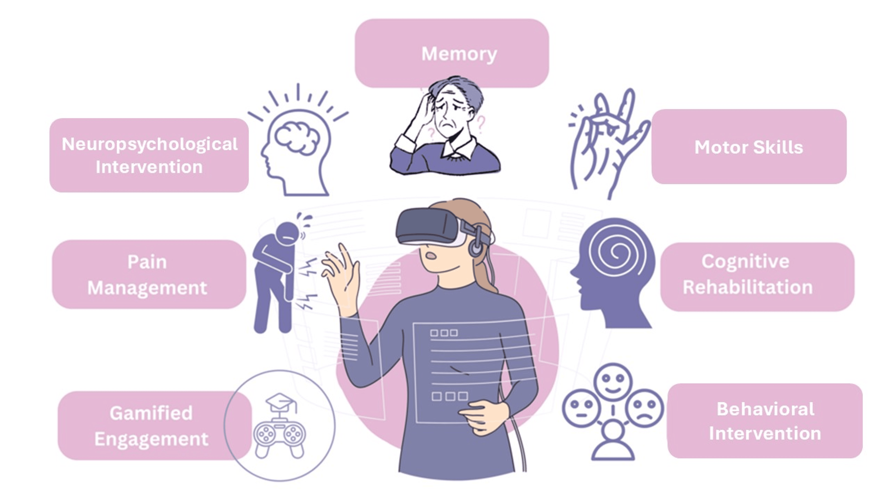
In recent years, extended reality (XR) technologies, encompassing virtual reality (VR), augmented reality (AR), and mixed reality (MR), have emerged as promising tools in neurological care. XR technologies differ from traditional therapies by offering immersive, sensory-rich environments that provide real-time feedback, enhancing therapeutic engagement and outcomes [4,5]. For example, VR is increasingly recognized for its role in cognitive research, evaluation, and rehabilitation. It allows patients to engage in realistic activities and receive precise performance measurements in a safe, controlled setting (Figure 1). Given the limitations of traditional treatments and the unique benefits XR technologies offer, this narrative review explores its potential, current applications, limitations, and future directions of XR technologies in managing chronic neurological conditions. In examining how XR can complement or enhance traditional therapies, this review aims to shed light on the evolving landscape of neurological rehabilitation and care.

Sunday, June 23, 2024

Treatment for intracerebral hemorrhage: Dawn of a new era

 This is why we need survivors in charge, we wouldn't give up. You better wait a couple of decades before you have your intracerebral hemorrhage stroke

Treatment for intracerebral hemorrhage: Dawn of a new era

Abstract

Intracerebral hemorrhage (ICH) is a devastating disease, causing high rates of death, disability, and suffering across the world. For decades, its treatment has been shrouded by the lack of reliable evidence, and consequently, the presumption that an effective treatment is unlikely to be found. Neutral results arising from several major randomized controlled trials had established a negative spirit within and outside the stroke community. Frustration among researchers and a sense of nihilism in clinicians has created the general perception that patients presenting with ICH have a poor prognosis irrespective of them receiving any form of active management. All this changed in 2023 with the positive results on the primary outcome in randomized controlled trials showing treatment benefits for a hyperacute care bundle approach (INTERACT3), early minimal invasive hematoma evacuation (ENRICH), and use of factor Xa-inhibitor anticoagulation reversal with andexanet alfa (ANNEXa-I). These advances have now been extended in 2024 by confirmation that intensive blood pressure lowering initiated within the first few hours of the onset of symptoms can substantially improve outcome in ICH (INTERACT4) and that decompressive hemicraniectomy is a viable treatment strategy in patients with large deep ICH (SWITCH). This evidence will spearhead a change in the perception of ICH, to revolutionize the care of these patients to ultimately improve their outcomes. We review these and other recent developments in the hyperacute management of ICH. We summarize the results of randomized controlled trials and discuss related original research papers published in this issue of the International Journal of Stroke. These exciting advances demonstrate how we are now at the dawn of a new, exciting, and brighter era of ICH management.

Intracerebral hemorrhage—the deadly sibling of ischemic stroke

Intracerebral hemorrhage (ICH) is caused by the rupture of cerebral vessels which results in bleeding within the brain parenchyma and/or ventricles.1 Overall, ICH comprises approximately 10–15% of all strokes worldwide, but the rates are higher in low- and middle-income countries.2 Compared to ischemic stroke, the incidence of ICH has increased in recent years and the prognosis remains poor.3,4 Current estimates predict a significant increase in the incidence of ICH in Europe related to aging and greater use of anticoagulants, with major implications for health care systems and societies.5

Treatment of ICH before 2023—widespread frustration and nihilism

For decades, treatment of ICH has been overshadowed by limited evidence and a presumed lack of effective treatment options reflected by neutral and restrictive guideline recommendations.6,7 Several randomized controlled trials of surgical treatment (i.e. different approaches to evacuation of parenchymal or intraventricular hematoma),811 blood pressure (BP) control,12,13 and hemostatic therapies,1416 resulted in either borderline significant or neutral results. The evidence was persuasive from INTERACT2,12 and stronger when pooled with other trials as part of an individual patient data meta-analysis,17 for a beneficial effect of early intensive BP lowering. Although a study-level meta-analysis of hematoma evacuation also found a potential benefit,18 there is ongoing uncertainty over which patients have the most to gain from neurosurgery along with the optimal timing and technique of intervention. Collectively, these efforts have contributed to somewhat of a negative spirit within (and outside) the stroke community, and in turn degrees of frustration and nihilism regarding treatment approaches and the perception of a uniformly poor prognosis for patients with ICH.19
 
More at link.

Saturday, June 8, 2024

Effect of Pink Noise on EEG and Memory Performance in Memory Task

 Since 30% of survivors have sleep problems, has your doctor done ONE DAMN THING about it in the last 4 years? NO? So you don't have a functioning stroke doctor, do you?

Is your doctor suggesting either of these? Never mind, way too soon for your doctor to read, understand and implement these interventions. Maybe 50 years from now. Of course your doctor needs to get this testing done on stroke survivors if they are competent at all!

Effects of saffron on sleep quality in healthy adults with self-reported poor sleep: A randomized, double-blind, placebo-controlled trial June 2020

Pink Noise Machines Improve Sleep & Fight Dementia  June 2020

 The latest here:

Effect of Pink Noise on EEG and Memory Performance in Memory Task


Abstract:

Recently, improvement of memory ability by listening noise in deep sleep has attracted extensive attention for its promising potential to prevent older adults from degrading cognitive function such as mild cognitive impairment and dementia. In particular, pink noise with 1/f-characteristics was frequently used to facilitate synchronization of neural oscillatory activities. However, the effect of pink noise on short-term memory task including memory encoding and recall during awake state has not been well investigated. We conducted a simple memory task consisting of memorization of ten fish names during which pink noise was presented and recalling them before and after mental arithmetic for seven healthy young males. Memory recall performance and amplitudes of electroencephalogram (EEG) for noise-added trials were compared with those for control trials without additive noise. The beta and gamma wave amplitudes in the noise-added trials for participants with higher memory performance in the noise-added trials than the control trials were larger than the amplitudes in the control trials by about 10 points, whereas the former amplitudes for participants with lower memory performance in the noise-added trials were smaller than the latter amplitudes. There is a possibility that pink noise could affect the memory ability through the enhancement of beta and gamma wave.
Date of Conference: 12-15 October 2021
Date Added to IEEE Xplore: 01 December 2021
ISBN Information:
Print on Demand(PoD) ISSN: 2378-8143

Monday, May 20, 2024

Functional Outcomes Associated With Blood Pressure Decrease After Endovascular Thrombectomy

 Leaders would have demanded a protocol come out of this research. But then there are NO LEADERS anywhere in stroke! Don't plan on having a stroke for at least 50 years.

Functional Outcomes Associated With Blood Pressure Decrease After Endovascular Thrombectomy

JAMA Netw Open. 2024;7(4):e246878. doi:10.1001/jamanetworkopen.2024.6878
Key Points

Question  Is a medication-induced blood pressure (BP) decrease (systolic BP <100 mm Hg) during the 24 hours after successful endovascular thrombectomy associated with poor outcomes in patients with ischemic stroke?

Findings  In a cohort study of 302 patients after successful endovascular thrombectomy, those experiencing medication-induced BP decreases exhibited a significantly lower odds of functional independence at 3 months (31.9%) compared with the no BP decrease group (49.1%), a significant difference. However, the odds of functional independence with spontaneous BP decrease did not significantly differ from those with no BP decrease.

Meaning  The findings of this study suggest that a medication-induced BP decrease during the first 24 hours after successful reperfusion with endovascular thrombectomy may be harmful for patients with acute ischemic stroke.

Abstract

Importance  The associations between blood pressure (BP) decreases induced by medication and functional outcomes in patients with successful endovascular thrombectomy remain uncertain.

Objective  To evaluate whether BP reductions induced by intravenous BP medications are associated with poor functional outcomes at 3 months.

Design, Setting, and Participants  This cohort study was a post hoc analysis of the Outcome in Patients Treated With Intra-Arterial Thrombectomy–Optimal Blood Pressure Control trial, a comparison of intensive and conventional BP management during the 24 hours after successful recanalization from June 18, 2020, to November 28, 2022. This study included 302 patients who underwent endovascular thrombectomy, achieved successful recanalization, and exhibited elevated BP within 2 hours of successful recanalization at 19 stroke centers in South Korea.

Exposure  A BP decrease was defined as at least 1 event of systolic BP less than 100 mm Hg. Patients were divided into medication-induced BP decrease (MIBD), spontaneous BP decrease (SpBD), and no BP decrease (NoBD) groups.

Main Outcomes and Measures  The primary outcome was a modified Rankin scale score of 0 to 2 at 3 months, indicating functional independence. Primary safety outcomes were symptomatic intracerebral hemorrhage within 36 hours and mortality due to index stroke within 3 months.

Results  Of the 302 patients (median [IQR] age, 75 [66-82] years; 180 [59.6%] men), 47 (15.6%)were in the MIBD group, 39 (12.9%) were in the SpBD group, and 216 (71.5%) were in the NoBD group. After adjustment for confounders, the MIBD group exhibited a significantly smaller proportion of patients with functional independence at 3 months compared with the NoBD group (adjusted odds ratio [AOR], 0.45; 95% CI, 0.20-0.98). There was no significant difference in functional independence between the SpBD and NoBD groups (AOR, 1.41; 95% CI, 0.58-3.49). Compared with the NoBD group, the MIBD group demonstrated higher odds of mortality within 3 months (AOR, 5.15; 95% CI, 1.42-19.4). The incidence of symptomatic intracerebral hemorrhage was not significantly different among the groups (MIBD vs NoBD: AOR, 1.89; 95% CI, 0.54-5.88; SpBD vs NoBD: AOR, 2.75; 95% CI, 0.76-9.46).

Conclusions and Relevance  In this cohort study of patients with successful endovascular thrombectomy after stroke, MIBD within 24 hours after successful recanalization was associated with poor outcomes at 3 months. These findings suggested lowering systolic BP to below 100 mm Hg using BP medication might be harmful.


More at link.