Changing stroke rehab and research worldwide now.Time is Brain! trillions and trillions of neurons that DIE each day because there are NO effective hyperacute therapies besides tPA(only 12% effective). I have 523 posts on hyperacute therapy, enough for researchers to spend decades proving them out. These are my personal ideas and blog on stroke rehabilitation and stroke research. Do not attempt any of these without checking with your medical provider. Unless you join me in agitating, when you need these therapies they won't be there.

What this blog is for:

My blog is not to help survivors recover, it is to have the 10 million yearly stroke survivors light fires underneath their doctors, stroke hospitals and stroke researchers to get stroke solved. 100% recovery. The stroke medical world is completely failing at that goal, they don't even have it as a goal. Shortly after getting out of the hospital and getting NO information on the process or protocols of stroke rehabilitation and recovery I started searching on the internet and found that no other survivor received useful information. This is an attempt to cover all stroke rehabilitation information that should be readily available to survivors so they can talk with informed knowledge to their medical staff. It lays out what needs to be done to get stroke survivors closer to 100% recovery. It's quite disgusting that this information is not available from every stroke association and doctors group.

Wednesday, June 14, 2023

Predictors of Discharge Destination After Stroke

NO, NO, NO! Predicting discharge destination is fucking useless to survivors. They want recovery. DO THE GODDAMNED RESEARCH THAT DELIVERS RECOVERY. Not this useless crapola.I'd have you all fired for incompetence including your mentors and senior researchers.


Predictors of Discharge Destination After Stroke

Abstract

Background

Determining the discharge destination after acute stroke care is important to prevent long-term disabilities and improve cost efficiency.

Objective

The aim of this study was to investigate where stroke patients are discharged to after acute treatment and to identify personal, social, stroke-related, and clinical predictors of discharge destination.

Methods

The present study included a secondary exploratory analysis of a prospective observational study. Patients with acute ischemic stroke, transient ischemic attack, or intracerebral hemorrhage were recruited consecutively over a 15-month period. A hierarchical multinomial logistic regression was performed to identify predictors of the primary outcome of discharge destination.

Results

We included 1026 stroke patients (48.7% female) with a mean age of 73.3 years (standard deviation 12.9 years) in the analysis. Overall, 55% of the patients were discharged home, 33% to a rehabilitation center, 3% to a residential facility, and 8% to another acute care hospital. Predictors that statistically significantly influenced the odds of the discharge destination were age, living situation pre-stroke, living location pre-stroke, stroke type, stroke severity, treatment type, and length of stay. Higher stroke severity was associated with discharge to all four inpatient facilities.

Conclusions

In line with previous research, predictors such as stroke severity and living situation pre-stroke significantly influenced the odds of the discharge destination. In contrast, pre-existing conditions and functional impairment pre-stroke had no significant impact on the primary outcome. This discrepancy could be due to a rather functional study sample before stroke and the use of clinical and patient-reported outcome measures.

Introduction

People aged 25 and above have a 25% global lifetime risk of suffering from stroke which is associated with long-term consequences and causes disabilities such as motor control impairments, cognitive and language impairments, and emotional disturbances.15
Inpatient or outpatient rehabilitative care follows acute stroke care. As patients are affected to varying degrees by disabilities after stroke, the type of follow-up care needed differs.6 For follow-up care, timely discharge to rehabilitation facilities or home with outpatient care has been shown to improve patients’ chances of recovery.7,8 In contrast, a rapid discharge without previous discharge arrangements is often associated with discontinued care and a delay in discharge is associated with increased mortality.8,9 Additionally, finding appropriate follow-up care strongly impacts the time and cost-efficiency of stroke care, which is important considering limited time resources at the hospital and high stroke care costs.9,10
In their review, Thorpe et al11 revealed that a discharge home becomes more likely with a better outcome on scoring systems for the assessment of acute stroke such as the National Institutes of Health Stroke Scale (NIHSS).12 However, they concluded that outcome measures are not sufficient to predict discharge destination. Patients with low performance scores were more likely to be discharged to rehabilitation, and patients with high performance scores were more likely to be discharged home, but discharge destination could not be predicted well for patients with mid-range scores.11 In these cases, additional factors are needed to enable the prediction of the discharge destination. In another review, support at home, living with others, being married, and living at home before stroke onset indicated a greater likelihood of a discharge home.13 Moreover, while the impact of age and sex was less clear, a better pre-stroke functional and post-stroke cognitive status increased the likelihood of being discharged home.14 In conclusion, different reviews recommend further research on age, sex, type of stroke, patient-specific biopsychosocial factors, other stroke-specific outcome measures, and global socioenvironmental determinants.11,13-15
Therefore, we aimed to assess where stroke patients are discharged after acute treatment and to identify personal, pre-stroke, stroke-related, and clinical predictors of discharge destination.
 
More at link.

New Artificial Intelligence-Integrated Electromyography-Driven Robot Hand for Upper Extremity Rehabilitation of Patients With Stroke: A Randomized, Controlled Trial

 Wouldn't work on me, that control is now dead brain. So cherry picking higher functioning survivors to make the research look good.

New Artificial Intelligence-Integrated Electromyography-Driven Robot Hand for Upper Extremity Rehabilitation of Patients With Stroke: A Randomized, Controlled Trial

Abstract

Background

An artificial intelligence (AI)-integrated electromyography (EMG)-driven robot hand was devised for upper extremity (UE) rehabilitation. This robot detects patients’ intentions to perform finger extension and flexion based on the EMG activities of 3 forearm muscles.

Objective

This study aimed to assess the effect of this robot in patients with chronic stroke.

Methods

This was a single-blinded, randomized, controlled trial with a 4-week follow-up period. Twenty patients were assigned to the active (n = 11) and control (n = 9) groups. Patients in the active group received 40 minutes of active finger training with this robot twice a week for 4 weeks. Patients in the control group received passive finger training with the same robot. The Fugl-Meyer assessment of UE motor function (FMA), motor activity log-14 amount of use score (MAL-14 AOU), modified Ashworth scale (MAS), H reflex, and reciprocal inhibition were assessed before, post, and post-4 weeks (post-4w) of intervention.

Results

FMA was significantly improved at both post (P = .011) and post-4w (P = .021) in the active group. The control group did not show significant improvement in FMA at the post. MAL-14 AOU was improved at the post in the active group (P = .03). In the active group, there were significant improvements in wrist MAS at post (P = .024) and post-4w (P = .026).

Conclusions

The AI-integrated EMG-driven robot improved UE motor function and spasticity, which persisted for 4 weeks. This robot hand might be useful for UE rehabilitation of patients with stroke.
Clinical Trial Registry Name: The effect of robotic rehabilitation using XMM-HR2 for the paretic upper extremity among hemiparetic patients with stroke.
Clinical Trial Registration-URL: https://jrct.niph.go.jp/
Unique Identifier: jRCTs032200045.

Get full access to this article

Autonomous Control of Music to Retrain Walking After Stroke

So no protocols from this research, useless for survivors to get 100% recovery on walking.

Autonomous Control of Music to Retrain Walking After Stroke

Abstract

Background

Post-stroke care(We need RECOVERY AND RESULTS instead of this lazy 'care'!) guidelines(WE NEED EXACT PROTOCOLS, not useless guidelines!) highlight continued rehabilitation as essential; however, many stroke survivors cannot participate in outpatient rehabilitation. Technological advances in wearable sensing, treatment algorithms, and care delivery interfaces have created new opportunities for high-efficacy rehabilitation interventions to be delivered autonomously in any setting (ie, clinic, community, or home).

Methods

We developed an autonomous rehabilitation system that combines the closed-loop control of music with real-time gait analysis to fully automate patient-tailored walking rehabilitation. Specifically, the mechanism-of-action of auditory-motor entrainment is applied to induce targeted changes in the post-stroke gait pattern by way of targeted changes in music. Using speed-controlled biomechanical and physiological assessments, we evaluate in 10 individuals with chronic post-stroke hemiparesis the effects of a fully-automated gait training session on gait asymmetry and the energetic cost of walking.

Results

Post-treatment reductions in step time (Δ: −12 ± 26%, P = .027), stance time (Δ: −22 ± 10%, P = .004), and swing time (Δ: −15 ± 10%, P = .006) asymmetries were observed together with a 9 ± 5% reduction (P = .027) in the energetic cost of walking. Changes in the energetic cost of walking were highly dependent on the degree of baseline energetic impairment (r =− .90, P < .001). Among the 7 individuals with a baseline energetic cost of walking larger than the normative value of healthy older adults, a 13 ± 4% reduction was observed after training.

Conclusions

The closed-loop control of music can fully automate walking rehabilitation that markedly improves walking after stroke. Autonomous rehabilitation delivery systems that can safely provide high-efficacy rehabilitation in any setting have the potential to alleviate access-related care gaps and improve long-term outcomes after stroke.

Introduction

Post-stroke neuromotor deficits result in gait asymmetries and an increased energetic cost of walking.1-3 Such deficits in walking quality often persist beyond standard rehabilitation efforts and result in reduced physical activity.4,5 Care guidelines highlight continued rehabilitation as essential to improve mobility and function and mitigate costly future morbidities6; however, most stroke survivors do not participate in outpatient rehabilitation, in part due to limited access to skilled care and disparities in transportation availability and time.7
Technological developments in high fidelity wearable sensing, automated treatment algorithms, and novel care delivery interfaces8,9 have created new opportunities for high-efficacy interventions to be delivered autonomously in any setting (ie, clinic, home, or community). We present the closed-loop control of music (Figure 1) as a novel approach to leveraging such advancements to autonomously provide highly salient and targeted, patient-tailored. Here, “closed-loop” refers to the autonomous control of music, driven by input from foot sensors as the participant walks. More specifically, the autonomous rehabilitation system integrates real-time gait sensing with music-based treatment algorithms to enable the application of auditory-motor entrainment for enhancing post-stroke walking.10
Figure 1. Patient-tailored walking rehabilitation fully automated via the closed-loop control of music. Real-time gait sensing and assessment enable music modulation algorithms to apply the mechanism-of-action of auditory-motor entrainment to fully automate patient-tailored walking rehabilitation after stroke. Continuous gait assessments individualize the rehabilitation to the user’s baseline walking abilities and facilitate safe progression.
Plainly, auditory-motor entrainment is why people can effortlessly synchronize their movements (ie, entrain) to the beat of an external rhythm.11-15 Indeed, auditory-motor neural circuits cause rhythmic motor output to be attracted to, and eventually lock on to, the frequency of an external rhythmic auditory signal.16-19 Our autonomous rehabilitation system applies the neurobiological process of auditory-motor entrainment to induce targeted and personalized changes in the post-stroke gait pattern. More specifically, the rhythm of music is purposefully modulated based on continuous, real-time gait assessments, resulting in gait training autonomously tailored to each user’s gait.
The profound effect that rhythmic auditory stimuli have on neuromotor control12,15,20-23 has led to development and study of numerous clinical interventions centered on auditory-motor entrainment. The efficacy of these interventions has been extensively studied in people post-stroke, with their ability to improve walking well-established,24-36 resulting in their recommendation37 and inclusion38 in published clinical practice guidelines. However, as with any skilled intervention, clinical benefits can only be realized if the intervention is readily accessible. The objective of this foundational study is to determine if gait training autonomously delivered by way of the closed-loop control of music has potential to improve walking quality after stroke, as measured by improvements in the energetic cost of walking and gait asymmetry.
 
More at link.

New Evidence Links Calcium Channel Blockers to Increased Risk of Glaucoma

I'll have to talk to my doctor since I'm doing Nifedipine ER  and Losartan

Calcium channel blockers include:
  • Amlodipine (Norvasc®)
  • Diltiazem (Cardiazem®, Tiazac®, Tiazac® XC )
  • Felodipine (Plendil®)
  • Nifedipine XL (Adalat XL®)
  • Verapamil (Isoptin®, Isoptin® SR, Verelan®)

New Evidence Links Calcium Channel Blockers to Increased Risk of Glaucoma

Meta-analysis shows highest glaucoma risk with CCB monotherapy, reduced IOP with beta-blockers

A photo of a man’s eye bulging from glaucoma.

Use of calcium channel blockers (CCBs), particularly cardioselective agents, had a modest but statistically significant association with glaucoma, a large meta-analysis showed.

Overall, patients with a history of CCB treatment had a 23% higher likelihood of developing glaucoma as compared with individuals who never used the antihypertensives. The likelihood almost doubled among patients who received single-agent cardioselective CCBs. In contrast, beta-blocker therapy was associated with modestly reduced intraocular pressure (IOP), which is associated with a reduced risk of glaucoma.

A number of other commonly used medications had no clear associations with glaucoma or IOP, including lipid-lowering drugs, antidepressants, and diabetes medications, reported Anthony Khawaja, MD, PhD, of University College London, and colleagues in the European Eye Epidemiology (E3) Consortiumopens in a new tab or window.

"While our novel findings require further studies to determine whether the associations are causal, these findings will be of interest to physicians caring for glaucoma patients with systemic comorbidities," the authors stated in Ophthalmologyopens in a new tab or window.

"A potentially harmful association of CCBs for glaucoma is particularly noteworthy, as this is a commonly prescribed class of medication," they added. "If further studies confirm a casual nature for this association, this may inform alternative treatment strategies for hypertensive patients with, or at risk of, glaucoma."

Adds to Existing Evidence

The findings add to evidence from previous studies showing that systemic beta-blockers lower IOP, and at least one prior study has shown an association between CCBs and glaucoma, said Roma Patel, MD, MBA, of Baylor College of Medicine and Ben Taub Hospital in Houston, and a clinical spokesperson for the American Academy of Ophthalmology.

"The study's contribution is one of power," Patel told MedPage Today via email. "The dataset used for this analysis represented over 143,000 individuals with glaucoma. Because of the size of population studied, this study was able to find that an even stronger association between CCBs with direct cardiac effects and glaucoma versus analysis of all CCBs and glaucoma. Perhaps this will help researchers figure out if there is a causal link. We know that glaucoma is ultimately related to dysfunction and death of the retinal ganglion cells. We don't concretely know the effects of calcium blockade at that level so it poses a good research question."

With respect to implications for clinical practice, Patel added, "I believe it's fair to tell patients there is a known association for calcium channel blockers and glaucoma but we don't fully understand why and there is no evidence of a causal relationship. If the patient chooses to discuss with their PCP [primary care physician] or cardiologist about a different treatment regimen for their cardiac issue, that is up to the patient. But I am not encouraging that conversation with my patients."

"I do encourage my patients to talk to their PCP or cardiologist about avoiding evening anti-hypertensive medications because we do know that low blood pressure overnight can lead to glaucoma progression," she said. "It's important to remember that the eye lives within the human body and all of our systems are connected. But if the cardiologist feels that a certain medicine is warranted, I recommend we follow that recommendation because we all know that the eye can't live without the heart."

Glaucoma is the leading cause of irreversible vision impairment worldwide, and IOP currently is the only modifiable risk factor for glaucoma onset and progression, Khawaja and co-authors noted. Several types of medication are known or suspected of modulating glaucomaopens in a new tab or window risk by affecting optic nerve head perfusion, retinal ganglion cell survival, and aqueous humor outflow facility.

A study based on U.S. health claimsopens in a new tab or window suggested that selective serotonin reuptake inhibitors were associated with a reduced risk of primary open-angle glaucoma and CCBs with an increased risk. Other medications that may influence glaucoma riskopens in a new tab or window include beta-blockers, metformin, statins, and bupropion. Additionally, some medications have been associated with higher IOPopens in a new tab or window, including angiotensin converting enzyme (ACE) inhibitors, angiotensin receptor blockers (ARBs), statins, and sulfonylureas.

"For many of the cited associations, there have been inconsistent findings between studies, and few studies have accounted for polypharmacy or important confounders," Khawaja and colleagues noted.

Clarifying Study

To continue the line of research, investigators in the E3 Consortium performed a meta-analysis of 11 European cohort studies involving a total of 143,240 participants, all of whom were included in analyses of glaucoma associations. Analyses of IOP included 47,177 participants. Medication use encompassed multiple types of antihypertensives, lipid-lower medications, antidepressants, and antidiabetic medications (limited only to participants with diagnosed diabetes). For antihypertensives, the investigators distinguished monotherapy from combination therapy.

Multivariable analyses showed that any CCB use was associated with a glaucoma OR of 1.23 (95% CI 1.08-1.39) versus no use. Patients who received CCB monotherapy had a glaucoma OR of 1.96 (95% CI 1.23-3.12). No other medications were clearly associated with glaucoma, the authors reported. Systemic beta-blockers were associated with marginally lower IOP (-0.33 mmHg, 95% CI -0.57 to -0.08). Monotherapy with selective beta-blockers was associated with a 0.45 mmHg reduction in IOP (95% CI -0.74 to -0.16) and non-selective agents with a 0.54 mmHg reduction in IOP (95% CI -0.94 to -0.15).

High-ceiling diuretics (such as furosemide and torasemide/torsemideopens in a new tab or window) had a "suggestive" association with lower IOP (-0.30 mmHg, 95% CI -0.47 to -0.14), but not when used as monotherapy.

None of the other medications included in the analysis had associations with IOP.

  • author['full_name']

    Charles Bankhead is senior editor for oncology and also covers urology, dermatology, and ophthalmology. He joined MedPage Today in 2007. Follow

Disclosures

The study was supported by multiple governmental, nonprofit, and philanthropic organizations.

Khawaja disclosed relationships with AbbVie, Aerie, Google Health, Novartis, Reichert, Santen, and Thea.

Patel disclosed no relevant relationships with industry.

Primary Source

Ophthalmology

Source Reference: opens in a new tab or windowVergroesen JE, et al "Association of systemic medication use with glaucoma and intraocular pressure: The E3 Consortium" Ophthalmol 2023; DOI: 10.1016/j.ophtha.2023.05.00

Impact of orally administered cannabidiol (CBD) on fitness and health among healthy adult individuals

If you have a functioning stroke doctor and hospital research on this will be initiated by them to test this on stroke survivors.  But I doubt that will occur.

Impact of orally administered cannabidiol (CBD) on fitness and health among healthy adult individuals

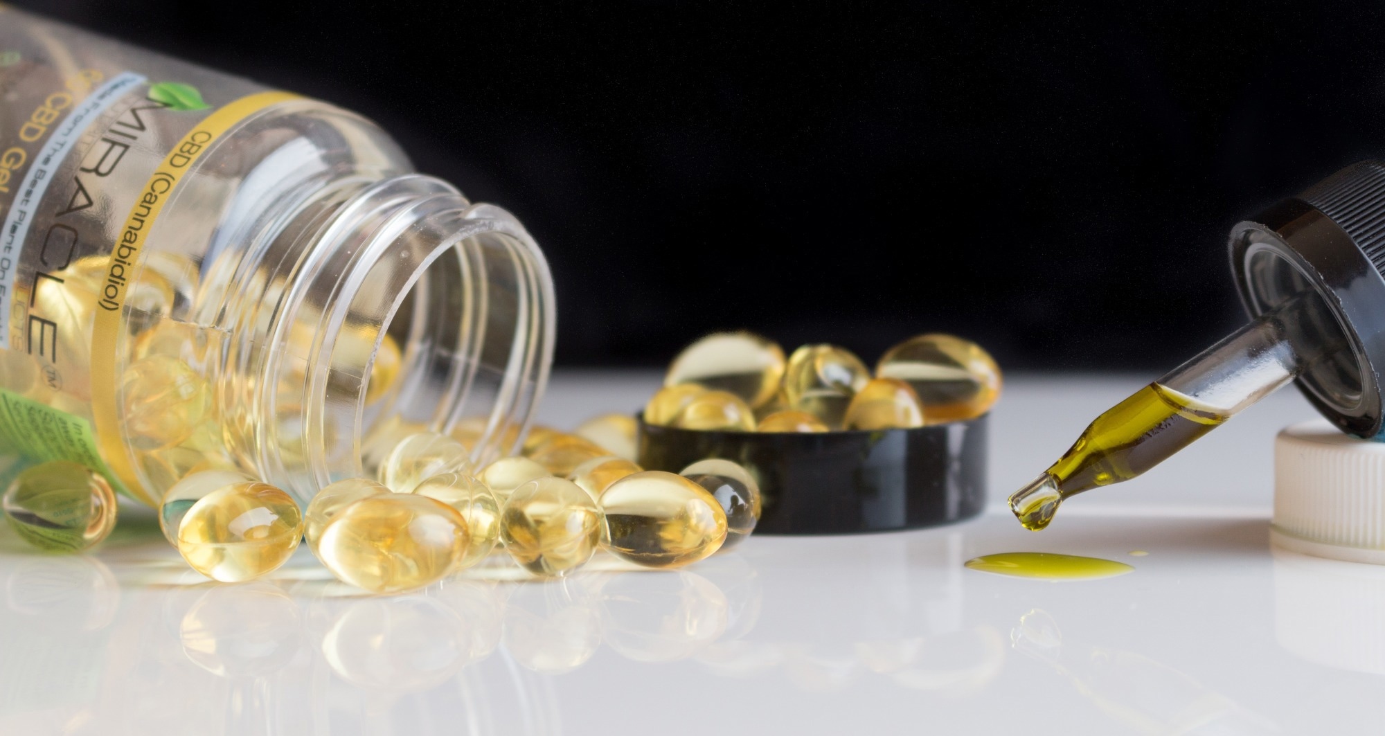
In a recent study published in the Nutrients Journal, researchers evaluated the effects of oral cannabidiol (CBD) administration on the health and fitness of healthy adults.

Study: Effects of Oral Cannabidiol on Health and Fitness in Healthy Adults: An 8-Week Randomized Trial. Image Credit: KimberlyBoyles/Shutterstock.comStudy: Effects of Oral Cannabidiol on Health and Fitness in Healthy Adults: An 8-Week Randomized Trial. Image Credit: KimberlyBoyles/Shutterstock.com

Background

CBD, a non-intoxicating phytocannabinoid extracted from Cannabis sativa L., has shown significant therapeutic benefits in managing neuroinflammatory disorders, including anxiety, depression, Alzheimer's disease, and Parkinson's disease.

Regular CBD usage has been encouraged to improve fitness. Studies have reported that CBD may alter body composition by impacting glucose metabolism and can increase maximal oxygen consumption (VO2 max) with no impact on other parameters of cardiovascular health.

CBD use has also improved learning and memory and increased blood flow in the cerebrum of individuals with cognitive decay.(This sounds very useful to us.) CBD can also reduce interleukin-6 (IL-6) levels among adults with cocaine usage disorders and lower pro-inflammatory gene expression and C-reactive protein (CRP) levels among individuals infected with the human immunodeficiency virus (HIV).

However, contradictory findings have been reported, warranting further research, including randomized controlled trials (RCTs).

About the study

In the present double-blinded, placebo-controlled RCT, researchers investigated the impact of oral CBD use over eight weeks on the health of adult individuals, particularly on health-associated fitness, cognitive health, body composition, physical exercise patterns, psychological well-being, and C-reactive protein (CRP) levels.

The trial included 48 individuals aged 18 to 50 years, who were randomly allocated to receive oral capsules of 50.0 milligrams of CBD (n=23) or 225.0 milligrams of medium-chain triglyceride (MCT) as a placebo (n=25) daily over eight weeks post-dinner and before sleeping.

The individuals completed four visits before and after the intervention, and assessments, including eight-hour fasted blood draws, fitness, body composition, physical exercise, and self-documented surveys, were performed.

All individuals were provided wearable activity trackers (Fitbits) to assess physical activity. The peak value for uptake of oxygen (VO2 peak) in relative terms was calculated on the second visit to evaluate aerobic fitness; a 30.0-second Wingate test was performed to assess power output and evaluate anaerobic fitness on the third visit; and bench press and back squat one-repetition maximums (1RMs) were performed during the fourth visit to assess muscle strength.

Adverse events (AEs) were documented. Primary study outcomes included relative VO2 peak, body fat percentage (BF%), body mass (BM) and lean BM (LBM), peak power (PP) and relative PP (RPP), anaerobic fatigue (AF), mean power (MP), relative MP (RMP), bench press and back squat 1RM, a one-week mean daily step count, self-documented psychological well-being and cognitive function scores, and serological CRP concentrations.

Individuals who abstained from cannabis [CBD and/or tetrahydrocannabinol (THC)] use for six weeks before study initiation and had no history of chronic alcohol consumption or drug usage were included.

Individuals diagnosed with metabolic, neurological, cardiovascular, or mood disorders; pregnant or lactating mothers; and individuals who could not adhere to the intervention for eight weeks were excluded from the analysis.

Cognitive function was evaluated using the National Institute of Health (NIH) Patient-Reported Outcomes Measurement Information System (PROMIS) Cognitive Function-Abilities-Short Form 8a.

Serological CRP levels were determined using enzyme-linked immunosorbent assays (ELISA). Muscular strength was assessed using the National Strength and Conditioning Association (NSCA) guidelines.

Results

The mean values for participant age, height, and BM were 25 years, 171 cm, and 73 kg, respectively. No significant differences were observed between groups regarding body composition, aerobic fitness, muscular strength, physical activity, cognitive health, psychological well-being, or serological resting CRP concentrations.

However, the placebo group experienced a 9.6% reduction in PP and a 6.6% reduction in RPP compared to the intervention group participants.

In contrast, CBD recipients experienced no changes in PP or RPP. No severe adverse events were reported. Combining both groups showed LBM ranging between 37 and 84 kg, BF% ranging between 8.2% and 40%, relative VO2 peak ranging between 28 and 62 mL/kg/min, bench press 1RM ranging between 25 and 161 kg, and back squat 1RM ranging between 39 and 166 kg.

Further, PP ranged between 372 and 1148 W, RPP ranged between 6.0 and 13 W/kg, MP ranged between 288 and 804 W, RMP ranged between 4.2 and 8.3 W/kg, and AF ranged between 43 and 71%. The one-week mean daily step count was 11,415 steps. Cognitive function T-scores ranged between 30 and 64, and cognitive function ability T-scores ranged between 37 and 67.

Concerning psychological well-being, autonomy scores ranged between 8.0 and 21.0; environmental mastery scores ranged between 7.0 and 21.0; personal growth scores ranged between 15.0 and 21.0; scores for positive relations with others ranged between 10.0 and 21.0; purpose in life scores ranged between 11.0 and 21.0; and self-acceptance scores ranged between 8.0 and 21.0. The mean CRP level in serum was 1.40 mg per liter, ranging between 0.10 and 8.8 mg per liter.

Conclusions

Overall, the study findings showed that the regular intake of 50.0 mg of CBD for eight weeks may prevent reductions in anaerobic power with time, likely due to an increase in the levels of antioxidants such as glutathione peroxidase and superoxide dismutase and a decrease in free radicals such as reactive oxygen species (ROS).

However, long-term CBD use did not improve aerobic fitness, body composition, mental health, physical activity, inflammation, or psychological well-being among adults.

Journal reference:

Level of fat within the body's muscle may lead to risk of cognitive decline while aging


What is your confidence level that your doctor has this equipment and the protocols to measure this and then the protocols to reduce muscle fat to healthy levels? I would say zero!

How is muscle fat composition measured?

A DXA or DEXA scan is the most accurate and advanced form of body composition analysis available. It uses x-ray technology to scan your body and provides a detailed assessment of how much muscle mass and fat mass you have (down to the pound), and exactly where fat and muscle is stored on your body.Mar 23, 2021

Level of fat within the body's muscle may lead to risk of cognitive decline while aging

New research reveals that the level of fat within the body's muscle-;or muscle adiposity-;may indicate a person's likelihood of experiencing cognitive decline as they age.

In the study published in the Journal of the American Geriatrics Society, 5-year increase in fat stored in the thigh muscle was a risk factor for cognitive decline. This risk was independent of total weight, other fat deposits, and muscle characteristics (such as muscle strength or mass) and also independent of traditional dementia risk factors.

Investigators assessed muscle fat in 1,634 adults 69–79 years of age at years 1 and 6 and evaluated their cognitive function at years 1, 3, 5, 8, and 10. Increases in muscle adiposity from year 1 to year 6 were associated with faster and more cognitive decline over time. The findings were similar for Black and white men and women.

Our data suggest that muscle adiposity plays a unique role in cognitive decline, distinct from that of other types of fat or other muscle characteristics. If that is the case, then the next step is to understand how muscle fat and the brain 'talk' to each other, and whether reducing muscle adiposity can also reduce dementia risk."

Caterina Rosano, MD, MPH, Study Corresponding Author, School of Public Health, University of Pittsburgh

Source:
Journal reference:

Rosano, C., et al. (2023) Increase in skeletal muscular adiposity and cognitive decline in a biracial cohort of older men and women. Journal of the American Geriatrics Society. doi.org/10.1111/jgs.18419.


Unlocking the fountain of youth: Diet and exercise have a remarkable impact on cognition in older adults

Still pretty useless, just guidelines; NOT EXACT PROTOCOLS.

Unlocking the fountain of youth: Diet and exercise have a remarkable impact on cognition in older adults

Given the vast number of people in the United States who are approaching 65 years of age, there is a need for interdisciplinary research on factors influencing the trajectory of cognition and brain aging in older adults. In a recent review published in Nutrients, researchers examine the collective effect of diet and exercise interventions on age-related cognition and brain health changes. 

Study: Impact of Diet and Exercise Interventions on Cognition and Brain Health in Older Adults: A Narrative Review. Image Credit: Isarat / Shutterstock.com Study: Impact of Diet and Exercise Interventions on Cognition and Brain Health in Older Adults: A Narrative Review. Image Credit: Isarat / Shutterstock.com

Age-related cognitive decline

Reduced processing speed is one of the major cognitive deficits observed in older adults, in addition to impaired semantic and episodic memory. Likewise, working memory that actively maintains information in the short term to enable goal-directed decision-making also declines because of aging.

Reduced working memory results in a corresponding reduction in executive functions in advanced age. Due to structural and functional changes in the brain, aging also affects an individual's crystallized and fluid intelligence.

Structural changes in the brain associated with aging include reduced gray matter volume and cortical thickness. Gray matter volume, a measure of neuronal and glial cell bodies, declines in volume within multiple brain regions, including the medial temporal lobe of the hippocampus and entorhinal cortex during aging. Specifically, age-related reduction of hippocampal volume, resulting from neuronal cell loss and a decrease in neurogenesis, is associated with decreased cognitive performance on memory, spatial learning, and emotional regulation tasks. 

Effect of diet and exercise on aging 

It is crucial to understand the relationship between nutrient consumption and neuronal function, neurometabolic processes, and cognitive decline. 

There is growing evidence that nutrients from various foods across multiple food groups have synergistic effects beyond the effects of individual nutrients. For example, the absorption of vitamins from green salad improves when served with olive oil and vinegar rather than a fat-free ranch dressing. 

To date, the Mediterranean diet (MeDi), Dietary Approaches to Stop Hypertension (DASH), and Mediterranean-DASH Intervention for Neurodegenerative Delay (MIND) diets are most frequently referenced in the literature. Each of these diets appears to improve neurological signs of aging, including cell membrane and vascular integrity, inflammation, resolution, and oxidation, as well as lipid and glucose metabolism.

Considerable evidence suggests that the MeDi and MIND diets are associated with both general and domain-specific facets of cognitive ability. For example, several studies have associated the MeDi diet with attention and long-term memory. Likewise, adherence to the MIND diet is positively associated with visuospatial ability, perceptual speed, and executive function.

The impact of the ketogenic diet (KD) and intermittent fasting (IF) on cognition and brain function have also been widely studied, as weight management diets appear to improve cognitive functioning. Likewise, weight loss achieved through bariatric surgery enhances attention, memory, and executive function.

In addition to diet, exercise positively affects cognition, as demonstrated by a previous study in which overweight and obese adults were enrolled in a one-year behavioral weight loss intervention. These patients were on an energy-restricted diet, an energy-restricted diet with 150 minutes of moderate-intensity exercise every week, or an energy-restricted diet with 250 minutes of exercise every week. 

Post-intervention, weight markedly decreased across both groups. More specifically, the high-exercise group improved their performance on Iowa Gambling Task (IGT) reward relative to the other two study groups. Overall, more exercise with behavioral weight loss regimens had an additional benefit on executive functioning, even with no weight loss benefits.

Endurance exercise typically encompasses walking, jogging, running, swimming, and cycling, with walking being the most practiced form among older adults. Higher endurance fitness levels are associated with less age-related brain volume decline. 

Randomized controlled trials examining the role of endurance exercise on cognition have led to mixed results. However, all evidence suggests that endurance exercise in older adults improved cognitive performance, visual attention, and memory, promoted brain plasticity, and weakened hippocampal atrophy.

Similarly, a recent systematic review found that resistance training positively affected older adults' executive and global cognitive functions. This form of exercise also positively impacted memory, albeit weakly, and did not significantly improve attention. Additionally, tri-weekly, as compared to biweekly resistance training, positively affected general cognitive abilities.

No evidence of an interference effect of aerobic and resistance training has been reported. However, literature comparing resistance or combined exercise to a non-exercise control is limited. Thus, it remains unclear which exercises should be prescribed to maintain and enhance cognition and brain health among older adults.

Behavioral interventions like yoga appear to mitigate age-related and neurodegenerative decline. In one review examining the effects of practicing yoga on brain structures, function, and cerebral blood flow, yoga positively affected the structure and function of the hippocampus, prefrontal and cingulate cortex, amygdala, and neuronal networks. 

A recent literature review evaluating the effect of tai chi, a traditional Chinese martial art, on brain structure and neurobehavior changes found that this form of exercise also increased cortical grey matter volume, improved neural activity and homogeneity, and increased neural connectivity in the frontal, temporal, and occipital lobes, cerebellum, and thalamus.

Conclusions

All diets evaluated in this review addressed factors associated with aging and neurodegenerative diseases like Alzheimer's disease and related dementias (ADRD). Depending on the type and intensity, exercises were found to positively affect brain vascularization, neurotransmitter regulation, growth factors, and neurogenesis.

Journal reference:
  • Key, M. N., & Szabo-Reed, A. N. (2023). Impact of Diet and Exercise Interventions on Cognition and Brain Health in Older Adults: A Narrative Review. Nutrients 15(11);2495. doi:10.3390/nu15112495

Tuesday, June 13, 2023

Antihistamine Sparks Hope for Myelin Repair in Multiple Sclerosis

So the implication is that myelin is damaged in your stroke so your doctor needs protocols to repair that damage. Will this do it?

Didn't your doctor already have this done 6 years ago?

Clemastine rescues myelination defects and promotes functional recovery in hypoxic brain injury December 2017

 Individuals with higher levels of physical activity after stroke show comparable patterns of myelin to healthy older adults

The latest here:

 Antihistamine Sparks Hope for Myelin Repair in Multiple Sclerosis

Summary: A new study paves the way for potential treatments for Multiple Sclerosis (MS) by identifying the OTC antihistamine, clemastine, as an effective drug for brain repair. Utilizing a newly-developed MRI scan technique, scientists were able to observe and measure the impact of clemastine on brain myelin levels.

The study offers the first documented example of brain repair for a chronic neurological condition through MRI. These results are expected to set a standard for future research into myelin-rebuilding therapies.

Key Facts:

  1. Clemastine, an over-the-counter antihistamine, has been identified as a potential therapy for MS, with observed increases in myelin water fraction indicating myelin repair.
  2. A unique MRI technique was used to measure changes in myelin levels before and after the drug was administered, setting a new standard for tracking myelin recovery.
  3. Beyond its potential impact on MS, clemastine is also being considered for use in treating brain injury in premature infants, who often experience myelin damage.

Source: UCSF

A decade after UC San Francisco scientists identified an over-the-counter antihistamine as a treatment for multiple sclerosis, researchers have developed an approach to measure the drug’s effectiveness in repairing the brain, making it possible to also assess future therapies for the devastating disorder.

The researchers, led by physician-scientist Ari Green, MD, who together with neuroscientist Jonah Chan, Ph.D., first identified clemastine as a potential MS therapy, used MRI scans to study the drug’s impact on the brain of 50 participants in a clinical study.

In MS, patients lose myelin, the protective insulation around nerve fibers. This myelin loss triggers delays in nerve signals, leading to weakness and spasticity, vision loss, cognitive slowing and other symptoms.

First in Carolinas: MUSC Health treats stroke patient with vagus nerve stimulation

 Wow, admitting incompetence in how long it took to bring vagus nerve stimulation into their stroke practice. And you haven't fired the board of directors for incompetence yet?

First in Carolinas: MUSC Health treats stroke patient with vagus nerve stimulation

June 13, 2023
A man wearing a long sleeved red shirt and glasses sits across a table from a woman wearing a white striped sweater. He has a device attached to his right hand. She has a computer screen in front of her.
Occupational therapist Alanna Herman watches as stroke survivor Billy Orbach tries to use his hand to grasp and release items during his therapy session at MUSC Health Neurologic Rehabilitation Institute. Photo by Sarah Pack

Billy Orbach, a former president of a steel parts company, is still hard at work. It’s just that his focus is narrower these days. Orbach, the first person in the Carolinas to receive a Vivistim implant to stimulate his vagus nerve to help him recover from a stroke, wants his right hand to work properly again.

“I used to golf three times a week, and I have a goal of getting back to that,” he said.

There’s no question about his determination. When Orbach woke up one morning in 2019 feeling like he couldn’t keep his balance, he didn’t let it stop him, his wife Carolyn said. He was in their home in Warren, Ohio, while she was in Ravenel, South Carolina, supervising construction of the home to which they planned to retire.

“What did he do? He took a shower, drove to work, went to a meeting. His secretary kept saying, ‘Billy, something's wrong. Something's wrong.’ And so at about 10 o'clock, he sat down and was eating an apple, and she just came in there and said, ‘You're going to the hospital.’ A coworker drove him to the hospital, and when they got there, he went to the bathroom and collapsed, clocked out. And that was it,” she said. 

Closeup of a man's hands. His right hand is wearing a black glove. His left, ungloved, is holding a wooden block up to the right hand. 
The fingers of Orbach's right hand tend to curve inward since his stroke. He's using vagus nerve stimulation to try to get a better range of motion – and maybe, one day soon, hold a golf club again. Photo by Sarah Pack

Her husband had suffered a stroke on the left side of his brain that affected his right arm and leg. Four years later, some of the effects linger. But that hasn’t kept the Orbachs from enjoying life. They now live in Ravenel with their kids and four grandchildren nearby. 

Since their arrival, Billy has been a patient at MUSC Health as he works to regain some of what the stroke cost him. That’s where he connected with neurosurgeon Nathan C. Rowland, M.D., Ph.D., and the rehab team that deemed Orbach the top candidate for receiving the first vagus nerve stimulation device for a stroke patient in the Carolinas.

So what is the vagus nerve? “It’s one of 12 cranial nerves,” Rowland said.

Four men wearing blue scrubs smile while standing together. 
Dr. Nathan Rowland, second from right, and from left: Deron Allen, Rob Hadley and Lionel Dacpano of Vivistim. Photo provided

“In antiquity, the vagus was known as the wandering nerve because it travels all the way from the brainstem down to other internal organs. When the brain needs to send messages to the heart and the gut, for instance, it uses the vagus nerve to do so. However, if you artificially stimulate the vagus nerve itself, you can have beneficial effects the other way around, that is, on the brain. That is what this form of vagus nerve stimulation is attempting to do.”

Orbach was eager to give it a try. “They put the coil in and wrapped the nerve six times,” he said, pointing to his neck. An incision mark showed where Rowland and his team inserted the electrode that connects to a battery-operated square shaped device under the skin of his chest. 

After the surgery with Rowland, Billy began working with a team of occupational therapists at the MUSC Health Neurologic Rehabilitation Institute. They have special training to operate the device. 

Illustration of a man. It highlights his brain. The words motor cortex are beside his head. A closeup on the right side shows the vagus nerve and the spiral cuff lead. Illustration also shows the lead and a battery operated device in his chest. 
Illustration of the vagas nerve stimulation system for chronic stroke patients. Image courtesy of Vivistim

During a recent morning appointment, Alanna Herman, one of Billy’s occupational therapists, worked with him on grasp and release using a stroke therapy glove, as well as electrical stimulation to his forearm, to allow for greater movement. As Orbach worked on straightening his fingers, Herman stimulated his vagus nerve to drive neuroplasticity, the ability of the brain to promote neural recovery. 

Each time the device is activated, it stimulates the patient’s brain to create new pathways, bypassing the damaged stroke area. Herman said by using this combination of techniques, early data has shown modest improvements in the functional scales and assessments that occupational therapists use to measure performance in chronic stroke patients undergoing rehabilitation. 

“The glove helps to keep his fingers extended so he can do more of the repetitive movements. We're trying to isolate his ability to grasp and release,” Herman said. “We are excited to see how beneficial Vivistim becomes as we use it as a supplemental tool to the evidence-based principles of neurorehabilitation performed as part of an occupational therapy plan.”

For additional practice, Orbach does exercises at home, activating the device himself. 

Man wearing a red shirt and glasses raises his gloved right hand. A woman sits across from with a computer screen in front her and wires connecting the man's glove with a monitoring system. 
Orbach tries to pick up and release a golf ball during a vagus nerve stimulation session. His 2019 stroke has had lingering effects. Photo by Sarah Pack

This new option to help stroke survivors is huge, Rowland said. “Over 800,000 strokes occur every year in this country, and at any one time, there are about 6.5 million survivors with what is considered chronic stroke, or people who survive six or more months after the initial stroke. And, unfortunately, the chronic period is when rehabilitation experts believe the brain is less prone to experience improvements.”

That’s why he was happy to be able to offer vagus nerve stimulation to Orbach. 

Nathan Rowland Neurosurgery 
Dr. Nathan Rowland

The neurosurgeon, who is also an associate professor at the Medical University of South Carolina, said the new technology is a good fit for MUSC Health, which has been recognized by The Joint Commission and the American Heart Association/American Stroke Association as a Comprehensive Stroke Center.

That means it can treat patients with the most complex strokes, including offering advanced imaging capabilities, 24/7 availability of specialized treatments and staff with the training and competencies to provide the most advanced level of stroke care achievable.

MUSC Health also offers the latest stroke rehabilitation options, such as the vagus nerve implant. “Most patients have never heard of this until we mention it as an option. They've been told by other physicians or care providers that there's nothing else that can be done. The team at MUSC wants to make sure they understand that, yes, there is hope,” Rowland said.

His team asks stroke patients who show potential for vagus nerve stimulation about medications they’re on, how long it’s been since they had a stroke and what kind of therapies they’ve had. The team also looks at hand and leg function. The implant is covered by insurance in South Carolina.

Orbach encouraged other stroke patients to do everything they can to get better. He’s thrilled to have the vagus nerve implant. “I feel blessed that I was given the first opportunity. It’s pretty amazing. Anything to help me get better.”

Rowland has high hopes for him. “I have no doubt whatsoever he’ll be able to swing a golf club again. And, when he does, we will be first in the crowd to cheer him on."


7 Depression Treatments You Might Not Know About by Debbie Hampton

 Before your doctors goes down one of these routes ask them why they don't have the best solution for post stroke depression. Namely 100% recovery protocols. If they haven't figured out that is the best way to prevent depression you don't have a functioning stroke doctor.

RUN AWAY!

7 Depression Treatments You Might Not Know About