Use the labels in the right column to find what you want. Or you can go thru them one by one, there are only 33,563 posts. Searching is done in the search box in upper left corner. I blog on anything to do with stroke. DO NOT DO ANYTHING SUGGESTED HERE AS I AM NOT MEDICALLY TRAINED, YOUR DOCTOR IS, LISTEN TO THEM. BUT I BET THEY DON'T KNOW HOW TO GET YOU 100% RECOVERED. I DON'T EITHER BUT HAVE PLENTY OF QUESTIONS FOR YOUR DOCTOR TO ANSWER.
Changing stroke rehab and research worldwide now.Time is Brain!trillions and trillions of neuronsthatDIEeach day because there areNOeffective hyperacute therapies besides tPA(only 12% effective). I have 523 posts on hyperacute therapy, enough for researchers to spend decades proving them out. These are my personal ideas and blog on stroke rehabilitation and stroke research. Do not attempt any of these without checking with your medical provider. Unless you join me in agitating, when you need these therapies they won't be there.
What this blog is for:
My blog is not to help survivors recover, it is to have the 10 million yearly stroke survivors light fires underneath their doctors, stroke hospitals and stroke researchers to get stroke solved. 100% recovery. The stroke medical world is completely failing at that goal, they don't even have it as a goal. Shortly after getting out of the hospital and getting NO information on the process or protocols of stroke rehabilitation and recovery I started searching on the internet and found that no other survivor received useful information. This is an attempt to cover all stroke rehabilitation information that should be readily available to survivors so they can talk with informed knowledge to their medical staff. It lays out what needs to be done to get stroke survivors closer to 100% recovery. It's quite disgusting that this information is not available from every stroke association and doctors group.
Tuesday, August 26, 2025
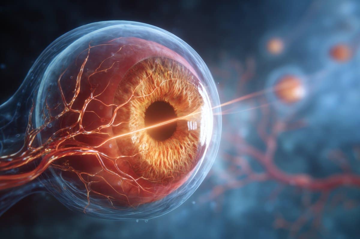
Eye Exams Could Spot Alzheimer’s Decades Before Symptoms Appear
Do you really think your competent? doctor
has enough functioning brain cells to implement this and create EXACT
PROTOCOLS to prevent Alzheimer's post stroke? I don't.
Your doctor knew about this almost a year ago. WAS ANYTHING DONE? NO? So, your doctor is fucking incompetent?
Summary: Eye exams could help detect Alzheimer’s
disease years before symptoms arise. By studying mice with a common
genetic mutation, researchers found abnormal changes in retinal blood
vessels that paralleled brain changes linked to dementia risk.
Because
the retina functions as an accessible extension of the brain, these
findings highlight its potential as a powerful biomarker. The research
paves the way for routine eye exams to aid in early diagnosis and
intervention for Alzheimer’s and related conditions.
Key Facts
Retina as Brain Window: The retina shares tissue with the brain, making it a reliable site to track neurological health.
Genetic Risk Factor: Mice with the MTHFR677C>T mutation showed early retinal vessel abnormalities tied to dementia.
Clinical Potential: Routine eye exams could one day detect Alzheimer’s risk 20 years before symptoms appear.
Source: Jackson Laboratory
Within
the next few years, doctors may be able to spot signs of Alzheimer’s
disease and other dementias using routine eye exams well before symptoms
appear, a new study suggests.
The research, recently published in Alzheimer’s & Dementia,
links abnormal changes in the tiny blood vessels of the retinas of mice
with a common genetic mutation known to increase Alzheimer’s disease
risk.
Even
though more studies are needed to gain a deeper understanding of how
vascular health in the retina affects the risk of dementia, the new
insights have important implications for people with this genetic
factor, Reagan said. Credit: Neuroscience News
The findings build on previous work from the same group at The
Jackson Laboratory (JAX), which found similar vascular changes in mice’s
brains and linked abnormalities in specific retinal cells to early
dementia risk, strengthening the case that the retina is a powerful
biomarker for Alzheimer’s disease and other dementias.
“If you’re
at an optometrist or ophthalmologist appointment, and they can see odd
vascular changes in your retina, that could potentially represent
something that is also happening in your brain, which could be very
informative for early diagnostics,” said Alaina Reagan, a neuroscientist
at The Jackson Laboratory (JAX) who led the work with Gareth Howell,
professor and Diana Davis Spencer Foundation Chair for Glaucoma Research
at JAX who spearheaded the previous study.
Because the retina
is part of the central nervous system, scientists often see it as an
extension of the brain that shares essentially the same tissue. That’s
why changes in retinal blood vessels can offer early clues about brain
health and diseases like Alzheimer’s, Reagan said.
“Your retina is
essentially your brain, but it’s much more accessible because your
pupil is just a hole, and we can see tons of stuff,” Reagan explained.
“All
the cells are very similar, all the neurons are quite similar, all the
immune cells are quite similar, and they behave similarly under pressure
if you’ve got a disease.”
The team studied mice with a mutation called MTHFR677C>T,
which is found in up to 40% of people. They found that the mice’s
retinas had twisted vessels, narrowed and swollen arteries, and less
vessel branching as early as six months of age.
This reflects
similar changes in the brain linked to poor blood flow and increased
risk of cognitive decline. Vessels that appear more twisted and looped
than normal can signal problems with hypertension, as the narrowing
tissue limits nutrient and oxygen transport, Reagan said.
“We
can see these wavy vessels in the retinas, which can occur in people
with dementia,” Reagan said. “That speaks to a more systemic problem,
not just a brain- or retina-specific problem. It could be a blood
pressure problem affecting everything.”
In 2022, a study by the same group revealed similar vascular changes in the brains of mice with the same MTHFR677C>T mutation, highlighting the link between vascular health in the retina that resembles human disease.
“These
mice have fewer vessels in their cortex and reduced blood flow to their
brains,” Reagan said. “These changes are subtle, but they are there.”
The
team also discovered changes in protein patterns in both the brain and
retina. They found disruptions in how cells produce energy, remove
damaged proteins, and maintain the structure and support of blood
vessels, offering important clues about how the MTHFR677C>T mutation affects the eye.
The
results also support a growing theory that blood vessel health plays a
central role in neurodegenerative diseases, Reagan said.
“A lot of
these molecular changes are happening in conjunction, which suggests
these systems in brain and retinal tissue are working in tandem,” she
said.
Even though more studies are needed to gain a deeper
understanding of how vascular health in the retina affects the risk of
dementia, the new insights have important implications for people with
this genetic factor, Reagan said.
For example, the study also
captured the influence of sex and age, with female mice showing worse
outcomes. By 12 months, they showed reduced vessel density and
branching, highlighting progressive vascular changes. Similarly, women
develop dementia more often than men, according to the World Health
Organization.
To see if the link between the mutation and
vascular changes occurs in humans, as well as whether the new insight
could be used in eye exams, the team is partnering with clinicians and
dementia care specialists at Northern Light Acadia Hospital in Bangor,
Maine.
The idea is to study not
just one cause or solution for Alzheimer’s and other dementias, as these
conditions depend on many different genetic and environmental factors,
but to learn more about how eye health adds to overall risk for these
diseases. If the clinicians know which signs to look for, they could
communicate those risk factors to patients and recommend further tests.
“Most people over 50 have some kind of vision impairment and get checked annually for prescription changes,” Reagan said.
“Are
they more at risk if they have these vascular changes, and is that a
point when doctors could start mitigating brain changes? That could be
20 years before cognitive damage becomes noticeable to patients and
their families.”
Other authors are Michael MacLean, Travis L. Cossette, and Gareth R. Howell from The Jackson Laboratory.
About this visual neuroscience and Alzheimer’s disease research news
Author: Roberto Molar Source: Jackson Laboratory Contact: Roberto Molar – Jackson Laboratory Image: The image is credited to Neuroscience News

No comments:
Post a Comment