Changing stroke rehab and research worldwide now.Time is Brain! trillions and trillions of neurons that DIE each day because there are NO effective hyperacute therapies besides tPA(only 12% effective). I have 523 posts on hyperacute therapy, enough for researchers to spend decades proving them out. These are my personal ideas and blog on stroke rehabilitation and stroke research. Do not attempt any of these without checking with your medical provider. Unless you join me in agitating, when you need these therapies they won't be there.

What this blog is for:

My blog is not to help survivors recover, it is to have the 10 million yearly stroke survivors light fires underneath their doctors, stroke hospitals and stroke researchers to get stroke solved. 100% recovery. The stroke medical world is completely failing at that goal, they don't even have it as a goal. Shortly after getting out of the hospital and getting NO information on the process or protocols of stroke rehabilitation and recovery I started searching on the internet and found that no other survivor received useful information. This is an attempt to cover all stroke rehabilitation information that should be readily available to survivors so they can talk with informed knowledge to their medical staff. It lays out what needs to be done to get stroke survivors closer to 100% recovery. It's quite disgusting that this information is not available from every stroke association and doctors group.

Thursday, May 31, 2018

Brains grow brand new neurons after experimental drug injection

You'll have to hope your doctor subscribes to New Scientist.
Brains grow brand new neurons after experimental drug injection

By Jessica Hamzelou
For the first time, a cocktail of drugs has been used to make new neurons in the brain. If the research, in mice, translates to humans, it could give us ways to repair the brain in Parkinson’s and Alzheimer’s disease, or after a stroke or brain injury.
The brain is notoriously bad at regenerating lost tissue. Although many other tissues and organs renew themselves throughout our lives, adult brains simply do not grow new neurons – or so we used to think. We now know that some brain regions do seem to …
log in with your email address to link your account access.

Brain Structure Covariance Associated with Gait Control in Aging

Your doctor should have an analysis as to how various stroke damage effects gait control. And stroke protocols to fix such damage. That's asking for a lot from your doctor, but you are paying them for such recovery information, Aren't you?  Then why are you left with the feeling that YOU have to do all the research to come up with recovery ideas on your own? Missing something? Like stroke rehab protocols?
https://academic.oup.com/biomedgerontology/advance-article-abstract/doi/10.1093/gerona/gly123/5011084?redirectedFrom=fulltext
Gilles Allali, MD, PhD
Department of Neurology, Geneva University Hospital and University of Geneva, Switzerland
Department of Neurology, Division of Cognitive & Motor Aging, Albert Einstein College of Medicine, Yeshiva University, Bronx, New York, USA
Corresponding author: Gilles Allali, MD, PhD, Department of Neurology, Geneva University Hospitals, 4 rue Gabrielle-Perret-Gentil, 1211 Geneva, Switzerland; Tel : ++ 41 22 372 83 18; Fax : ++ 41 22 372 83 33; E-mail: gilles.allali@hcuge.ch
Search for other works by this author on:
Gilles Allali, MD, PhD
Maxime Montembeault, BSc
Centre de recherche de l’Institut universitaire de gériatrie de Montréal, Montréal, Quebec, Canada
Département de psychologie, Université de Montréal, Montréal, Quebec, Canada
Search for other works by this author on:
Maxime Montembeault, BSc
Simona M Brambati, PhD
Centre de recherche de l’Institut universitaire de gériatrie de Montréal, Montréal, Quebec, Canada
Search for other works by this author on:
Simona M Brambati, PhD
Louis Bherer, PhD
Centre de recherche de l’Institut universitaire de gériatrie de Montréal, Montréal, Quebec, Canada
Département de Médecine, Institut de cardiologie de Montréal et centre EPIC, Université de Montreal, Quebec, Canada
Search for other works by this author on:
Louis Bherer, PhD
Helena M Blumen, PhD
Departments of Neurology and Medicine, Albert Einstein College of Medicine, Bronx, NY, USA
Search for other works by this author on:
Helena M Blumen, PhD
Cyrille P Launay, MD, PhD
Division of Geriatric Medicine and Geriatric Rehabilitation, Department of Medicine, Lausanne University Hospital
Search for other works by this author on:
Cyrille P Launay, MD, PhD
Teresa Liu-Ambrose, PhD, PT
Aging, Mobility and Cognitive Neuroscience Laboratory, University of British Columbia, Vancouver, British Columbia, Canada
Search for other works by this author on:
Teresa Liu-Ambrose, PhD, PT
Jorunn L Helbostad, PhD
Department of Neuromedicine and Movement Science, Faculty of Medicine and Health Sciences, Norwegian University of Science and Technology, Trondheim, Norway
Search for other works by this author on:
Jorunn L Helbostad, PhD
Joe Verghese, MBBS
Departments of Neurology and Medicine, Albert Einstein College of Medicine, Bronx, NY, USA
Search for other works by this author on:
Joe Verghese, MBBS
Olivier Beauchet, MD, PhD
Department of Medicine, Division of Geriatric Medicine, Sir Mortimer B. Davis - Jewish General Hospital and Lady Davis Institute for Medical Research, McGill University, Montreal, Quebec, Canada
Dr. Joseph Kaufmann Chair in Geriatric Medicine, Faculty of Medicine, McGill University, Montreal, Quebec, Canada
Centre of Excellence on Aging and Chronic Diseases of McGill integrated University Health Network, Quebec, Canada
Search for other works by this author on:
Olivier Beauchet, MD, PhD
The Journals of Gerontology: Series A, gly123, https://doi.org/10.1093/gerona/gly123
Published:
26 May 2018
Article history
Received:
08 December 2017

Abstract

Background
Structural and functional brain imaging methods have identified age-related changes in brain structures involved in gait control. This cross-sectional study aims to investigate gray matter networks associated with gait control in aging using structural covariance analysis.
Methods
Walking speed were measured in 326 non-demented older community-dwellers (age 71.3±4.5; 41.7% female) under three different walking conditions: normal walking and two challenging tasks: motor (i.e.; fast speed) and an attention-demanding dual task (i.e.; backward counting).
Results
Three main individual gray matter regions were positively correlated with walking speed (i.e.; slower walking speed was associated with lower brain volumes): right thalamus, right caudate nucleus and left middle frontal gyrus for normal walking, rapid walking and dual-task walking condition, respectively. The structural covariance analysis revealed that prefrontal regions were part of the networks associated with every walking condition; the right caudate was associated specifically with the hippocampus, amygdala and insula for the rapid walking condition and the left middle frontal gyrus with a network involving the cuneus for the dual-task condition.
Conclusion
Our results suggest that brain networks associated with gait control vary according to walking speed and depend on each walking condition. Gait control in aging involved a distributed network including regions for emotional control that are recruited in challenging walking conditions.

CDC: Outpatient rehab rates suboptimal for stroke survivors

Who gives a shit about participation rates? Survivors want to know about efficacy and results of stroke rehab. Maybe if results were better you would have higher participation. The goal is 100% recovery, start measuring that.  
https://medicalxpress.com/news/2018-05-cdc-outpatient-rehab-suboptimal-survivors.html
(HealthDay)—In 2015, 35.5 percent of adult stroke survivors used outpatient rehabilitation, up from 31.2 percent in 2013, according to research published in the May 25 issue of the U.S. Centers for Disease Control and Prevention's Morbidity and Mortality Weekly Report.
Carma Ayala, Ph.D., from the CDC in Atlanta, and colleagues analyzed 2013 and 2015 data from the Behavioral Risk Factor Surveillance System to update estimates of participation in outpatient rehabilitation after hospital discharge for adult stroke survivors.
The researchers found that outpatient rehabilitation use was 31.2 percent overall in 20 states and the District of Columbia in 2013 and 35.5 percent in four states in 2015. There were disparities in use based on sex, race, Hispanic origin, and education level.
"Although estimates of stroke outpatient rehab referral might be high, participation in stroke outpatient rehab remains suboptimal," the authors write. "Barriers to participation in outpatient rehab are evident, but focused attention on system-level interventions that ensure participation is needed, especially among populations with lower levels of participation."
More information: Abstract/Full Text

1-Hour Exercise, 3 Times a Week Boosts Cognition in Older Adults

How is your doctor making sure you can accomplish this post stroke? This is your doctors' responsibility, don't let them weasel their way out of it.
https://www.medpagetoday.com/neurology/dementia/73179?


But no improvement in memory


  • by Contributing Writer, MedPage Today
Exercising for 52 hours over a 6-month period may be an optimal dose for cognitive improvement in older adults, a systematic review of 98 randomized clinical trials suggested.
Interventions that averaged 52 hours over a span of 6 months -- averaging about an hour, 3 times a week -- were linked to specific cognitive improvements in adults with and without cognitive impairment, reported Joyce Gomes-Osman, PT, PhD, of the University of Miami Miller School of Medicine, and colleagues in Neurology: Clinical Practice
.
"The constructs of cognition that were most amenable to exercise were processing speed and executive function," Gomes-Osman told MedPage Today. "This is an encouraging result because those two constructs are among the first that start to go with the aging process. "This is evidence that you can actually turn back the clock of aging in your brain by adopting a regular exercise regimen."
Interestingly, statistical associations did not hold for memory improvement, noted Art Kramer, PhD, of Northeastern University in Boston, who was not involved in the study. "Despite the fact that animal studies have found robust memory benefits from exercise, memory benefits were not consistently observed in the human studies that were reviewed."
Gomes-Osman's group searched medical databases in December 2016 for randomized controlled trials that tested the effect of exercise on cognition. After a review of 4,612 relevant studies, they included 98 trials with a total of 11,061 participants in their review. Participants had an average age of 73 and 67.58% were female. Of the total sample, 59.41% of participants were classified as older healthy adults, 25.74% had mild cognitive impairment (MCI), and 14.85% had dementia.
The clinical trials assessed exercises that included walking, biking, dancing, strength training, tai chi, and yoga over spans from 4 weeks to 1 year. Most participants (58.2%) did not exercise regularly before enrolling in a study. Most studies used either high (37.8%) or medium intensity (36.7%) exercise.
Aerobic exercise, strength training, mind-body exercises like yoga and tai-chi, and combinations of exercises all were linked to improved cognitive skills in both healthy individuals and those with MCI. Only the total length of time over a 6-month period was linked to improved cognitive skills, not weekly exercise minutes.
"Although half of the exercise in the studies we assessed was in support of aerobic exercise, it doesn't mean that aerobic exercise necessarily was more effective," said Gomes-Osman. "It just means that more trials have actually studied aerobic exercise."
Within aerobic exercise interventions, the most common exercise was walking, Gomes-Osman noted. "It's encouraging to know that you don't need to be running. If you start walking, you're going to get benefit. But this is not window-shopping; this is walking. It's physical exercise, not just physical activity."
Since most participants did not exercise regularly before joining a trial, this data also "strongly supports that decreasing sedentary behavior is something associated with brain health," Gomes-Osman said.
The effect of exercise on overall cognition is not clear because so few studies have assessed this, she added. And it's possible that future trials -- ones that compare different types of exercise, or evaluate exercise in both physically fit and sedentary people -- may show different results.
Nonetheless, some cognitive benefit is clear. "I believe in giving people knowledge about outcomes," Gomes-Osman said. "If you tell people to be active, they may be less interested overall than if you say 'You can do this, this, this, or this, and you need to keep it up a couple times a week for about 6 months, and then you should get a benefit.' I think that's a better sell for patients."
The study was supported by the Evelyn F. McKnight Institute at the University of Miami Miller School of Medicine.
Gomes-Osman and co-authors disclosed relevant relationships with Neosync, Starlab, Neuronix, Neuroelectrics, Constant Therapy, Cognito, and Novavision.

Alcohol Consumption and Incident Stroke Among Older Adults

You'll have to absorb this on your own since your doctor will likely reflexively tell you no alcohol. 

Alcohol Consumption and Incident Stroke Among Older Adults

The Journals of Gerontology: Series B, Volume 73, Issue 4, 16 April 2018, Pages 636–648, https://doi.org/10.1093/geronb/gbw153
Published:
10 February 2017
Article history

Abstract


Objectives
This study examines the relationship between alcohol consumption and incident stroke among older adults and tests whether alcohol consumption contributes to observed race and sex differences in stroke.
Method
Data are from a U.S. national cohort of black and white adults aged 45 and older, the REasons for Geographic And Racial Differences in Stroke (REGARDS) study. Current and past drinking levels were reported at baseline (2003–2007). Participants who had never had a stroke were followed for adjudicated stroke events through September 2015 (n = 27,265). We calculated Cox proportional hazard models for stroke, adjusting for demographic, socioeconomic, behavioral, and health characteristics.
Results
Participants, mean age 64.7 years, consumed on average 2.2 drinks/week and experienced 1,140 first-time stroke events over median 9.1 years follow-up. Nondrinkers had a 12% higher risk of stroke than current drinkers; the risk of stroke among nondrinkers largely reflected high risks among past drinkers; these differences were explained by socioeconomic characteristics. Among current drinkers, light drinkers had significantly lower stroke risks than moderate drinkers after accounting for demographic, socioeconomic, behavioral, and health characteristics. Implications of alcohol did not differ between blacks and whites but did differ by sex: Especially among women, nondrinkers, and specifically past drinkers, had higher risks; these differences were largely explained by health characteristics and behaviors. Alcohol did not explain race and sex differences in stroke incidence.
Discussion
Among older adults, those who used to, but no longer, drink had higher risks of stroke, especially among women; current light drinkers had the lowest risk of stroke.

Writing during the prohibition era, Raymond Pearl concluded that moderate consumption of alcohol was not harmful to one’s health (Pearl, 1926). Since then, several studies have indicated that moderate consumption of alcohol is associated with better health and lower mortality risks. The relationship between alcohol consumption and mortality has been described as U shaped, with higher mortality for abstainers and heavy drinkers and lower mortality for moderate drinkers (Marmot, Rose, Shipley, & Thomas, 1981). Moderate alcohol consumption has been found to be inversely associated with coronary heart disease morbidity and mortality across several populations (Marmot, 1984). Some scholars have argued that the robustness of these associations across methods and populations indicates protective effects of alcohol consumption against heart disease and mortality (Bovet & Paccaud, 2001; Marmot, 2001).
Few studies have examined the health implications of alcohol consumption for older adults, but there is some evidence that moderate alcohol consumption is also linked with lower mortality at older ages (Goldberg, Burchfiel, Reed, Wergowske, & Chiu, 1994; Thun et al., 1997). For adults, especially at older ages, stroke is a major cause of morbidity and mortality (Howard & Goff, 2012; Mozaffarian et al., 2015). This study examines the relationships between alcohol consumption among older adults and their risk of stroke.

Alcohol Consumption Patterns

In the United States in 2012, 71% of adults aged 18 years and older reported drinking in the past year, and 51.3% of adults were current regular drinkers, defined as 12 or more drinks in the past year (Schiller, Lucas, Ward, & Peregoy, 2012). Older adults tend to decrease their total alcohol intake after retirement (Ferreira & Weems, 2008); in a 2008 national survey, approximately 40% of adults aged 65 years and older reported that they drank alcohol (Jardim-Botelho et al., 2014).
Alcohol consumption patterns differ by sex and race (Petrea et al., 2009; Rosamond et al., 1999). Men tend to drink more frequently and in larger amounts than women, and women are more often lifetime abstainers (Wilsnack, Vogeltanz, Wilsnack, & Harris, 2000); these patterns are similar for adults aged 65 years and older (Ferreira & Weems, 2008). Whites are more likely to drink alcohol, but blacks who drink have higher volume of intake and frequency of heavy drinking (Chartier & Caetano, 2010; Fesahazion, Thorpe, Bell, & LaVeist, 2012; Kerr, Patterson, & Greenfield, 2009). Fewer black than white men are heavy drinkers; however, those black men who are heavy drinkers tend to maintain heavy drinking practices to older ages (Chartier & Caetano, 2010).

Alcohol Consumption and Health

Several studies have shown links between alcohol consumption, morbidity, and mortality. Even after controlling for numerous possible confounders, such as age, employment, race, smoking, blood pressure, body mass index (BMI), fat consumption, and cholesterol, the relationship between alcohol consumption and mortality is U shaped (Fuller, 2011; Liao, McGee, Cao, & Cooper, 2000; Marmot et al., 1981; Paganini-Hill, Kawas, & Corrada, 2007; Thun et al., 1997; Wannamethee & Shaper, 1997). Studies have found U-shaped relationships between alcohol consumption and cardiovascular disease (CVD) mortality in several populations (Fuller, 2011; Marmot, 1984; Thun et al., 1997). Data from the Cancer Prevention II study of adults aged 30 years and older showed that risk of death from coronary heart disease and other circulatory diseases was lower for men and women who drank one to three drinks per day compared with those who did not drink (Thun et al., 1997). Nationally representative U.S. data indicated that moderate drinkers had lower all-cause and coronary heart disease mortality than nondrinkers, even when adjusting for age, race, education, marital status, employment, smoking, income, self-reported health, and previous diagnosis of heart problems (Fuller, 2011). In the Whitehall Study of male civil servants aged 40–64 years in England, CVD mortality was higher among nondrinkers than among drinkers (Marmot et al., 1981).
Biological mechanisms could underlie cardioprotective effects of moderate drinking. Specifically, moderate drinking increases levels of high-density lipoprotein cholesterol, which can prevent clots and reduce platelet aggregation and so can protect against CVD and stroke (Agarwal, 2002).
At the same time, inverse associations between alcohol intake and morbidity and mortality could be spurious (Fillmore, Stockwell, Chikritzhs, Bostrom, & Kerr, 2007; Thun et al., 1997). For example, in the United States, nondrinkers often are from poorer socioeconomic circumstances and have lower levels of education than drinkers, and socioeconomic status and education are positively associated with health; thus, the poorer health outcomes of nondrinkers may be due to their socioeconomic disadvantage rather than their avoidance of alcohol (Fekjaer, 2013; Fillmore et al., 2007; Naimi et al., 2005; Naimi, Xuan, Brown, & Saitz, 2013). Therefore, it is important to examine associations using adequate controls for socioeconomic status and to not rely on cross-sectional associations. Another concern is that people may stop drinking precisely because they are experiencing health problems, entailing possible reverse causation. Thus, former drinkers may be at higher risk for adverse health outcomes; occasional drinkers (less than 12 drinks/year) may also include individuals who reduced their alcohol intake due to health problems. Therefore, former drinkers, occasional drinkers, and lifetime abstainers should be separated in analyses, as combining them may show artificially high risks for nondrinkers. Some of the documented protective effects of alcohol may disappear when former drinkers and occasional drinkers are separated from lifetime abstainers (Fillmore et al., 2007). In addition, consequences may differ for those drinking seven drinks over the course of a week or over the course of one day, so patterns of drinking should be considered (Marmot, 2001; Thun et al., 1997). Nonetheless, several studies showed that moderate alcohol consumption was associated with better outcomes, even after controlling for socioeconomic status and distinguishing former and occasional drinkers from lifetime abstainers. They have shown a U-shaped relationship between alcohol consumption and all-cause mortality, coronary heart disease mortality, and intracerebral hemorrhage (Thrift, Donnan, & McNeil, 1999).

Patterns of Stroke and Associatons With Alcohol

There are two major types of stroke: (a) ischemic stroke accounts for the majority of strokes in the United States and occurs as a result of an obstruction in a blood vessel supplying blood to the brain; (b) hemorrhagic stroke occurs when a weakened blood vessel ruptures. The association between alcohol consumption and stroke may vary with type of stroke (Klatsky, 2015).
Across case–control and cohort studies, ischemic stroke morbidity and mortality had J-shaped relationships with alcohol consumption (Camargo, 1996; Patra et al., 2010). A meta-analysis of 35 studies found lower risks of stroke for drinkers who consumed ≤12 g of alcohol per day compared with abstainers (Reynolds et al., 2003): light drinkers had a 17% lower risk of total stroke and 20% lower risk of ischemic stroke compared with abstainers; moderate drinkers (12–23 g/day) also had a 25% lower risk of ischemic stroke compared with abstainers.
Hemorrhagic stroke morbidity and mortality increased with alcohol use (Camargo, 1996; Patra et al., 2010), with a positive linear relationship between alcohol consumption and hemorrhagic stroke (Reynolds et al., 2003). Although heavy drinking is associated with hemorrhagic stroke, the relationships between light-to-moderate alcohol consumption and hemorrhagic stroke have been conflicting, likely because of small numbers of hemorrhagic strokes in most studies (Owolabi & Agunloye, 2013; Patra et al., 2010; Thrift et al., 1999).
Another meta-analysis of 27 prospective studies found that light drinkers had a lower risk of total stroke, ischemic stroke, and stroke mortality but not hemorrhagic stroke; heavy drinkers had higher risk of total stroke but not of hemorrhagic stroke, ischemic stroke, or stroke mortality (Zhang et al., 2014). In the ARIC study of older adults across four U.S. communities, light and moderate drinkers did not have a lower incidence of ischemic stroke than abstainers, whereas heavy drinkers had higher incidence (Jones et al., 2015).
In a prospective cohort of Swedish older adults, those who had been very light drinkers (<0.5 drink/day) in middle age had significantly lower risks of stroke during the following four decades than did heavy drinkers (>2 drinks/day) and nondrinkers; the risk of stroke among nondrinkers increased with age, whereas the risks associated with heavy drinking decreased (Kadlecová, Andel, Mikulík, Handing, & Pedersen, 2015).
Stroke incidence and mortality differ between blacks and whites in the United States (Gillum, 1999; Go et al., 2014; Howard et al., 2011; Kleindorfer et al., 2010; Sacco et al., 1998): black men have the highest age-adjusted rate of incident stroke (4.4/1,000 person-years), black women the second highest (3.1/1,000 person-years), and white men (1.8/1,000 person-years) and women the lowest (1.2/1,000 person-years; Rosamond et al., 1999). Stroke mortality rates follow similar patterns (Gillum, 1999).
Whether the relationship between alcohol consumption and stroke outcomes differs for men and women remains uncertain. Some studies have reported lower risks of incident stroke among drinkers for both men and women, but different patterns with respect to stroke mortality (Ikehara et al., 2008; Zheng et al., 2015). Others have reported protective effects only for women (Hansagi, Romelsjo, Gerhardsson de Verdier, Andreasson, & Leifman, 1995); still others have reported that women experience a J-shaped relationship rather than linear relationship for hemorrhagic stroke (Jimenez et al., 2012).
This study examined the relationships between alcohol consumption among older adults and their risk of experiencing a stroke. As previous research identified higher risk of stroke among blacks compared with whites and among men compared with women, as well as differences in alcohol consumption patterns across these groups (Fesahazion et al., 2012; Go et al., 2014; Howard et al., 2011; Kleindorfer et al., 2010; Petrea et al., 2009; Sacco et al., 1998), we explore whether differences in alcohol consumption explain some of the observed differences in stroke risks between blacks and whites and between men and women.

Method

Data

We used data from the REasons for Geographic and Racial Differences in Stroke (REGARDS) study, a national longitudinal study of black and white adults aged 45 years and older (n = 30,239). Stroke risks differ across regions of the United States (Borhani, 1965; Howard et al., 2011), and the REGARDS study was designed to measure and understand these differences (Howard et al., 2005). Therefore, a stratified random sample was conducted with oversampling in the region dubbed the “stroke belt” (Alabama, Arkansas, Georgia, Louisiana, Mississippi, North Carolina, South Carolina, Tennessee; Lanska & Kuller, 1995). Twenty-one percent of the sample was randomly selected from the “buckle” of the stroke belt (coastal plain region of North Carolina, South Carolina, and Georgia), 35% from the rest of the stroke belt states (remainder of North Carolina, South Carolina, and Georgia plus Alabama, Mississippi, Tennessee, Arkansas, and Louisiana), and the remaining 44% from the other 40 contiguous U.S. states. Blacks were oversampled to characterize racial differences in stroke.
Participants were recruited between January 2003 and October 2007. Each participant was first mailed a letter and brochure explaining the study and then telephoned to recruit and obtain verbal consent. Written consent was obtained during the subsequent in-person evaluation. Using a computer-assisted telephone interview (CATI), trained interviewers obtained information on demographic and socioeconomic characteristics, medical history, and lifestyle risk factors. A brief physical exam, including blood pressure measurements, blood samples, and anthropometry, was conducted in-person 3–4 weeks after the CATI. Participants were contacted every 6 months by telephone to document self- or proxy-reported suspected stroke. The institutional review boards of participating institutions approved the study. Additional details on REGARDS are provided elsewhere (Howard et al., 2005).
REGARDS cooperation and response rates at baseline were 49% and 33%, respectively, comparable with other cardiovascular cohort studies (Morton, Cahill, & Hartge, 2006). During follow-up, more than 80% of participants completed at least 75% of follow-up. The REGARDS study is still ongoing, but, for this analysis, we used follow-up data through September 2015.
Respondents who had reported having ever experienced a stroke at baseline were excluded from this analysis (n = 1,930), as were those missing in-person data (n = 56), those who only participated in the baseline questionnaire (n = 447), and those who did not respond to questions about alcohol (n = 541), resulting in an analytic sample of 27,265.

Alcohol Determination

Data on alcohol use were collected through questions at baseline. The first was “Do you presently drink alcoholic beverages, including beer, wine, and other drinks made with hard liquor, even occasionally?” If answered affirmatively, the following question was asked: “How many alcoholic beverages do you presently drink? For example, one per day, three per week, and so on. Please include beer, wine and hard liquor.” If participants answered “no” to the first alcohol question, the follow-up question was “Have you ever drunk alcoholic beverages, including beer, wine, and other drinks made with hard liquor on a regular basis? By regular, we mean at least 1 drink per month for 1 year.”
We created three measures of alcohol consumption. The simplest was current drinking status: drinker or not drinker. A second measure was drinking history: current drinker, lifetime abstainer, or past regular drinker. The third measure was consumption level, which additionally categorized current drinkers, in line with National Institute on Alcohol Abuse and Alcoholism definitions, as occasional drinkers (current drinkers who did not drink in an average week), light drinkers (up to 1 drinks/week on average for both men and women), moderate drinkers (1–7.5 drinks/week for women and 1–15 drinks/week for men), and heavy drinkers (≥7.5 drinks/week for women and ≥15 drinks/week for men; National Institute on Alcohol Abuse and Alcoholism, 2016).

Stroke Events Determination

The outcome of interest was the occurrence of any adjudicated stroke event in a person who reported never having had a stroke at baseline. During telephone interviews at each follow-up contact, a report of possible stroke, transient ischemic attack, death, hospitalization or emergency department visit for brain aneurysm, brain hemorrhage, stroke symptoms, or unknown reason generated a request for retrieval of medical records. A stroke nurse conducted an initial review to exclude events that were obviously not strokes. Then, medical records of suspected strokes were centrally adjudicated by physicians. For deaths with no medical records, death certificates and/or proxy interviews were used. Stroke was defined using the World Health Organization (WHO) definition of focal neurologic symptoms lasting more than 24 hr or those with neuroimaging data consistent with stroke. Details of this method are described elsewhere (Howard et al., 2011). The outcome variable combined clinical and WHO stroke definitions. Strokes were classified as ischemic or hemorrhagic whenever the type could be determined.

Covariates

Self-reported characteristics at baseline were used as covariates in models. Demographic characteristics were: age, race (black, white), and sex (female, male). Social and economic variables were urbanicity of residence (urban, rural, or mixed—county-level category from the 2000 U.S. Census), annual household income (<$20,000, $20,000–$34,000, $35,000–$74,000, >$75,000), education level (less than high school, high school graduate, some college, college graduate and above), and marital status (married, divorced, widowed, single, other). Stroke belt residence is a socioeconomic indicator as well as a sampling criterion (stroke belt, stroke buckle, non-belt). Health and health behaviors measures were smoking (yes, no), physical activity (≥4 times/week, 1–3 times/week, 0 times/week—in response to a question about frequency of engaging in intense physical activity sufficient to work up a sweat), BMI category (underweight, normal, overweight, obese—based on measured height and weight compared with standard CDC cut points), diabetes (yes, no—based on fasting glucose ≥ 126 mg/dL, nonfasting ≥ 200 mg/dL, or self-report of glucose control medication), and hypertension (yes, no—based on systolic blood pressure ≥ 140 mmHg, diastolic blood pressure ≥ 90 mmHg, or self-reported current hypertension medication use).

Statistical Analysis

The follow-up period for analysis was from recruitment until September 30, 2015. Follow-up time for each participant was calculated from date of in-home visit to date of first stroke, death, or last telephone contact. Similarly, attained age was calculated from date of birth to date of first stroke, death, or last telephone contact. Demographic, social, economic, health, and behavior characteristics at baseline were examined for the entire cohort and then compared across current drinkers and nondrinkers. Continuous variables were summarized as means and standard deviations, and statistical differences were detected using t tests. Categorical variables were summarized as proportions and tested for significant differences using chi-square tests.
Cox proportional hazard models were used to estimate the associations between incident stroke and the three measures of alcohol exposure (Table 2), first unadjusted (Model 0) and then sequentially adding demographic characteristics (Model 1), social and economic characteristics (Model 2), and health characteristics and behaviors (Model 3). Attained age was used as the time variable in models.
To determine whether the implications of alcohol consumption were different for blacks compared with whites and for men compared with women, we tested two-way and three-way interaction terms between race and sex and the three measures of alcohol use. Significance of interaction terms was examined using an a priori level of α < .10, which indicates heterogeneity in risk justifying stratified models. Stratified results are presented in Table 3.
As previous research has identified higher risk of stroke among blacks compared with whites and among men compared with women, as well as differences in alcohol consumption patterns across these groups, we examined whether patterns of alcohol consumption explained these differences in risks of stroke (Table 4).
Finally, because the relationships between alcohol and stroke type may differ, we also present stratified models examining relationships between alcohol consumption and ischemic and hemorrhagic stroke (Table 5).
Analyses were conducted using SAS 9.3. The proportional hazards assumption was tested by including the cross-product of log-transformed age and each of the covariates in the final Cox models.

Results

Drinking patterns are shown in Table 1. Nearly half (48.2%) of the participants were not current drinkers; most nondrinkers (63.3%) were lifetime abstainers. Among current drinkers, more than a third were moderate drinkers, 28.6% were light drinkers, 26.5% were occasional drinkers, and less than 8% were heavy drinkers.

More at link. 

Analyzing the Behavior of Neuronal Pathways in Alzheimer's Disease Using Petri Net Modeling Approach

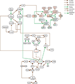
This seems to be a complicated computer simulation of AD. The stroke medical world should be able to create a similar model for the neuronal cascade of death. If we can objectively describe what is going on we can ask researchers to come up with ways to stop that death cascade.  But that would require innovation, leadership and someone with brains. That combination won't be found anywhere in the stroke medical world.
https://www.frontiersin.org/articles/10.3389/fninf.2018.00026/full?
  • 1Research Center for Modeling and Simulation, National University of Sciences and Technology, Islamabad, Pakistan
  • 2Atta-Ur-Rahman School of Applied Biosciences, National University of Sciences and Technology, Islamabad, Pakistan
  • 3Dr. Panjwani Center for Molecular Medicine and Drug Research, International Center for Chemical Sciences, University of Karachi, Karachi, Pakistan
Alzheimer's Disease (AD) is the most common neuro-degenerative disorder in the elderly that leads to dementia. The hallmark of AD is senile lesions made by abnormal aggregation of amyloid beta in extracellular space of brain. One of the challenges in AD treatment is to better understand the mechanism of action of key proteins and their related pathways involved in neuronal cell death in order to identify adequate therapeutic targets. This study focuses on the phenomenon of aggregation of amyloid beta into plaques by considering the signal transduction pathways of Calpain-Calpastatin (CAST) regulation system and Amyloid Precursor Protein (APP) processing pathways along with Ca2+ channels. These pathways are modeled and analyzed individually as well as collectively through Stochastic Petri Nets for comprehensive analysis and thorough understating of AD. The model predicts that the deregulation of Calpain activity, disruption of Calcium homeostasis, inhibition of CAST and elevation of abnormal APP processing are key cytotoxic events resulting in an early AD onset and progression. Interestingly, the model also reveals that plaques accumulation start early (at the age of 40) in life but symptoms appear late. These results suggest that the process of neuro-degeneration can be slowed down or paused by slowing down the degradation rate of Calpain-CAST Complex. In the light of this study, the suggestive therapeutic strategy might be the prevention of the degradation of Calpain-CAST complexes and the inhibition of Calpain for the treatment of neurodegenerative diseases such as AD.

1. Introduction

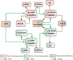
Alzheimer's disease (AD) is a neurodegenerative disorder which has impacted nearly 44 million1 people around the world and this number is still increasing. AD is the leading cause of dementia in the old age (Ashford, 2004). Unfortunately, it is diagnosed only in one out of four people living with the disease1. Clinical characterization of AD includes memory loss and cognitive impairment which further lead to damaged behavioral activities and render a person completely dependent on an external aid (Budson and Price, 2005). AD establishes over time with the appearance of pathological emblems which are senile plaques and neurofibrillary tangles. These lesions comprise of extracellular deposits of Amyloid beta () (Selkoe, 2000; Golde, 2005; Tam and Pasternak, 2012) and intracellular self-gathered clumps of tau proteins (Lee et al., 2001), respectively. is a 40–42 amino-acids long peptide which is formed after the proteolytic cleavage of Amyloid Precursor Protein (APP) (Selkoe, 2000; Golde, 2005; Tam and Pasternak, 2012). Previous studies have shown that monomers are initially non-toxic but their conversion to oligomers makes them toxic (Volles and Lansbury, 2002; Walsh and Selkoe, 2004). Eventually, the abnormal accumulation of oligomers form plaques (Walsh et al., 2002) that deposit into neuronal Endoplasmic Reticulum (ER) (Cuello, 2005) and in extracellular space (Trojanowski and Lee, 2000; Walsh et al., 2000). Aggregation of senile plaques and neurofibrillary tangles cause neuronal cell death and synaptic failure (Tiraboschi et al., 2000; Selkoe, 2002). During the last two decades, several lines of studies have pointed toward the imbalance between production and its clearance plays a central role in pathogenesis of AD. Since 1992, this hypothesis has earned acquiescence (Hardy and Higgins, 1992) and is known as “Amyloid cascade hypothesis (ACH)”. It suggests that and processing of APP are crucial in neuro-degeneration. In AD, aggregation of is the first step leading toward the formation of senile plaques (Hardy and Selkoe, 2002; Vassar, 2005). APP is a type1 trans-membrane protein produced in ER (Greenfield et al., 1999; Roussel et al., 2013). In neurons, production and metabolism of APP occurs rapidly which makes it a crucial element in neuro-pathogenesis (Lee et al., 2008). The main APP proteolytic processing steps occur at the cell surface and Trans-Golgi networks (TGNs). Proteolysis of APP can occur through the so-called non-amyloidogenic and amyloidogenic Pathways (Figure 1). The first step of non-amyloidogenic pathway is carried out by the enzyme alpha (α)-secretase that breaks down APP into soluble Amyloid precursor protein alpha (sAPPα) and alpha C-terminal fragment (αCTF / CTF83). The catalysis by α-secretase is imperative as it cuts APP within domain which blocks formation (Lichtenthaler, 2011). This initial step can also be driven by the beta (β)-secretase / β-site APP-cleaving enzyme (BACE), a transmembrane aspartyl protease (Vassar et al., 1999; Haass, 2004) (Figure 1), which constitute amyloidogenic pathway. BACE is a crucial enzyme, that acts as a rate limiting protein in generation. It breaks down the APP into soluble Amyloid precursor protein beta (sAPPβ) and beta C-terminal fragments (βCTF / CTF99) (Cai et al., 2001). The CTFs are intermediate products of the first step in both pathways which remain attached to the membrane and they are further cleaved by gamma (γ)-secretase (Zhang et al., 2011). In non-amyliodogenic pathway, the fragment α-CTF is cut down by γ-secretase into p38 and the Amyloid Precursor Protein Intracellular Cytoplasmic / C-terminal Domain (AICD). While in amyloidogenic pathway, γ-secretase degrades the βCTF into and AICD (O'Brien and Wong, 2011) (Figure 1).
FIGURE 1
www.frontiersin.orgFigure 1. APP and processing products: APP is synthesized in the ER and then transported to the trans-Golgi-network (TGN) where it is cleaved by secretases. In non-amyloidogenic pathway (left), cleavage of APP by α-secretase results in the generation of sAPPα and C-terminal fragments CTF83 which is further cleaved by γ-secretase into p3 and AICD. Proteolysis by α-secretase prevents production as the cleavage site in APP is within the domain. In amyloidogenic pathway (right), APP is cleaved into sAPPβ and CTF99 by β−secretase / BACE activity. Furthermore, CTF99 breaks down into AICD and by γ-secretase activity. fragments oligomerize and fibrillize into plaques.
The Biological Regulatory Networks (BRN) of APP processing, depicted in Figure 2, is also built from Figure 1. APP processing depends on sequential cleavage by three secretases (α/β-secretase and γ-secretase). In normal conditions, α-secretase residing at the plasma membrane is constitutively active for APP coming to the cell surface and thus favoring non-amyloidogenic pathway (De Strooper and Annaert, 2000). Though there is an interesting fact about APP proteolysis that none of the secretases show special substrate specificity toward APP. There are several transmembrane proteins such as cell surface receptors and ligand, growth factors and cytokines besides APP which undergo ectodomain shedding by enzymes with α-secretase activity (Annaert and Saftig, 2009). In the same manner, BACE shows low affinity toward APP and it is not its exclusive physiological substrate (DeStrooper et al., 2006; Hu et al., 2006). Many observations highlight that in healthy cells APP is frequently processed through non-amyloidogenic pathway to resist amyloid generation while it is altered in pathological conditions (De Strooper and Annaert, 2000). Abnormal processing of APP is stated to be the first and fundamental step in plaques formation in AD pathogenesis (Jonsson et al., 2012). In neuropathological conditions, BACE affinity toward APP increases two folds which leads to enhanced production (Yang et al., 2003; Li and Südhof, 2004). Recent studies on transgenic mice model have shown that BACE activity is modulated by Calpain activation in AD pathology (Liang et al., 2010). Calpain-Calpastatin system also plays a key role in neurodegeneration. Transgenic mice models have shown that over expression of APP, increased production of , inhibition of Calpastatin (CAST) and activation of Calpain increase neuronal degeneration in AD (Higuchi et al., 2012).
FIGURE 2
www.frontiersin.orgFigure 2. APP processing pathways BRN derived from Figure 1.
Calpains are protein clan of cysteine/ thiol proteases and their activity depends on Ca2+ concentration (Ferreira, 2012). The most studied Calpains, mu(μ)-Calpain (Calpain1) and m-Calpain (Calpain2) are present abundantly in neurons, central nervous system (CNS) and glial cells. Though their distribution differs, Calpain1 is ubiquitous and expressed more in neurons while Calpain2 is present in glial cells (Ono and Sorimachi, 2012; Santos et al., 2012). Calpain1 requires micro-molar concentration of Ca2+ (10–50μM), while Calpain2 is activated by mili-molar concentration of Ca2+ (250–350μM) in vitro (Goll et al., 2003; Ryu and Nakazawa, 2014). Ca2+ plays important role in ensuring the cell's vital functions. In addition to calcium, Calpain is tightly regulated in the cell by CAST which is also ubiquitous and solely a specific endogenous inhibitor for both Calpains (Melloni et al., 2006).
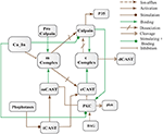
CAST is reported as an explicit suicide substrate for Calpain (Yang et al., 2013). The proportion of CAST in a cell is normally larger than Calpain, its ratio with location is crucial in controlling the extent of activation of Calpain within a cell (Todd et al., 2003). CAST interacts with Calpain at different stages i.e., first it constrains Calpain at the membrane where pro-Calpain is attached then it interacts with active Calpain inside cytosol (Hanna et al., 2008). CAST forms a reversible complex with Calpain at both the sites. At membrane, the reversible complex breaks down when Ca2+ influx increases to release Calpain. Inside cytosol, Calpain undergoes autolysis to attain active conformation. In response, CAST changes its cellular distribution to make itself widely available in the cytoplasm to counter active Calpain (Todd et al., 2003). Both active Calpain and CAST rejoin in a reversible complex to resist persistent activity of Calpain (De Tullio et al., 1999). Active Calpain modulates CAST by slowly digesting it into small inactive fragments which results in plethora of Calpain in cell leading to pathological condition (Averna et al., 2001b; Tompa et al., 2002) (Figure 3). It has been reported that in AD CAST becomes depleted from different regions of the brain as compared to healthy aged brain (Rao et al., 2008). It has also been observed that by controlling Calpain, CAST is indirectly preventing cell membrane damages induced by high Ca2+ and peptide (Vaisid et al., 2008).
FIGURE 3
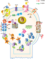
www.frontiersin.orgFigure 3. Different calcium channels work in harmony to establish homeostasis in neurons. Calcium influx is controlled by voltage gated (VGCCs) or receptor-ligand based (NMDAR, GPCR) channels. ER also release Ca2+ into the cytoplasm through inositol-1,4,5-trisphosphate (IP3R) and ryanodine receptors. Calcium efflux is carried out by energy (ATP) dependent channels such as plasma membrane calcium ATPase (PMCA), sodium-potassium ATPase (NKA) and sodium-calcium exchanger (NCX) channels. Calcium homeostasis influences Calpain-CAST system. At membrane, Calpain is bound to CAST to form mComplex at low Ca2+ level. At high Ca2+ concentration, Calpain is released into cytoplasm and autolysed to active form ACalp that again forms complex with CAST (cComplex). Gradually the complex breaks down and releases ACalp which enhances Plaque accumulation and LTP events.
CAST pool is regulated by reversible phosphorylation via PKC, which is a Ca2+-activated phospholipid dependent kinase. Moreover, it is de-phosphorylated by protein phosphatases (ppase) (Melloni et al., 2006). Phosphorylation control CAST inhibitory efficiency in brain (Averna et al., 2001a) to regulate its availability for calpain inhibition. Reversible protein phosphorylation regulates many neuronal functions and is important for neuronal signal transduction (Wu and Lynch, 2006). Inactive PKC is converted to Ca2+-bound activated form in the presence of diacylglycerol (DAG) which in turn is activated by receptor based hydrolysis of phosphoinositides 3 (IP3) (Courjaret et al., 2003). The N-terminal region of CAST which is responsible for the function of the protein has a site for phosphorylation by PKC. CAST is phosphorylated by PKC to decrease its inhibitory efficiency toward calpain (Averna et al., 2001a) (Figure 3). It has been observed that PKC also regulates APP processing by activating α-secretase (Rossner et al., 2001; Racchi et al., 2003), it promotes non-amylodogenic pathway over β-secretase (Lanni et al., 2004). In vivo studies show that in the presence of PKC, secretion of sAPPα increases and secretion declines (Chen and Fernandez, 2004). Other studies about AD found that PKC has substantial role in AD pathology (Etcheberrigaray et al., 2004; Alkon et al., 2007). Active Calpain also interacts with PKC and converts it into constitutive active enzyme (Yamakawa et al., 2001; Goll et al., 2003). Calpain1 directly starts depletion of PKC from cell by converting it into protein kinase M (PKM) (Yamakawa et al., 2001; Liu et al., 2008). The whole mechanism is also depicted in the form of Calpain-CAST system BRN in Figure 4.
FIGURE 4
www.frontiersin.orgFigure 4. Calpain-CAST system BRN derived from Figure 3.
FIGURE 5
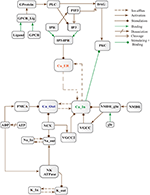
www.frontiersin.orgFigure 5. Calcium Influx Efflux BRN derived from Figure 3.
The dysregulation of Calcium homeostasis contributes in aging and neurodegeneration (Mattson, 2004; Smith et al., 2005; Stutzmann, 2005). A tremendous deal of work by calcium is tightly regulated in time, space and intensity by intracellular stores, influx and efflux channels (Stutzmann, 2005). At resting stage, extracellular Ca2+ concentration ranges from 1.5 to 2.0 mM (Orrenius et al., 2003). While magnitude of Ca2+ inside a cell is very low (between 50–100/ 50–300 nM) (LaFerla, 2002; Orrenius et al., 2003) and after activation it can rise to several micromoles. On contrary, inside ER, the level of Ca2+ is in the range 100-500μM (LaFerla, 2002) which is approximately 1000 times higher than cytosol concentration at the resting phase. Persistent alteration of Ca2+ homeostasis affects production and digestion of pathological proteins such as Calpain, and tau protein. Dysregulation of cellular Ca2+ level is an early and main feature of AD (Mattson et al., 2000; LaFerla, 2002; Small, 2009).
Cytosolic Ca2+ is maintained at very low level as compared to extracellular space through several homeostatic mechanisms, working both temporally and spatially (Figure 3). These equilibrating apparatuses include voltage-operated channels (VOCs) and receptor operated channels (ROCs) for Ca2+ inclusion, Ca2+ storage in organelles e.g., ER (Wojda et al., 2008) and Ca2+ extrusion to extracellular space. Different ATP-dependent membrane pumps such as plasma membrane calcium ATPase channel (PMCA) and sodium-calcium exchanger (NCX) which are dependent on sodium-potassium ATPase (NKA) (Wojda et al., 2008; Brittain et al., 2012) are used for Ca2+ efflux. In different physiological processes, elevation of Ca2+ is necessary to switch-on respective proteins. Ca2+ inclusion is administered by several routes such as N-methyl-D-aspartate receptor (NMDAR), an imperative type of ROCs, which switch into open conformation after binding of endogenous glutamate (glu) as ligand. Another important influx gateway is voltage gated Ca2+ channel (VGCC) which is in closed conformation when neuronal membrane is polarized (Schmolesky et al., 2002; Cain and Snutch, 2011). The VGCC adopts open conformation as plasma membrane depolarizes due to Ca2+/ sodium (Na+) influx through ROCs or ion channels (Weber, 2012). Ca2+ influx also increases from intracellular stores in ER through store-operated channels. There are two calcium channels in ER which are IP3-sensitive and ryanodine (RyRs)-sensitive Ca2+ stores (Berridge, 2009). IP3 driven release of Ca2+ starts by binding of G-protein coupled receptor (GPCR) on plasma membrane which induces Phospholipase C (PLC) mediated cleavage of phosphatidylinositol-4,5-bisphosphate (PIP2) on cell membrane into DAG and IP3. IP3 binds to its receptor on ER membrane and stimulate Ca2+ release into the cytoplasm (Berridge, 2009; Krebs et al., 2015). Furthermore, depletion of ER stores mediate influx of extracellular Ca2+ through store-operated channels (SOCs) (Emptage et al., 2001; Weber, 2012). The mechanism for lowering Ca2+ from cell is controlled by PMCA and NCX. Both PMCA and NCX are energy dependent while, NCX is also Na+ gradient dependent (Wojda et al., 2008). The BRN of Calcium channels, Figure 4, is also helpful in understanding the mechanism underlying the Ca2+ homeostasis.
To comprehend the above mentioned neuronal pathways, models are constructed to understand their dynamics. Stochastic approaches describe the randomness of biological system accurately as compared to ordinary differential equations. In BRNs, the activation or inhibition processes take place with random time delays, therefore, stochastic modeling frameworks are more suitable for their modeling. Petri nets provide complementary approach for both qualitative and quantitative modeling and simulation of the dynamical behavior of large systems in an intuitive way (Mounts and Liebman, 1997; Tsavachidou and Liebman, 2002; Tareen and Ahmad, 2015). The study (Tsavachidou and Liebman, 2002) shows that the Petri net models predict the experimental findings which support the soundness of these models. Stochastic petri nets (SPNs) have emerged as a promising tool for modeling and analyzing BRNs in the field of molecular biology (Goss and Peccoud, 1998). The dynamic behaviors of a variety of BRNs have been studied using stochastic simulations (Mura and Csikász-Nagy, 2008; Lamprecht et al., 2011; Castaldi et al., 2012; Marwan et al., 2012).
In this study, we have modeled and analyzed the neuronal physiological system constituting Ca2+ channels maintaining homeostasis, CAST regulating Calpain system and APP processing pathways separately and collectively at molecular level using SPNs to understand the AD progression mechanism. Particularly, we have analyzed neuronal patho-physiological dynamic behaviors causing the development of hallmark lesions in brain to answer many question e.g., how dysregulation of Ca2+ triggers AD? When CAST, the sole inhibitor of Calpain, depletes from the brain cells? how production increases? and when the accumulation of plaques start? The answers to these questions lie in the modeling of the combined BRN Figure 6. The model predicts that Calpain is the main cause of dysregulation, which start with the rise in Ca2+ levels in the cytosol. Calpain activates different pathways through which production and accumulation increases. Plaques start building with time at the age of forty and older. Plaques first enter lag phase and then into rapid growth phase. Calpain slowly degrades CAST which depletes from the cell and eventually neuronal degradation progresses. These results suggest that patho-physiological events such as dysregulation of Ca2+ homeostasis, Calpain hyper-activation, CAST degradation and abnormal digestion of APP, all are inter-connected and a cumulative study of these processes through SPN was needed.
FIGURE 6
www.frontiersin.org
Many pages to go yet and haven't even gotten to the calculations.