Changing stroke rehab and research worldwide now.Time is Brain! trillions and trillions of neurons that DIE each day because there are NO effective hyperacute therapies besides tPA(only 12% effective). I have 523 posts on hyperacute therapy, enough for researchers to spend decades proving them out. These are my personal ideas and blog on stroke rehabilitation and stroke research. Do not attempt any of these without checking with your medical provider. Unless you join me in agitating, when you need these therapies they won't be there.

What this blog is for:

My blog is not to help survivors recover, it is to have the 10 million yearly stroke survivors light fires underneath their doctors, stroke hospitals and stroke researchers to get stroke solved. 100% recovery. The stroke medical world is completely failing at that goal, they don't even have it as a goal. Shortly after getting out of the hospital and getting NO information on the process or protocols of stroke rehabilitation and recovery I started searching on the internet and found that no other survivor received useful information. This is an attempt to cover all stroke rehabilitation information that should be readily available to survivors so they can talk with informed knowledge to their medical staff. It lays out what needs to be done to get stroke survivors closer to 100% recovery. It's quite disgusting that this information is not available from every stroke association and doctors group.

Monday, October 31, 2016

Did you get your money’s worth today?

A valid question to ask about your doctor. Did you receive ANY concrete steps to take to get recovered? Or was it all try this? Exercise some more? I know nuthin!

Did you get your money’s worth today?

How Repetition Can Heal Your Brain Faster during Stroke Recovery

But wouldn't incorrect movement actually make you learn faster? Why doesn't your therapist know that?

We know that learning from your mistakes is one of the best ways to learn,

How Repetition Can Heal Your Brain Faster during Stroke Recovery


You’ve heard us preach about the importance of neuroplasticity for stroke recovery before. It’s the #1 thing all stroke survivors should know about.
Neuroplasticity is the healing you need, and repetition is the tool for accessing that healing and speeding your recovery along.
In order to explain why repetition matters so much, we will give a brief overview of neuroplasticity first.

Neuroplasticity – The King of Rehab

Neuroplasticity is the mechanism that your brain uses to rewire itself.
There are two ways that neuroplasticity works: the creation of new connections between neurons (brain cells), and the deletion of old connections.
After stroke, a chunk of the brain is damaged and those brain cells are unable to carry out their tasks. For example, arm movement can become difficult after stroke if the part of the brain responsible for arm movement is affected.
In order to heal from this damage, the surrounding areas of the brain can pick up the slack. Meaning, they can learn the tasks that the damaged parts once controlled. This is neuroplasticity.
But neuroplasticity can only happen through repetitive practice.

Repetition & Neuroplasticity

So if you want to regain arm function after stroke, then you need to repeat arm rehab exercises over and over and over until it sticks and your brain has successfully rewired itself.
Because each time you move your arm, you begin to form and strengthen the connections between the neurons responsible for that arm movement.
That’s why learning a language is so difficult, for instance. We have to repeat new words over and over and over again until they stick. Similarly, that’s why learning how to play a new sport requires lots of practice.
You’re forming new connections in the brain, and it takes time.
So when you grow frustrated during rehab (which is perfectly normal and understandable – here’s an article on how to deal with it), remind yourself of the intricate work that you’re doing.
You’re rewiring your brain!
You’re calling upon your brain the same way that professional athletes call upon their brain. You’re forming new neural networks – and it’s phenomenal!

Repetition & Consistency

So you’ve got the repetition part down now, so you’re good, right? All your bases are covered…
Well, not quite.
The neurons in your brain need good repetition in order to strengthen themselves, but they also need consistency in order to stick.
For example, if you’re really good about repeating your rubber band hand exercises 50 times each, but you only do that once a week, then you’re in trouble.
Because 50 repetitions is great! But the time in between rehab sessions will cause those new connections to weaken.
So if you want to maximize your healing, you need to be repetitious and consistent with your rehab exercises.

Repetition & Motivation

All the examples we’ve been using so far have revolved around movement after stroke – and for good reason. It’s important for your quality of life, safety, and independence.
But another way that you can use neuroplasticity to improve your quality of life is by using it to hardwire motivation into your brain.
This is especially important for those who procrastinate on their rehab exercises or can’t find the willpower to keep going.
Because when you feel like giving up, there’s a thought in your brain telling you to give up. And if that’s a pattern, then your brain is really good at telling you to give up! (Repetitive practice still works even if you’re not aware of it.)
So if you tell yourself to keep going – to keep taking one small step each and every day – and you do that over and over and over, then you will hardwire persistence into your brain.
If you tell yourself that you’ve got this – that all your hard work will pay off even if you can’t see results yet – and you repeat that to yourself multiple times a day, then you will hardwire confidence into your brain.
You have the power to become whatever you want to become.
It’s just a matter of letting go of old connections (like self-doubt and fear) and forming new ones (like persistence and balance).
So, what will you use neuroplasticity for?
Will you repeat those rehab exercises consistently from here on out?
Will you talk kindly to yourself consistently, too?
We always encourage both 🙂

WHY SEX IS BETTER FOR YOUR BRAIN THAN SUDOKU

You have to go 1/3 down in the link to get to this section. Your doctor is probably not prescribing sex as part of your recovery, What the hell is the matter with her/him?
http://www.dailymail.co.uk/health/article-3887974/Food-brain-buzzing-easy-meals-boost-memory-sex-better-sudoku-delicious-diet-ll-boost-mood.html?ITO=1490&ns_mchannel=rss&ns_campaign=1490

The best non-food sources of nourishment for your brain are good sleep, regular exercise . . . and great sex.
However, according to the statistics, we seem to be having less of it — on average we have sex fewer than five times a month, compared to six-and-a-half times 20 years ago, according to the National Survey of Sexual Attitudes and Lifestyles.
Yet numerous studies have linked regular sexual activity to emotional well-being, reduced migraine pain and even a lower risk of prostate cancer.

As a form of exercise, an enthusiastic session between the sheets is roughly equivalent to 20 minutes of doubles tennis or walking uphill, 33 minutes of golf on a driving range, 40 minutes of yoga or 19 minutes of light rowing.
In fact, the British Heart Foundation suggests 30 minutes of sex daily is as good for you as walking the dog.
And research is now showing that sex provides a ‘triple-whammy’ of benefits by combining a workout for the heart and lungs, the release of hormones that could lower stress and the production of new brain cells.
Then — for women — there’s the added plus of a toning effect on the muscles in the pelvic floor.
Certainly, orgasm triggers increased blood flow around the body, which keeps the tissues and organs healthy, giving the brain an extra boost of oxygen and nutrients. Although younger couples might get a real exercise hit from active sex, generally speaking few of us would engage in this kind of sex regularly enough to glean aerobic benefits.
However, in today’s fast-paced world, it may be that the calming, soothing effect and feeling of connection that occurs between two loving people during sex is better for your brain than almost anything else.

Self-Paced Reaching after Stroke: A Quantitative Assessment of Longitudinal and Directional Sensitivity Using the H-Man Planar Robot for Upper Limb Neurorehabilitation

It would seem to be much simpler and faster to use wearable sensors to determine base measurements. But that makes the assumption that our researchers are reading research in their field.

Self-Paced Reaching after Stroke: A Quantitative Assessment of Longitudinal and Directional Sensitivity Using the H-Man Planar Robot for Upper Limb Neurorehabilitation

Asif Hussain1*, Aamani Budhota1,2, Charmayne Mary Lee Hughes1,3,4, Wayne D. Dailey1,5, Deshmukh A. Vishwanath6, Christopher W. K. Kuah6, Lester H. L. Yam6, Yong J. Loh6, Liming Xiang7, Karen S. G. Chua6, Etienne Burdet5 and Domenico Campolo1*
  • 1Robotics Research Centre, School of Mechanical and Aerospace Engineering, Nanyang Technological University, Singapore, Singapore
  • 2Interdisciplinary Graduate School, Nanyang Technological University, Singapore, Singapore
  • 3Department of Kinesiology, San Francisco State University, San Francisco, CA, USA
  • 4Health Equity Institute, San Francisco State University, San Francisco, CA, USA
  • 5Department of Bioengineering, Imperial College of Science, Technology and Medicine, London, UK
  • 6Centre for Advanced Rehabilitation Therapeutics, Tan Tock Seng Hospital Rehabilitation Centre, Singapore, Singapore
  • 7School of Physical and Mathematical Sciences, Nanyang Technological University, Singapore, Singapore
Technology aided measures offer a sensitive, accurate and time-efficient approach for the assessment of sensorimotor function after neurological insult compared to standard clinical assessments. This study investigated the sensitivity of robotic measures to capture differences in planar reaching movements as a function of neurological status (stroke, healthy), direction (front, ipsilateral, contralateral), movement segment (outbound, inbound), and time (baseline, post-training, 2-week follow-up) using a planar, two-degrees of freedom, robotic-manipulator (H-Man). Twelve chronic stroke (age: 55 ± 10.0 years, 5 female, 7 male, time since stroke: 11.2 ± 6.0 months) and nine aged-matched healthy participants (age: 53 ± 4.3 years, 5 female, 4 male) participated in this study. Both healthy and stroke participants performed planar reaching movements in contralateral, ipsilateral and front directions with the H-Man, and the robotic measures, spectral arc length (SAL), normalized time to peak velocities (TpeakN), and root-mean square error (RMSE) were evaluated. Healthy participants went through a one-off session of assessment to investigate the baseline. Stroke participants completed a 2-week intensive robotic training plus standard arm therapy (8 × 90 min sessions). Motor function for stroke participants was evaluated prior to training (baseline, week-0), immediately following training (post-training, week-2), and 2-weeks after training (follow-up, week-4) using robotic assessment and the clinical measures Fugl-Meyer Assessment (FMA), Activity-Research-Arm Test (ARAT), and grip-strength. Robotic assessments were able to capture differences due to neurological status, movement direction, and movement segment. Movements performed by stroke participants were less-smooth, featured longer TpeakN, and larger RMSE values, compared to healthy controls. Significant movement direction differences were observed, with improved reaching performance for the front, compared to ipsilateral and contralateral movement directions. There were group differences depending on movement segment. Outbound reaching movements were smoother and featured longer TpeakN values than inbound movements for control participants, whereas SAL, TpeakN, and RMSE values were similar regardless of movement segment for stroke patients. Significant change in performance was observed between initial and post-assessments using H-Man in stroke participants, compared to conventional scales which showed no significant difference. Results of the study indicate the potential of H-Man as a sensitive tool for tracking changes in performance compared to ordinal scales (i.e., FM, ARAT).

Introduction

The rehabilitation of neurological disorders such as stroke and cerebral palsy is a labor -intensive process that requires daily one-on-one interactions with therapists. The significant burden placed on the health care providers and the overall health care system have stimulated particular interest in technology assisted systems for neurorehabilitation (Maciejasz et al., 2014), with the underlying objective of decreasing the workload of the therapist and to facilitate training with minimal supervision at an affordable cost. A significant amount of this work has focused on the development of robotic devices to train upper extremity (UE) task-related movements (Riener et al., 2005; Prange et al., 2006; Brewer et al., 2007; Balasubramanian et al., 2010). The advantages of robot-assisted therapy include the ability to actively assist or resist human motions, to acquire accurate measurements of the dynamic and kinematic performance of participants during training using integrated sensors, and to administer repetitive task-specific training with limited supervision from a therapist. To date, clinical studies have shown that robot-assisted therapy of the UE is at least as effective as conventional rehabilitation therapy in terms of reducing motor impairments over a short-term period (Prange et al., 2006; Kwakkel et al., 2008; Lo et al., 2010; Norouzi-Gheidari et al., 2012) and thus can effectively complement conventional therapy. Although conventional therapy, itself is not very productive/ efficient, Duncan et al. reported that only 33–70% of the stroke patients recover useful arm ability, and initial paresis severity remains the best predictor of arm function recovery over 6 months (Duncan et al., 1992; Huang and Krakauer, 2009). It is possible that the limited recovery success for UE dysfunction after stroke is hampered by the limited amount of training offered to the affected population. As such, increasing the frequency and intensity of training could significantly improve performance (Harvey, 2009). However, an arguably equal, if not more important factor contributing to this limited improvement can be attributed to the partial understanding and incomplete assessment of the disability itself, which in technology intervention systems has been explored less thoroughly. Clear knowledge of the level of sensorimotor deficits is required for devising a comprehensive and efficient training regime (Balasubramanian et al., 2012a).
Conventionally, assessment of motor functions is carried out by therapists by means of ordinal clinical scales to examine specific aspects of a subject's motor behavior and devise an appropriate treatment strategy accordingly (Fugl-Meyer et al., 1974; Lyle, 1981; Gladstone et al., 2002). For example, the Action Research Arm Test (ARAT) scores performance on various tasks using a 4-point scale, where 0 indicates no movement and 3 indicates the task is completed with normal performance (Lyle, 1981). Although the ARAT and other post-stroke motor assessments are widely accepted and have high test-retest and interrater reliability, their reliance on ordinal scoring renders them insensitive to subtle differences in deficit and changes over the rehabilitation lifespan. Furthermore, the additional time required to perform manual assessment discourages their regular use in clinical practice to track and understand motor recovery in the affected population.
It is apparent that stroke rehabilitation would benefit if clinicians had a complete understanding of the specific sensorimotor deficits exhibited by the patient (Balasubramanian et al., 2012a). Robotic technology has the potential to augment the assessment process by using integrated sensors to record continuous, high-resolution data. These sensory measurements are collected during normal use of the system and do not require additional time for a discrete assessment protocol. These systems are (semi-) autonomous, potentially more objective than functional assessments, and less prone to human error/subjectivity (Bosecker et al., 2010; Lambercy et al., 2010). However, this form of assessment has yet to be fully established and validated when compared to the gold standards, and is expensive due to the high cost of (most) robotic systems for use in standard clinical practice.
At Nanyang Technological University (NTU) we have designed a novel low-cost, planar, table-top robot for decentralized neurorehabilitation (hereafter called H-Man) (Campolo et al., 2014; Hussain et al., 2015a). It can benefit participants with limited access to a therapist for rehabilitation, and with properly validated assessment protocols, can provide continuous updates about motor progress to the patient, their caregivers, and the therapy team. In this study, we evaluated the ability of the H-Man to detect differences in planar self-paced reaching as a function of neurological status (stroke, age-matched healthy control), direction (front, ipsilateral, contralateral), and movement segment (outbound, inbound). In addition, we investigated the longitudinal sensitivity of these performance metrics to capture motor performance changes in stroke patients, and examined the relationship between robotic measures and conventional scales. Multiple studies have previously addressed variations in performance metrics on workspace. However, due to variations in protocols/task definitions (for example point to point vs. path reaching, free reaching vs. supported movements) and varying outcomes, the reliability, and validity of reaching movements as measures of upper limb motor functionality is still limited (Levin, 1996; Archambault et al., 1999; Kamper et al., 2002; Sukal et al., 2007). Moreover, most of these studies focus on developing relations to clinical scales and/or inter-relationships between performance metrics. In this paper, we focus on a more fundamental question: the distribution/variation of performance outcomes within a control group and across stroke participants for different directions, and for different segments of movements [outbound movements (i.e., away from the body) and inbound movement (i.e., toward the body)]. Multiple papers briefly address this question but not as a major focus of study for-example, Kamper et al. and Levin presented studies on free reaching in 3D and planar supported reaching tasks, respectively (Levin, 1996; Kamper et al., 2002), which showed modest variations across directions but pre-dominantly focused on results (of all directions) to establish relationships with performance matrices and or clinical scales. Here, we report variations for all directions and performance matrices, for both control and stroke participants, along with comparisons between inbound and outbound movement segments. These results help build a clearer understanding of the characteristics of reaching movements and how they differ across stroke and healthy participants. Further, we also show the sensitivity of selected performance measures compared to clinical scales by analysing longitudinal changes in metrics by assessing performance over a 2-week period, which adds weight to the potential of the H-Man as an effective assessment tool.

More at link

3 ways Brigham and Women's engages patients, improves care

How is your hospital engaging patients to improve care? If your hospital doesn't even realize that 10% full recovery or 12% tPA full efficacy from a stroke is a problem then that hospital is hopelessly incompetent.
http://www.fiercehealthcare.com/patient-engagement/brigham-and-women-s-3-pronged-strategy-to-engage-patients-improve-care?utm_medium=nl&
Hospitals that make an effort to gain a greater understanding of what a patient experiences during a hospital stay can significantly improve care quality and outcomes.
That’s the experience at Brigham and Women’s Hospital in Boston, which has worked for nine years with a patient and family advisory council to better understand what it’s like to be an inpatient and how to improve the patient experience.
And the engagement effort goes beyond basic service improvements, such as better food. Ultimately, the approach could “elevate healthcare to the exemplary level of service seen in today’s consumer industries,” Maureen Fagan, associate chief nurse for OB-GYN at Brigham and Women’s Hospital and the executive director for its Center for Patients and Families, writes in a post for Hospitals & Health Networks.
Fagan shares three tips to make the most of patient and family advisers:
1. Seek out a diverse group of patients and caregivers who represent the hospital’s patient populations. To find 8 to 12 advisers, Brigham & Women’s asked questions such as: “Tell us something that went wrong and what you would have preferred to have happened,” Fagan writes.
2. Don’t expect one group of advisers to represent all patients. The hospital has more than 85 patients and family members serving on 16 different councils to offer advice on individual service lines. Individual councils focus on LGBTQ healthcare, ethics, emergency care and obstetrics, for example.
3. Give the councils the ability to help influence decisions on care delivery. Once trained, Fagan writes, it’s vital the organization give patient and family advisers the authority to make decisions. The hospital's advisers are “meaningful contributors to policy and process changes.”

Omega-3 fatty acids could promote clearance of metabolites in the brain, research shows

Would this help in the aftermath of a stroke? I bet no followup will ever occur. Don't try this on your own, you know how dangerous fish oil is if your doctor didn't prescribe it.

Omega-3 fatty acids could promote clearance of metabolites in the brain, research shows

New research published online in The FASEB Journal suggests that omega-3 polyunsaturated fatty acids, which are found in fish oil, could improve the function of the glymphatic system, which facilitates the clearance of waste from the brain, and promote the clearance of metabolites including amyloid-β peptides, a primary culprit in Alzheimer's disease.
To make this discovery, scientists first used transgenic fat-1 mice, which express high endogenous omega-3 polyunsaturated fatty acids (PUFAs) in the brain, to investigate the effect of omega-3 PUFAs on the clearance function of the glymphatic system. Compared to the wild-type mice, the fat-1 mice with enriched endogenous omega-3 PUFAs significantly promote the clearance function of the lymphatic system, including the Aβ clearance from the brain. Wild-type mice were supplemented with fish oil, which contains high concentrations of omega-3 PUFAs, and found that fish oil-supplemented mice also improved the clearance function of the glymphatic system compared to the control mice without fish oil supplementation. Omega-3 PUFAs help maintain the brain homeostasis, which may provide benefits in a number of neurological diseases, such as Alzheimer's disease, traumatic brain injury, and sleep impairment, among others.
"These now-famous fatty acids have been the subject of major studies both in academia and industry. Just when we thought we had heard everything, here is something new, and it is provocative indeed," said Thoru Pederson, Ph.D., Editor-in-Chief of The FASEB Journal. "This study should not turn attention away from the roles of these substances in maintaining vascular health, but neither should they restrict our view. The brain is an extremely vascularized organ, while we might also bear in mind that omega-3 fatty acids may impact neurons, glia, and astrocytes themselves."
Source:
Federation of American Societies for Experimental Biology

Novel Alzheimer's treatment uses microscopic droplets of fat to carry drugs into the brain

If we ever get a drug developed that would help stroke recovery this could be used to deliver it to the brain. As long as we have someone keeping track of a stroke strategy and all the moving parts that need to be remembered.  Or maybe these 13 nasal delevery options? I bet no one in stroke is keeping track of this.
http://www.news-medical.net/news/20161020/Novel-Alzheimers-treatment-uses-microscopic-droplets-of-fat-to-carry-drugs-into-the-brain.aspx
New Alzheimer's treatment could be delivered as nasal spray
Researchers have developed a novel treatment that could block the development of Alzheimer's disease using microscopic droplets of fat to carry drugs into the brain. This treatment approach, which is used to target drugs to cancer cells, has been successfully applied to Alzheimer's disease for the first time, restoring memory loss in mice.
The study, which was led by researchers at Lancaster University and funded by Alzheimer's Society, is published  in the journal Nanomedicine: Nanotechnology, Biology and Medicine.
The treatment uses tiny droplets of fat, called nanoliposomes, which are coated in protein fragments that are able to stop amyloid protein accumulating into plaques, even at low concentrations. Amyloid plaques are the toxic clumps of protein that cause damage to cells in the brains of people with Alzheimer's disease.
Mice that were genetically altered to develop Alzheimer's disease were injected with the nanoliposomes for three weeks. Those which received the drug recovered their long-term memory and could recognise familiar objects after a 24-hour period. In comparison, mice which received a placebo injection had no memory of objects seen the day before.
Lead researcher, Professor David Allsop, commented: "Following results this summer, there is renewed optimism for antibody drugs - treatments that harness the body's immune system to target amyloid plaques. However if these prove successful, treatments will have to be administered in a clinic by an IV drip and could have some potentially harmful side effects.
"Using nanoliposomes offers an alternative way to inhibit the toxic build-up of amyloid plaques without activating an immune response in the brain. Our hope is that this could one day be administered by something as simple and non-invasive as a nasal spray, which patients could use in the comfort of their own home."
Nanoliposomes are already used to better target toxic chemotherapy drugs to cancer cells. Recent studies have also shown that the fat droplets can pass directly into the brain through the nose, opening up the possibility of using a nasal spray to administer treatments for brain diseases, such as Alzheimer's.
Commenting on the need for innovative approaches to dementia treatments, Dr Doug Brown, Director of Research and Development at Alzheimer's Society, said: "With no new dementia drugs in nearly 15 years, we're at a critical time for dementia research. It's absolutely vital we continue to sniff-out new approaches to getting drugs into the brain. While we wait in anticipation for the results of ongoing clinical trials, Alzheimer's Society will continue to fund innovative research to tackle dementia head-on."
"Nanotechnology is promising great benefits to people with many different types of cancer, and it's exciting that it could one day offer the same hope to people with the most common form of dementia." 
There are 850,000 people in the UK living with dementia, and currently available drugs are only able to treat the symptoms of dementia, rather than slowing its progression. The research team at Lancaster University are now seeking investment from industry to take their novel treatment forward to be tested in people.
Source:
Lancaster University

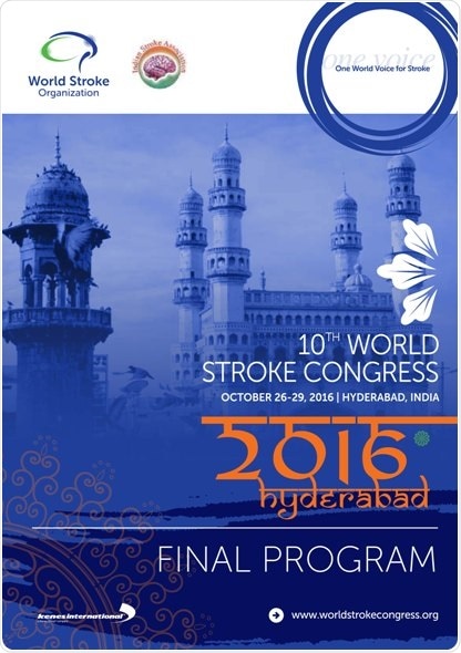
Experts meet at Hyderabad for four-day World Stroke Congress

And they will do fucking nothing because they aren't even acknowledging all the problems in stroke. Just 'happy talk' from this idiotic group. I didn't know 'experts' could be so dumb.
http://www.news-medical.net/news/20161031/Experts-meet-at-Hyderabad-for-four-day-World-Stroke-Congress.aspx
Stroke is devastating. Everyone can have a stroke.  For many people, the stroke happens suddenly and without warning. According to Stroke Association, when it happens, there is a little time to prepare. It can affect how one moves, feels and thinks.
Hyderabad is the host to World Stoke Congress.  Nearly 2200 stroke experts descended on the city to participate in a four-day conference.  The meeting begins just a few days before World Stroke Day, to be observed on October 29 every year.
What is a stroke? According to Dr. Jeyaraj Pandian, Co-Chair of the World Stoke Congress and a top stroke neurologist in India, a stroke is a brain attack.  It happens when the blood supply to part of the brain cuts off.
Blood carries essential nutrients and oxygen to the brain.  Without blood, brain cells can't work because of the damage.  They won't be able to perform their tasks.
The brain is a critical part of the body. It controls the entire body. A stroke can affect the way body functions.
The brain also controls how one thinks, learns, feels and communicates.
The blood flow to the brain can be cut off by two ways—due to Blockage or a Bleeding.  Because of which strokes occur, which are of two types—01. Blockage and 02. Bleeding.
In 85 per cent cases stroke happens by a Blockage and reaming 15 per cent cases by Bleeding.
According to Stroke Association, some of the things that increase the risk of stroke cannot be changed are Gender(in people under 75, Men have more strokes than women); Age(you are more likely to have a stroke if you are over the age of 65). Family History (If a close relative has had an attack, risk is likely to be higher) and Ethnic Background(South Asian or African-Caribbean origin are at an increased risk).
BP, High Cholesterol, Type 2 Diabetes, Atrial Fibrillation(irregular beating of the heart), Heart disease, Smoking are some of the medical problems that can increase a risk of having a stroke.
Smokers are twice as likely to have a stroke, and this risk will increase more with more smoking. Stopping smoking will reduce the danger of a stroke.
Drinking too much alcohol raises blood pressure, which in turn causes a stoke.
The common effects of stroke are weakness or paralysis; speech and language problems, unsteadiness, difficulty with swallowing, suffer from extreme tiredness, may have a problem with sensation and much more such problems.
According to experts at the ongoing World Stroke Congress, many strokes can be prevented.
The food that will help you save from stroke are fresh fruits, vegetables, and dried fruits and avoid junk food. But you don't list a food protocol, just generalities.
Choose low-fat proteins.  Eat plenty of fiber such as cereals, porridge, brown rice, whole wheat bread and pasta or grains such as couscous.  Cut down on the intake of salt. Watch your weight.
Keep active.  Do exercises. Warm up before and after exercise.  Find someone to exercise with you.
Stroke is the leading cause of disability and the second leading cause of death globally.
Stroke devastates lives around the world.  Over 17 million people around the world suffer a stroke every year and 6.5 million deaths happen.  5 million are permanently disabled.  Nearly 26 million inhabitants survive a stroke every year. Doesn't anyone vet these numbers?
In India alone, 1.7 million new stroke cases occur every year. Stroke affects young people in India during their productive period of life.  The reasons for this are due to unorganized lifestyles and poor food habits. After stroke care significantly reduces the risk of death.
According to Dr, Jayaraj Pandian, the prevalence of the problem is 350 to 400 people per lakh population in India.
In Ludhiana, more youth are getting the stroke. Nearly 23 percent of the youth population is suffering from a stroke. That is due to unhealthy eating habits, stressful life.  Also, it is because today we have more youth population.
Stroke care infrastructure is developed mainly in private hospitals, but the vast majority of public hospitals are ill-equipped to treat stroke patients. The treatment expenses are borne by the patients and relatives and hence there is a huge economic burden on the family. The neighboring countries in South Asia also face a similar problem of a huge burden of stroke with an inadequate infrastructure to tackle the situation stated Dr. Jeyaraj Pandian the Congress Co-Chair from India.
Dr. C. Laxma Reddy, Minister for Health, Government of Telangana, while addressing the inauguration of World Stoke Congress, said TS Govt has established ICUs exclusively for stroke patients in all govt hospitals across Telangana state. We want government hospitals in the state to be stroke-ready hospitals he said.
The right care makes a difference, but many people are not getting the stroke treatment they need. According to World Stroke Campaign by the World Stroke Organisation, there are six key facts about stroke treatment
1.Early recognition makes a big difference.  Knowing the signs of stroke and getting treatment saves lives and improves recovery.
2.  1 in 10 people makes an excellent recovery when cared for in a specialized stroke unit. That is an incredible failure rate - 90% failure, it has nothing to do with the specialized stroke unit at all.
3. Clot-busting drugs (tPA or thrombolysis) increase the chance of a good outcome by  30%. (Bullshit, tPA only fully works 12% of the time)Clot-busting drugs break up blood clots. This treatment can be administered up to 4.5 hours of symptom onset in many patients with ischaemic stroke. The earlier it is given, the greater the effect.
4. Clot retrieval treatment increases the chance of a good outcome by more than 50%.  Clot retrieval treatment (mechanical thrombectomy) involves removing a blood clot  and can improve survival rates and reduce disability. Notice they never talk about results, just better chance.
5. Recovery is a critical step in the treatment process. Rehabilitation starts in the hospital as soon as possible following a stroke.
6. One in four survivors will have another stroke. Treatments that prevent another stroke include drugs to lower blood pressure and cholesterol, antiplatelet therapies, anticoagulation for atrial fibrillation, surgery or stenting for selected patients with severe carotid artery narrowing.
Lifestyle changes can also substantially reduce the risk of another stroke. Changes include eating well, being physically active, being tobacco-free, managing stress, and limiting alcohol consumption.
Stroke is treatable.  One must gain more awareness to save from devastating stroke effect.

Sunday, October 30, 2016

6 Signs That You’re Going to Die Early

I'm going to die of something stupid after age 94.
http://www.cheatsheet.com/health-fitness/signs-youre-going-die-early.html/
If you were to just take a look at the most common ways people die in modern America, you can probably count on the fact that you’re going to die as a result of heart disease or cancer at some point down the line. And again, you can take steps to lower your odds of that happening starting today. If you’re serious about living a long and healthy life, see if any of these other signs apply to you and your present condition.
If they do, death may come knocking sooner than you’d like.

1. Your physical shape

Yeah, I'm 35 ponds over what I should be at. I'm not worrying about it, it will slowly be whittled away.

2. Gnarly nails

Nope.


3. A weak sense of smell

Nope, I can still smell the different red wine aromas and bouquets. Not that I can tell the wine type based on that. But I can spout wine terms with the best of them. It had hints of white peaches with an angular crossing of acids and tannins.

4. Grip strength

Right hand was 100 lbs plus in the hospital, left hand maybe made it to 20 pounds.

5. Educational attainment

Bachelor of Science degree, dropped out of MBA program 2 classes short. It became not important since I found out I hated management and I would have needed an A in one of those classes to match to the C I got earlier in order to maintain a B average for graduating. Sometimes you just have to give up, declare failure.

6. Bad breath

Nope.


How Alcohol Attacks The Brain

Or the positive side here: Your choice which to believe

5 Hidden Health Benefits of Alcohol

6 Ways Alcohol Can Be Unexpectedly Good For Your Mind And Brain

This is your brain on beer vs. coffee (8 Photos)


This Simple Diet Linked To Higher IQ

You mean I could become even smarter if I became a vegetarian? Or did I have to do this by age 30?
http://www.spring.org.uk/2016/10/higher-iq-diet.php?omhide=true
The reason people with a higher IQ also tend to be healthier could be down to their diet.
People with a higher IQ are more likely to be vegetarian, psychological research finds.
In fact, vegetarians could be up to 10% more intelligent than red meat eaters, according to some studies.
A higher IQ is also seen among those who describe themselves as vegetarian, but also eat chicken and fish.
The conclusion comes from a survey of 8,170 men and women whose IQ was tested when they were 10-years-old.
By age 30, 4.5% had become vegetarian, of these 2.5% were vegan and 33.6% said they were vegetarian, but still ate chicken and/or fish.
People with higher IQs at age 10, the analysis showed, were more likely to be vegetarians at age 30.
There was no difference between stricter vegetarians (ovo-lacto vegetarians) and those who ate some chicken and/or fish as well.
The findings could help to explain why more intelligent people are also healthier, since a vegetarian diet is better for the heart and for maintaining a healthy body mass.

Michael J. Fox Foundation newsletter, notice the patient council

Our fucking failures of stroke associations have nothing like this.
If you have time, look at the last page of the newsletter to see how MJFF is organized
Notice there is a Patient Council.

Do You Think You’re Too Young to Have a Stroke?

And when you get to the emergency room you better hope you have full blown classic stroke symptoms.

Pediatric Stroke Often Misdiagnosed, Treatment Delayed 

Doctors tell boy, 15, he had a migraine after rugby tackle - but he was actually suffering a paralyzing stroke which nearly killed him


Factors Associated With Misdiagnosis of Acute Stroke in Young Adults


Amy on her 36 hour wait for a diagnosis.

But the excuses here:

https://health.clevelandclinic.org/2016/02/think-youre-young-stroke/? 


If you’re a young or middle-aged adult, you may think strokes only happen to senior citizens. But experts say you’re never too young for a stroke, which occurs when a blood vessel in the brain becomes blocked or bursts.
“Although stroke incidence is lower for younger people, strokes can happen at any age,” says stroke specialist Gabor Toth, MD.
“People are beginning to recognize the symptoms of a stroke when they may not have before,” he says. And it’s a good idea to educate yourself on the risks, signs and symptoms of stroke.

What does current research say?

There is research, though conflicting, that suggests that strokes may now be diagnosed more frequently in people between the ages of 20 and 54.
Ischemic strokes occur when a clot blocks blood flow to the brain. A 2011 study found that ischemic stroke hospitalization rates in teens and young adults aged 15 to 44 increased up to 37 percent between 1995 and 2008.
In 2012, researchers found that stroke incidence for people under the age of 55 had increased from 12.9 percent to 18.6 percent between 1993 and 2005.
However, Dr. Toth says there are limitations in these studies and that other research does not support the notion that stroke rates are rising in the young — but he still thinks it’s important to note the findings.
“It certainly raises awareness of an significant problem. Hopefully, the public and even those in the medical field are becoming better educated on the subject, potentially leading to earlier and more timely diagnosis and treatment of this devastating disease,” he says.
RELATED: 5 Things You Don’t Know About Childhood Stroke

Who is at risk?

Stroke risk factors for younger people are largely the same as they are for older adults. There is research suggesting that the incidence of these risk factors may also be increasing in the young.
These include:
However, younger people may have some additional risk factors:
  • Congenital heart disease and blood clotting disorders. If not recognized and treated appropriately, these conditions may result in strokes in childhood, early adulthood, or even later in life.
  • Birth control. Women who are on birth control pills and who also smoke are also at an increased risk.
  • Tear in a blood vessel wall. Also called dissection, a tear in the blood vessel wall is also a risk factor that can be found in younger adults. “Dissection can be spontenous, but is often times caused by some sort of trauma, including car accidents or being jerked around by roller coaster rides,” says Dr. Toth. Even aggressive chiropractic neck manipulations have been suggested to sometimes cause dissection.
  • Drug use. “Younger people may be more likely to use drugs, which can increase their risk for stroke,” Dr. Toth says.
RELATED: Stressed at Work? You May Have a Higher Risk of Stroke

What if you think you’re having a stroke?

Seek help quickly because strokes are time-sensitive. The more time that passes, the greater the chance of increased brain damage and inability to reverse symptoms.
“If you get to the hospital quickly, you may qualify for early medical or interventional therapies that have the highest chance of reversing your symptoms, and result in a better outcome in the long term,” Dr. Toth says.
RELATED: Do You Know the Signs of a Stroke?

What if you think someone else is having a stroke?

If you think a friend, a family member or a co-worker is having a stroke, call 911 immediately.
Then, keep them safe while you’re waiting for the ambulance. If they feel weak, have them sit or lie down so they won’t fall, he says.
“If the person is stubbornly refusing to go to the hospital, do your best to convince them to go,” Dr. Toth says. “Even if you’re not sure the person is having a stroke, it’s best for them to get a quick checkup to make sure everything’s OK. Ask what’s the worst that can happen compared to what can happen if they don’t go.”

Don’t give aspirin or water

You don’t necessarily want to give a potential stroke victim an aspirin before a quick CT scan can be obtained, because it may increase the risk of bleeding in the brain if the actual stroke is hemorrhagic.
Likewise, you shouldn’t offer someone who is having a stroke a glass of water because there’s a danger of choking.


Breaking up sitting time after stroke (BUST-Stroke)

It will never happen. Our doctors don't read and implement information from stroke research. And we have NO stroke leadership pushing such information out to stroke hospitals. Incompetency run amok in the stroke world. But what the hell, it is just stroke survivors that are impacted. And since 90% of strokes are preventable, the problem is completely on the patient to solve. You caused it, you solve it.
http://wso.sagepub.com/content/early/2016/10/27/1747493016676616.abstract
  1. Heidi Janssen1,2,3,4
  2. David W Dunstan5
  3. Julie Bernhardt6,7
  4. Frederick R Walker2,4,8
  5. Amanda Patterson3
  6. Robin Callister2,4,8,9
  7. Ashlee Dunn9
  8. Neil J Spratt2,4,8,10
  9. Coralie English2,3,4
  1. 1Hunter Stroke Service, Hunter New England Local Health District, Newcastle NSW, Australia
  2. 2Centre for Research Excellence in Stroke Rehabilitation and Recovery, Hunter Medical Research Institute, Newcastle NSW, Australia
  3. 3School of Health Sciences, Faculty of Health, University of Newcastle, Newcastle NSW, Australia
  4. 4Priority Research Centre for Stroke and Brain Injury, University of Newcastle, Newcastle NSW, Australia
  5. 5Baker IDI Heart and Diabetes Institute, Melbourne VIC, Australia
  6. 6Centre for Research Excellence in Stroke Rehabilitation and Recovery, Florey Institute of Neuroscience and Mental Health, Heidelberg VIC, Australia
  7. 7University of Melbourne, Parkville VIC, Australia
  8. 8School of Biomedical Science and Pharmacy, Faculty of Health, University of Newcastle, Newcastle NSW, Australia
  9. 9Priority Research Centre for Physical Activity and Nutrition, University of Newcastle, Newcastle NSW, Australia
  10. 10Department of Neurology, John Hunter Hospital, Hunter New England Local Health District, Newcastle NSW, Australia
  1. Heidi Janssen, Hunter Medical Research Institute, Stroke Recovery Group, Level 3 East, Kookaburra Crt, New Lambton Heights, NSW 2305, Australia. Email: heidi.janssen@hnehealth.nsw.gov.au

Abstract

Rationale Prolonged sitting is associated with an increased risk of cardiovascular and all-cause mortality and morbidity. The metabolic and cardiovascular effects of breaking up sitting time in people with stroke are unknown.
Aims and hypotheses To determine the (i) metabolic and cardiovascular effects and (ii) safety and feasibility of an experimental protocol to break up uninterrupted sitting in people with stroke. We hypothesize that activity breaks will attenuate the effects of uninterrupted sitting on glucose and insulin metabolism, blood pressure, lipid profiles, and plasma fibrinogen and that it will be both safe and feasible.
Sample size estimate Based on previous estimates of population variability (SD 1% glucose and 30% insulin), 19 paired observations (i.e. participants) will achieve a power of 0.9 to detect a difference of 0.8% in glucose and 24% in insulin area under the curve (two-tailed testing, α = 0.05).
Methods and design People with stroke will complete three experimental conditions one week apart in randomized order: (a) uninterrupted sitting, (b) prolonged sitting with intermittent walking, and (c) prolonged sitting with intermittent standing exercises. Serial blood samples will be collected and blood pressure measured at 30 min intervals for 8 h.
Study outcomes Primary outcome will be postprandial glucose and insulin responses. Secondary outcomes will include fibrinogen concentrations, blood pressure, and adverse events and protocol feasibility.
Discussion This is the first important step in determining the cardiovascular effects of breaking up sitting time after stroke. Findings will guide future studies testing behavioral strategies to reduce sitting time for the purpose of lowering recurrent stroke risk.

Medtronic joins hands with WSO to build road map for quality stroke care

Why the WSO? The World Stroke Organization knows nothing and does nothing about stroke.
And to focus on care/awareness rather than results means they are not serious. Just words not action, survivors are screwed once again.
http://pharmabiz.com/NewsDetails.aspx?aid=98378&sid=2&fromNewsdog=1
Medtronic, a Dublin based medical device manufacturing company has joined hands with World Stroke Organisation (WSO) consecutively for the third year to raise awareness and build a road map for quality stroke control care at the World Stroke Conference held in Hyderabad.

As part of the framework, WSO launched the road map, for the implementation, monitoring and evaluation of stroke services globally. Medtronic is one of the sponsors of the road map. India has been chosen for the launch to coincide with the World Stroke Congress in Hyderabad and World Stroke Day.

The road map will provide guidance for local healthcare officials and stroke care clinical groups for the selection of evidence-based recommendations, approaches to implementations in clinical practice and the calculation of performance measures to create an environment of continuous quality improvement.

"India is facing a stroke epidemic. The road map’s introduction is timely and will help our country take positive steps to address the impact of this disease,” said Jeyaraj Durai Pandian, head of neurology, Christian Medical College (CMC), Ludhiana, Punjab, India.

Currently, the stroke incidence in India is much higher than Western industrialized countries. With a population of 1.2 billion, 1.8 million people suffer from stroke and one-third of those afflicted die annually. A 2013 study showed that approximately 23 percent of Indians are unaware of stroke warning symptoms.


Advances in stroke treatment have been significant in recent years and stroke treatment guidelines have begun to reflect those changes; however, many countries, including India, are still struggling to implement the latest guidelines and ensure that patients are getting the appropriate treatment within the recommended time frame. “Every day we see the impact that innovative treatment solutions can have on acute ischemic stroke patients. We are committed to working with our partners to ensure that stroke patients in India and across the world have access to the right treatment at the right time,” said Madan Krishnan, vice president of Indian Sub-continent at Medtronic.

In addition to improving stroke care, Medtronic through its partnerships with WSO and the American Heart Association/American Stroke Association is focused on raising awareness of the signs and symptoms of stroke. The signs of stroke may include drooping or numbness in the face; sudden weakness or numbness of one arm; trouble speaking; trouble seeing; loss of balance; or severe headache with no known cause. If someone is exhibiting any of these symptoms, even if the symptoms go away, it is important to seek medical attention immediately.

“The speed at which someone gets medical attention during a stroke is critical to the opportunity for complete recovery. On World Stroke Day and year-round, WSO works to improve awareness of this devastating disease,” said Professor Werner Hacke, president, World Stroke Organization.

Imaging stroke risk in 4D

Hell, we can't even get 3d diagnosis pictures for stroke, who the hell thinks this will ever come to fruition?
https://www.mdlinx.com/internal-medicine/top-medical-news/article/2016/10/26/6

Northwestern Medicine News

New MRI technique detects blood flow velocity to identify who is most at risk for stroke.
Markl, who is a professor of biomedical engineering in the McCormick School of Engineering and of radiology in the Feinberg School of Medicine, has developed a new imaging technique that can help predict who is most at risk for stroke. This breakthrough could lead to better treatment and outcomes for patients with atrial fibrillation.

The research was described online in the journal Circulation: Cardiovascular Imaging.

Atrial fibrillation is linked to stroke because it slows the patient’s blood flow. The slow, sluggish blood flow can lead to blood clots, which can then travel to the brain and initiate stroke. Markl’s cardiac magnetic resonance (CMR) imaging test can detect the blood’s velocity through the heart and body. Called “atrial 4D flow CMR,” the technique is non–invasive and does not require contrast agents. The imaging program, which images blood flow dynamically and in the three spatial dimensions, comes in the form of software that can also be integrated into current MRI equipment without the need of special hardware and scanners or equipment upgrades.

“We simply programmed the scanner to generate information differently — in a way that wasn’t previously available,” Markl said. “It allows you to measure flow, diffusion of molecules, and tissue elasticity. You can interrogate the human body in a very detailed manner.”

Markl’s 4D flow imaging technique can give a more precise assessment of who needs the medication, preventing physicians from over treating their patients. In a pilot study with 60 patients and a control group, Markl found that atrial fibrillation patients who would have been considered high risk for stroke by the traditional scoring system in fact had normal blood flow, while patients who were considered lower risk sometimes had the slow blood flow indicative of potential clotting.

World Stroke Day: Everything you should know about stem cell therapy for stroke

Way too early for using stem cells for stroke. Nothing has been proven yet.

The problems that can occur:

Donor-derived brain tumor following neural stem cell transplantation in an ataxia telangiectasia patient.


He went abroad for stem cell treatment. Now he’s a cautionary tale. Stroke patient Jim Gass


http://www.thehealthsite.com/diseases-conditions/world-stroke-day-stem-cell-therapy-for-stroke-b1016/

Stem cell therapy for stroke -- here's what Dr Pradeep Mahajan has to say.


A stroke is an event which takes place when there is a bleeding in the brain, spinal cord or nervous system, thereby damaging the area. It could be due to a clot that obstructs the blood supply to a particular area, thereby damaging a specific area. While there are many treatment approaches used to deal with it like physiotherapy and medications, the rate of improvement in overall living is only 5 – 15%. Moreover, unlike the conventional methods, steam cell therapy does not cause any adverse side effects and can show up to 100% improvement in a patient’s condition*. Our expert Dr Pradeep Mahajan, Neurogenerative medicine researcher, cell-based therapy, consultant neurosurgeon, Stemrx Hospital sheds light on some aspects related to steam cell-based therapy for stroke.
How is it done?

The stem cells present in the bone marrow are surgically removed and injected into the damaged area of the brain. As the process uses one’s own stem cells, breathing and living entities, which can be transformed into any specific area as these cells communicate with surrounding and turn into viable cells of that particular region. For example, if stem cells are placed in the brain, these cells acquire function of the brain cells, placed in the bone, become bone cells and so on. Known as living drugs, stem cells can help improve the overall function of the brain, which is affected due to stroke. Here are lifestyle tips to prevent stroke.
Who can undergo the treatment?
Anyone suffering from a stroke can undergo this treatment approach, and there are no specific criteria. However, it is advised that a person suffering from a stroke can undergo the therapy two weeks after an attack.
Is it common in India? What is the success rate?
The cellular therapy for stroke is not very common in India. However, people undergoing the treatment have shown a significant improvement in performing the activities of daily living, without being dependant on others. There are few hospitals in India where the therapy is done. As far as the cost is involved, it is less than that needed to undergo an organ transplant. The key is to consult a right expert, stem cell-based therapist to undergo the therapy.
*A person can show 100% improvement in the condition following the therapy. However, the success rate various based on various factors like
  • The type of the stroke
  • The duration of the stroke
  • The location of stroke
  • The age of the patient
Hence, the treatment approach involves a personalised and customised approach to deal with the condition. The initial treatment lasts for around 6 – 8 weeks and depending on how the person is responding and the improvement in the condition, adjustment in the treatment protocol is made by the experts.
Are there any contraindications?
Although the therapy is known to be comparatively safe, just like any other treatments, it also has some complications. However, the main ones include bleeding disorders and risk of infections. Hence, it is important o control risk factors like blood pressure, diabetes, abdominal fat and infection to improve the recovery and lower the risk of complications post therapy.
What about the after-care approach?
After the therapy, there are many things that the patient should consider. These include
It is important to undergo rehabilitation after the therapy as it helps in improving cognitive and physical function.
Also, one should be aware of the exact cause of stroke and make sure that it is taken care of. This is because there is a high chance that it might put you at risk of stroke again.
The patient can resume their day-to-day activities from day one after the therapy. It is important to be mobile and start things slowly.
Make sure that the person undergoes clinical evaluation from time to time as recommended by a doctor as it helps in knowing any key difficulty before hand and treat accordingly.
The risk factors like hypertension, diabetes, coronary artery disease should be taken care of as it can up the risk of a second stroke attack. Read more on how diabetes is linked with stroke.
Tests like MRI/CT scan and neurological and muscular assessment are required to be done as it helps in keeping a tab on the recovery process.
Image Source: Shutterstock
Published: October 28, 2016 12:02 pm
Disclaimer: TheHealthSite.com does not guarantee any specific results as a result of the procedures mentioned here and the results may vary from person to person. The topics in these pages including text, graphics, videos and other material contained on this website are for informational purposes only and not to be substituted for professional medical advice.

'Smart drug' clears fat from liver and blood

You wouldn't be reading this if the real clinical title was used.
Chemical Hybridization of Glucagon and Thyroid Hormone Optimizes Therapeutic Impact for Metabolic Disease
 
Technische Universität München News
Scientists from the Technical University of Munich (TUM) and Helmholtz Zentrum München have developed a ‘smart’ drug that safely clears the liver of fat and prevents blood vessels from clogging up. Similar to a trojan horse, the drug enters the liver with a trick: It uses a pancreatic hormone glucagon as a vehicle to transport a thyroid hormone to the liver while keeping it away from other organs. Once delivered, it improves both the cholesterol and the lipid metabolism while avoiding typical side effects of thyroid hormones.

The newly developed glucagon/T3 molecule delivered the T3 selectively to the liver and thereby safely improved within a few days cholesterol metabolism in diet–induced obese mice. The molecule further decreased body weight, corrected non–alcoholic fatty liver disease, and improved glucose metabolism without deleterious effects of T3 in the heart and bone. Notably, the molecule failed to improve metabolism in mice lacking either the glucagon receptor or which lack the thyroid hormone receptor in only the liver, demonstrating the liver–specific signal specificity of this new molecule.

“The next task is to see whether this drug candidate will reach the same level of targeted tissue–selectivity in clinical studies”, says diMarchi. “If the molecule shows equal efficacy and safety in humans, then this particular ‘smart’ drug design may indeed offer perspectives for metabolic precision medicine”, summarizes Tschöp.

The paper "Chemical Hybridization of Glucagon and Thyroid Hormone Optimizes Therapeutic Impact for Metabolic Disease" was published in the journal Cell.

Saturday, October 29, 2016

Stroke in Canada

At least Canada created the StrokEngine.
Heart and Stroke Foundation of Canada  is decent.
Canada has Stroke Report Cards for some provinces.
Canadian Stroke strategy for 2010
http://canadianstrokestrategy.com/

Poster and details here:
http://www.healthycanadians.gc.ca/publications/diseases-conditions-maladies-affections/stroke-accident-vasculaire-cerebral/index-eng.php

Scientists Take Big Step Toward Being Able To Repair Brain Injuries

I wouldn't call this a transplant, it seems more like a cross between stem cells and neurogenesis. Now WHOM is going to step up to the plate and get foundation grants to complete this research in humans? Our fucking failures of stroke associations won't lift a finger.
http://www.huffingtonpost.com/entry/brain-injuries-research_us_5813933de4b064e1b4b22e4c
Scientists have long been working toward a day when a traumatic injury or stroke doesn’t cause brain cells to be permanently lost.
Executing this extremely difficult task would involve figuring out how to transplant new neurons into brain tissue. But neurons form precise connections with each other, and are guided by physiological signals that are active during early brain development ― meaning that you can’t sow a fistful of new neurons into mature brain tissue and expect them to grow the way they should.
But scientists are making progress.
Embryonic neurons transplanted into the damaged brain of mice formed proper connections with their neighbors and restored function, researchers wrote in a study published Wednesday in the journal Nature.
By the fourth week, the transplanted young cells became the type of cells normally seen in that area of the brain. They were functional and responded to visual signals from the eyes. Moreover, the cells didn’t develop aberrant connections, something that could lead to epileptic seizures.
“What we did there is proof of concept,” said neuroscientist Magdalena Götz of Ludwig-Maximilians University and the Institute of Stem Cell Research at the Helmholtz Center in Munich, Germany.
“We took the best type of neurons, chosen at a specific time, and then we put them in the lesioned brain,” she said. “That was to find out how well can it work.”
The finding is an important step forward for someday repairing brain injury by using replacement neurons, other researchers said. Still, there are many challenges left. 
“I’m excited about this study,” said Sunil Gandhi of University of California, Irvine, who wasn’t involved with the research. “This is evidence that the brain can accept the addition of new neurons, which normally doesn’t happen. That’s very exciting for its potential for cell-based repair for brain.”


Sofia Grade
Transplanted cells formed long-range connections with thalamic cells (shown in black).

But with complicated human biology comes complicated questions. What if the new cells become cancerous? What if the trauma of brain surgery causes more harm than the good a transplant might bring?
“In the case of stroke, there are therapeutic avenues that involve behavioral rehabilitation that can help to some degree,” Gandhi said. “It is true that the options are limited and frustrating. But the alternative is that we may end up going too fast and have unwanted harmful side effects.”
Neuroscientist Zhiping Pang, of Rutgers Robert Wood Johnson Medical School, agreed.
“This is absolutely an interesting and exciting paper,” he said. “Nevertheless, translating this to human stroke patients, safety will be a concern. A lot more work needs to be done, like the current study, before we can realize this exciting cell-replacement strategy in restoring proper brain functions of a stroke patient.”
The new study is promising, Götz said, but acknowledged that things are a lot messier outside the lab. Injuries to the brain are not clean-cut. They can occur in various sites, involve different types of neurons, and are accompanied by inflammation and other meddling signals. But Götz is hopeful that these problems can be solved.
“We are doing this now in more realistic models, in models of traumatic and ischemic brain injury and all I can say is that it looks pretty good,” she said.
Another challenge is to account for glial cells in the brain, which form scar tissue when an injury happens. That’s why Götz and her team are exploring the potential for turning these glial cells into new neurons that can replace the lost ones.
That approach could also solve the problem of supply, as using cells from fetuses is not a practical option for human patients.
Some forms of neuron transplantation have been done before. People with Parkinson’s disease suffer from a death of dopamine-producing cells deep in the brain, and it’s possible to transplant into their brains new neurons that secrete dopamine and help with certain symptoms. These neurons, however, don’t need to become a part of the existing circuitry. They don’t even need to be human cells ― the first transplant of this kind was done using brain cells from pigs.
Other groups have turned to induced pluripotent stem cells, or adults cells ― from a patient’s skin, for example ― that can be reprogrammed to an embryonic state and then directed to grow into a desired type of neuron.
“What’s going to be important now is to demonstrate that neurons that are grown from pluripotent stem cells can be coaxed to wiring up into the brain,” Gandhi said.

Factors influencing the efficacy of aerobic exercise for improving fitness and walking capacity after stroke: a meta-analysis with meta-regression

Useless research since they don't tell you how to improve your fitness and walking capacity. Damn it all, write up some fucking stroke protocols. Be useful to stroke survivors.
https://www.mdlinx.com/internal-medicine/medical-news-article/2016/10/21/stroke-exercise-deconditioning-locomotion-aerobic-dosing/6902273/?news_id=387&newsdt=102916&subspec_id=4&
Archives of Physical Medicine and Rehabilitation, 10/21/2016
The center of this study was to evaluate the impact of dosing parameters and patient attributes on the viability of aerobic exercise (AEX) post–stroke. AEX essentially enhances aerobic capacity post–stroke, yet may need to be task particular to affect walking speed and endurance. Higher AEX intensity is connected with better results. Future randomized studies are expected to affirm these outcomes.
Go to PubMed Go to Abstract Print Article Summary Cat 2 CME Report