I happen to think it would be vastly more important to stop pericytes strangling capillaries in the first week during the neuronal cascade of death thus preventing a lot of damage in the first place. But what the hell do I know? I'm just a stroke-addled stroke survivor. And you know stroke survivors know absolutely nothing about stroke.
Pericytes as Cell Therapy for Locomotor Recovery
Current Tissue Microenvironment Reports (2020)
Purpose of Review
The purpose of this review is to explore the rationale and evidence for using pericytes in cell therapy applications to improve locomotor recovery post-injury.
Recent Findings
Pericyte adaptability in form and function aids in maintaining central nervous system homeostasis after injury but can also contribute to pathology. Current cell therapy techniques involve manipulation of endogenous pericytes to enhance their pro-recovery activities and manipulation of exogenous pericytes in vitro for injection post-injury. The mechanisms of pericyte-induced recovery of locomotor function are unclear but seem to involve stabilization of vascular functions and regulation of vascular and axonal growth. Injected exogenous pericytes, manipulated in vitro before injection, could provide neurotropic signals that promote recovery and/or differentiate and take the place of missing neuronal components.
Summary
Pericytes have emerged as novel candidates for cell therapy-mediated tissue recovery. Pericytes are harvestable cells that have the potential to be a readily translatable therapy for locomotor recovery. The field is in a state of infancy. Although promising, at this point only pre-clinical evidence exists. Choosing a consistent and reliable source of pericytes, characterizing them extensively, and then applying these principles in a large animal model of central nervous system injury could help to move the field towards clinical trials.
Introduction
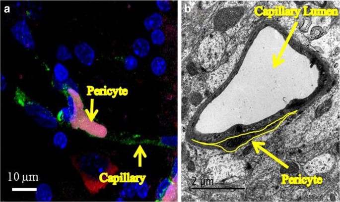
Pericytes have emerged as novel tools for cell-mediated recovery of tissue function [1]. How is it that pericytes, ubiquitous throughout the body, embedded in the basement membrane of capillary walls and often encircling a vessel (Fig. 1) [2, 3] have become a source for cell-based therapy applications? What is the evidence that they can be effective in treating clinical concerns, like enhancing locomotor recovery after brain or spinal cord injury? A review of the unique cell biology of pericytes and the evidence available to date will be helpful in answering these questions.
Pericytes are perivascular cells that are embedded in the basement membrane of capillaries throughout the body. a Immunohistochemical image of a capillary within the spinal cord of a neonatal rat pup. Capillaries (isolectin labeling) are green, smooth muscle cells (α-smooth muscle actin labeling) are red, pericytes (PDGFR-β labeling) are magenta, and nuclei (Hoechst labeling) are blue. b Transmission electron microscope image of a pericyte encircling a brain capillary

No comments:
Post a Comment