Changing stroke rehab and research worldwide now.Time is Brain! trillions and trillions of neurons that DIE each day because there are NO effective hyperacute therapies besides tPA(only 12% effective). I have 523 posts on hyperacute therapy, enough for researchers to spend decades proving them out. These are my personal ideas and blog on stroke rehabilitation and stroke research. Do not attempt any of these without checking with your medical provider. Unless you join me in agitating, when you need these therapies they won't be there.

What this blog is for:

My blog is not to help survivors recover, it is to have the 10 million yearly stroke survivors light fires underneath their doctors, stroke hospitals and stroke researchers to get stroke solved. 100% recovery. The stroke medical world is completely failing at that goal, they don't even have it as a goal. Shortly after getting out of the hospital and getting NO information on the process or protocols of stroke rehabilitation and recovery I started searching on the internet and found that no other survivor received useful information. This is an attempt to cover all stroke rehabilitation information that should be readily available to survivors so they can talk with informed knowledge to their medical staff. It lays out what needs to be done to get stroke survivors closer to 100% recovery. It's quite disgusting that this information is not available from every stroke association and doctors group.

Showing posts with label nanomaterials. Show all posts

Monday, February 16, 2026

Brain-saving shot after stroke? New IV therapy shows promise

 Why is this in India Times and not the top item in the WSO or ASA? This just proves once again that all we have for stroke is fucking failures of stroke associations.

Send me personal hate mail on this: oc1dean@gmail.com. I'll print your complete statement with your name and my response in my blog. Or are you afraid to engage with my stroke-addled mind? No excuses are allowed! You're medically trained; it should be simple to precisely state EXACTLY WHY you aren't working on 100% recovery protocols with NO EXCUSES! I've never received any communications from any stroke association. You'd think they would want to talk to their fiercest critic, but no, they are hiding under a rock someplace, probably don't even know I exist! Swearing at me is allowed, I'll return the favor. Don't even attempt to use the excuse that brain research is hard.

Brain-saving shot after stroke? New IV therapy shows promise

Scientists unveil an experimental IV therapy that not only restores blood flow after stroke but may also shield fragile brain cells from further damage—offering new hope in the fight against long-term disability.

When an ischaemic stroke strikes—the most common type, caused by a clot blocking blood flow to the brain—doctors race against time to restore circulation within the “golden hour” as rapid treatment can save brain tissue and lives.

Yet the sudden return of blood flow, known as reperfusion, can paradoxically trigger a damaging cascade of inflammation and cell death, worsening long-term disability.

Researchers at Northwestern University now report an experimental injectable nanomaterial designed to protect the brain during this fragile window. The therapy, delivered intravenously immediately after blood flow is restored, aims not just to reopen blocked vessels but to shield and repair vulnerable brain cells.

The global need is urgent. According to the World Health Organization, stroke was the third leading cause of death and disability worldwide in 2021, affecting an estimated 93.8 million people.

Lifetime risk has surged by 50 percent over the past two decades, with one in four adults projected to suffer a stroke.

In India alone, stroke accounts for 1.5 to 1.8 million cases annually and ranks as the second leading cause of death after ischemic heart disease, and the third leading cause of disability.

NOVEL PROMISE

Ischemic stroke occurs when a clot blocks a brain artery, starving tissue of oxygen. Hemorrhagic stroke, by contrast, results from a ruptured blood vessel that causes bleeding in the brain. Current treatments focus almost entirely on restoring blood flow through clot-busting drugs or mechanical clot removal.

But while reopening the artery is critical, it does little to directly repair injured brain cells.

The new study, published in Neurotherapeutics, tested a single intravenous dose in mice immediately after reperfusion or restoration of circulation.

Remarkably, the therapy crossed the blood-brain barrier—a protective shield that blocks many drugs from reaching brain tissue—and promoted tissue repair.

Treated mice showed significantly less brain damage, with no detectable toxicity in major organs.

The injectable is built on supramolecular therapeutic peptides – advanced drug delivery systems and functional materials formed by the spontaneous short peptides into organised nanostructures – developed by Northwestern researcher Samuel I. Stupp in 2021.

These engineered molecules are designed to assemble into regenerative structures that support cellular recovery.

Researchers say the approach could one day complement existing stroke therapies by limiting secondary injury and enhancing functional recovery.

Reducing disability is the ultimate goal. Severe strokes often leave patients with lasting physical, cognitive, and emotional impairments, affecting their ability to work and engage with family and society.

The financial and social burden on families and healthcare systems is immense.

A therapy that minimises damage and promotes repair could have a transformative long-term impact.

LONG WAY, STILL

Yet experts urge caution. Senior neurologist Dr Sudhir Kumar of Apollo Hospital, Hyderabad notes that while the findings are scientifically exciting, they remain early-stage and limited to animal models.

Many treatments that show promise in mice fail to replicate results in humans, particularly because real-world stroke patients are typically older and have multiple medical conditions that complicate recovery, he emphasised.

Calling the therapy a breakthrough would be premature, he also pointed out, noting that only well-designed human clinical trials can determine whether this innovative peptide-based strategy will truly change the future of stroke care.

For now, the brain-saving shot remains a promising step forward—one that could, with further testing, redefine how doctors protect the brain after stroke.

Friday, January 16, 2026

Injectable nanomaterial reduces secondary brain injury after ischemic stroke

I can almost guarantee your doctor and hospital will KNOW NOTHING AND DO NOTHING! 

No human research will occur; nothing will be done! That is how fucking incompetent the whole stroke medical world is. Hopefully comeuppance will hit them all with a stroke. And they can regret their incompetence in not solving stroke to 100% recovery!

Al this incompetence is a result of NO leadership firing the incompetent persons!

Injectable nanomaterial reduces secondary brain injury after ischemic stroke

When a person suffers a stroke, physicians must restore blood flow to the brain as quickly as possible to save their life. But, ironically, that life-saving rush of blood can also trigger a second wave of damage - killing brain cells, fueling inflammation and increasing the odds of long-term disability.

Now, Northwestern University scientists have developed an injectable regenerative nanomaterial that helps protect the brain during this vulnerable window.

In a new preclinical study, the team delivered a single intravenous dose, immediately after restoring blood flow, in a mouse model of ischemic stroke, the most common type of stroke. The therapy successfully crossed the blood-brain barrier - a major challenge for most drugs - to reach and repair brain tissue. The material significantly reduced brain damage and showed no signs of side effects or organ toxicity.

Published Jan. 7 in the journal Neurotherapeutics, the findings suggest the new therapy could eventually complement existing stroke treatments by limiting secondary brain injury and supporting recovery.

Current clinical approaches are entirely focused on blood flow restoration. Any treatment that facilitates neuronal recovery and minimizes injury would be very powerful, but that holy grail doesn't yet exist. This study is promising because it's leading us down a pathway to develop these technologies and therapeutics for this unmet need."

Dr. Ayush Batra, associate professor, neurology (neurocritical care) and pathology at Northwestern University Feinberg School of Medicine, co-director of the NeuroVascular Inflammation Laboratory at Northwestern and a neurocritical care physician with Northwestern Medicine

The injectable therapy is based on supramolecular therapeutic peptides (STPs), a platform developed by Northwestern's Samuel I. Stupp. A study published in 2021 in the journal Science demonstrated the use of an STP technology - nicknamed "dancing molecules" - because of the highly dynamic nature of its therapeutic agents that could reverse paralysis and repair tissue in mice after a single injection at the site of severe spinal cord injury. The new study found scientists can administer similar dynamic assemblies of molecules intravenously, without requiring surgery or an invasive injection directly into the brain.

"One of the most promising aspects of this study is that we were able to show this therapeutic technology, which has shown incredible promise in spinal cord injury, can now begin to be applied in a stroke model and that it can be delivered systemically," said Stupp, co-corresponding author and Board of Trustees Professor of Materials Science and Engineering, Chemistry, Medicine and Biomedical Engineering at Northwestern. "This systemic delivery mechanism and the ability to cross the blood-brain barrier is a significant advance that could also be useful in treating traumatic brain injuries and neurodegenerative diseases such as ALS."

Stupp also is founding director of the Center for Regenerative Nanomedicine. He has appointments in the McCormick School of Engineering, Weinberg College of Arts and Sciences and Feinberg School of Medicine.

Study mimicked real-world stroke treatment

Acute ischemic stroke, which accounts for 80% of all strokes in the U.S., is a devastating condition and is one of the leading causes of morbidity and mortality worldwide, Batra said. Ischemic strokes occur when a clot blocks blood flow to the brain. Physicians reopen the vessel by administering "clot-busting" drugs or using devices to surgically remove the clot.

Severe strokes can lead to permanent, significant disability that affects a patient's quality of life and their ability to return to work and engage with their family and society.

"It has not only a significant personal and emotional burden on patients, but also a financial burden on families and communities," he said. "Reducing this level of disability with a therapy that could potentially help in restoring function and minimizing injury would really have a powerful long-term impact."

The findings are highly relevant for future clinical applications because the scientists tested the approach in a mouse model that closely mimics real-world ischemic stroke treatment, Batra said. They first blocked blood flow to simulate a major ischemic stroke and then restored it (a process called reperfusion), just as doctors restore blood flow acutely for ischemic stroke patients. 

The scientists monitored the mice for seven days and didn't observe any significant side effects or biocompatibility issues such as toxicity or immune system rejection. They used advanced imaging techniques, such as real-time intravital intracranial microscopy seen in this video, to confirm the therapy localized to the stroke injury site. Compared to untreated mice, those treated with the "dancing molecules" had significantly less brain tissue damage, reduced signs of inflammation and reduced signs of excessive, damaging immune response.

Stupp said the therapy has pro-regenerative and anti-inflammatory properties, both of which contributed to the positive results.

"You get an accumulation of harmful molecules once the blockage occurs and then suddenly you remove the clot and all those 'bad actors' get released into the bloodstream, where they cause additional damage," Stupp said. "But the dancing molecules carry with them some anti-inflammatory activity to counteract these effects and at the same time help repair neural networks." 

Dynamic 'dancing molecules' can be dialed down in concentration

The secret behind Stupp's "dancing molecules" breakthrough therapeutic is tuning the collective motion of molecules, so they can find and properly engage constantly moving cellular receptors. The treatment sends signals that encourage nerve cells to repair themselves. For example, it can help nerve fibers (called axons) grow again and reconnect with other nerve cells, restoring lost communication. This process is called plasticity, which means the brain and spinal cord can adapt and rebuild connections after injury. 

In previous studies, scientists injected the dancing molecules as a liquid, and when used to treat spinal cord injury, the therapy immediately gels into a complex network of nanofibers that mimic the dense, extracellular matrix of the spinal cord. By matching the matrix's structure, mimicking the motion of biological molecules and incorporating signals for receptors, the synthetic materials are able to communicate with cells.

In the new study, the scientists dialed down the concentration of supramolecular peptide assemblies to prevent possible clotting as the therapy enters the bloodstream. Smaller aggregates of peptides easily crossed the blood-brain barrier. Once enough molecules cross, larger nanofiber assemblies can form in brain tissue to produce a more potent therapeutic effect, Stupp said.

"We chose for this stroke study one of the most dynamic therapies we had in terms of its molecular structure so that supramolecular assemblies would have a better probability of crossing the blood-brain barrier," Stupp said. 

Optimizing therapeutic targeting

The fact that seemingly effective therapies cannot cross the blood-brain barrier has plagued the neuroscience field for decades, Batra said. This new therapy could change that.

When a physician acutely restores blood flow to a region of the brain in a stroke patient, the blood-brain barrier permeability is locally increased, naturally creating a transient opening and opportunity for therapeutic intervention, Batra said.

"Add to that a dynamic peptide that is able to cross more readily, and you're really optimizing the chances that your therapy is going where you want it to go," Batra said. 

Next steps

Further studies will need to assess whether this treatment can support longer-term, functional recovery, Batra said. For instance, many stroke patients suffer from significant cognitive decline throughout the subsequent year after a stroke. The new therapy is primed to address that secondary injury, Batra said, but the studies will require a longer follow-up period and more sophisticated behavioral testing. 

In addition, the team is interested in testing whether additional regenerative signals could be incorporated into the therapeutic peptides to produce even better results.

The study is titled, "Toward Development of a Dynamic Supramolecular Peptide Therapy for Acute Ischemic Stroke." Graduate student Zijun Gao and postdoctoral researcher Luisa Andrade da Silva are co-first authors of the paper. 

Funding for this study was primarily provided by the SQI Synthesizer Grant Program at the Center for Regenerative Nanomedicine.

Monday, March 24, 2025

Nanomaterial Technologies for Precision Diagnosis and Treatment of Brain Hemorrhage

 But is it faster than this? Or don't you even know about this one?

And then this to rule out a bleeder.

New Device Quickly Assesses Brain Bleeding in Head Injuries - 5-10 minutes April 2017 

The latest here:

Nanomaterial Technologies for Precision Diagnosis and Treatment of Brain Hemorrhage

, ,
https://doi.org/10.1016/j.biomaterials.2025.123269
Get rights and content

Highlights

  • Nanomaterial-assisted imaging techniques that enhance hemorrhage detection accuracy through real-time, high-resolution assessments of blood-brain barrier integrity, cerebral perfusion, and hemorrhage progression were summarized.
  • Nanomaterial-based systems that enable precise drug delivery and support neural repair, including the control of primary/secondary injuries and enhancement of neuroprotection, were systematically reviewed.
  • The benefits, challenges, and future perspectives regarding the research and clinical translation of nanomaterial-based agents for brain hemorrhage were discussed.

Abstract

Brain hemorrhage events present complex clinical challenges due to their rapid progression and the intricate interplay of oxidative stress, inflammation, and neuronal damage. Traditional diagnostic and therapeutic approaches often struggle to meet the demands for timely and effective intervention. This review explores the cutting-edge role of nanomaterials in transforming cerebral hemorrhage management, focusing on both diagnostic and therapeutic advancements. Nanomaterial-assisted imaging techniques, such as optical imaging, magnetic resonance imaging, and magnetic particle imaging, significantly enhance the accuracy of hemorrhage detection by providing real-time, high-resolution assessments of blood-brain barrier (BBB) integrity, cerebral perfusion, and hemorrhage progression, which is critical for guiding intervention strategies. On the therapeutic front, nanomaterial-based systems enable the precise delivery of drugs and bioactive molecules, fostering neural repair and functional recovery while minimizing systemic side effects.(Then where exactly are the protocols delivering that?)  Furthermore, multifunctional nanomaterials not only address the primary injury but also offer precise control over secondary injuries, such as edema and oxidative stress. Their ability to enhance neuroprotection, prevent re-bleeding, and stimulate brain tissue regeneration provides a holistic approach and marks a significant advancement in brain hemorrhage therapy. As the field continues to advance, nanotechnology is set to fundamentally reshape the clinical management and long-term outcomes of brain hemorrhages, presenting a paradigm shift towards personalized and highly effective neurological care.

Introduction

Brain hemorrhages represent life-threatening medical emergencies characterized by bleeding within the cranial cavity, often resulting in severe neurological deficits and high mortality rates.[1], [2] The etiology of brain hemorrhage is diverse, encompassing traumatic brain injuries (TBI) sustained in accidents or falls, and non-traumatic events such as chronic hypertension, vascular malformations (e.g., arteriovenous malformations), aneurysm rupture, amyloid angiopathy, and the transformation of ischemic strokes into hemorrhagic events. Each of these etiological factors presents distinct challenges both in immediate management and long-term rehabilitation.[3], [4], [5]. Clinical outcomes are heavily dependent on the underlying causes, as well as the location and volume of the hemorrhage, which often lead to acute neurological dysfunction, elevated intracranial pressure, and potential long-term disability. Patients often suffer from impaired cognitive and motor functions, substantial reductions in quality-of-life, and extended rehabilitation periods.[6], [7], [8], [9] Given the complexity of brain hemorrhage, timely and accurate diagnosis is crucial for effective management and treatment.
In clinic practice, diagnostic tools such as computed tomography (CT) and magnetic resonance imaging (MRI) are essential for determining the extent, type, and impact of hemorrhages on surrounding brain structures.[10], [11], [12] Treatment strategies primarily focus on stabilizing the patient, managing intracranial pressure, and addressing underlying causes. These approaches typically include pharmacological interventions to control blood pressure and prevent further bleeding, alongside surgical options to evacuate hematomas and repair damaged blood vessels.[13], [14], [15], [16] Despite the advances in medical technology, significant challenges remain in diagnosis and treatment of brain hemorrhages. Accurate diagnosis is particularly complex due to the need to distinguish among various types of hemorrhages—subarachnoid, subdural, epidural, and intracerebral—each of which requires distinct management strategies. Clinical imaging techniques must be employed effectively to identify the location and extent of the hemorrhage, while also distinguishing it from other neurological conditions, such as intracerebral edema, calcification or tumors.[17], [18], [19] Treatment challenges involve addressing both primary and secondary injuries. Primary brain hemorrhage management entails controlling the hemorrhage, preventing re-bleeding, managing elevated intracranial pressure, and determining appropriate surgical interventions. Secondary injuries pose even greater challenges, requiring effective long-term care and rehabilitation to mitigate secondary inflammation and further damage. Addressing these concerns is critical for preventing neurological deficits and managing complications such as brain edema and infections, which are vital for recovery.[20], [21], [22] These complexities highlight the need for a coordinated, multidisciplinary approach to improve patient outcomes in brain hemorrhage management.
In recent decades, rapid advancements of nanotechnology have led to the development of engineered nanomaterials as innovative alternatives to traditional contrast agents and therapeutic drugs.[23], [24], [25], [26], [27], [28], [29] In the realm of hemorrhage medical imaging, nanomaterial-based imaging agents, due to their optimal hydrodynamic diameters, can remain confined within blood vessels and only extravasate at sites of vascular injury during bleeding.[30], [31] This enables precise depiction of the location and extent of hemorrhages. Moreover, the specific molecular targeting capability of nanomaterial-based agents allows for the spatiotemporal mapping of key molecules within the site of cerebral hemorrhage. In terms of treatment, nanomaterial-based therapeutic agents can improve the pharmacological and pharmacokinetic profiles of conventional drugs or enable targeted delivery through functionalized drug carriers.[32], [33], [34], [35] These approaches introduce novel strategies for treating hemorrhage, including advancements in neuroprotection, complication prevention, and nerve regeneration. By leveraging their unique properties, nanomaterial-based agents have been developed and explored for the diagnosis and treatment of brain hemorrhages across different etiologies.[36], [37], [38] These agents hold significant promise in addressing the specific clinical needs related to cerebral hemorrhage.
Despite this progress made, there still leaves considerable room for improvement in nanomaterials-based agents for brain hemorrhage, with the potential to further enhance the clinical efficacy of both diagnosis and treatment. However, significant challenges continue to impede the widespread implementation of nanomaterial-based approaches in managing cerebral hemorrhage.
In this review, the current status of nanomaterial-based strategies for diagnosing and treating various types of brain hemorrhages is summarized. It encompasses imaging techniques for stroke and TBI-related hemorrhages, as well as therapeutic approaches aimed at resisting oxidative stress, alleviating inflammation, and promoting brain tissue repair and functional recovery (Scheme 1). By presenting effective examples of in vivo imaging and treatment, the benefits and opportunities offered by nanomaterial-based agents for these conditions are highlighted. Additionally, the challenges and future perspectives related to both fundamental research and clinical translation of nanomaterial-based agents for brain hemorrhage are discussed. The overarching aim of this review is to provide a comprehensive understanding of recent advancements in nanomaterial-based strategies for high-resolution, high-sensitivity diagnosis and effective treatment of brain hemorrhage, which holds significant promise for improving clinical outcomes and reducing morbidity and mortality associated with this disease.

Access through your organization

Monday, June 19, 2023

Revolution in neuroscience: 2D nanomaterials propel advances in brain repair, treatment, and diagnosis

Are there any ways this could be used to treat stroke? Ask your doctor and not politely.

Revolution in neuroscience: 2D nanomaterials propel advances in brain repair, treatment, and diagnosis

Different types of nanomaterials have been used in neuroscience, including two-dimensional (2D) nanomaterials widely known for their unique structures and physicochemical properties. In a recent Journal of Nanobiotechnology review, scientists discuss the applications of 2D nanomaterials in neuroscience.

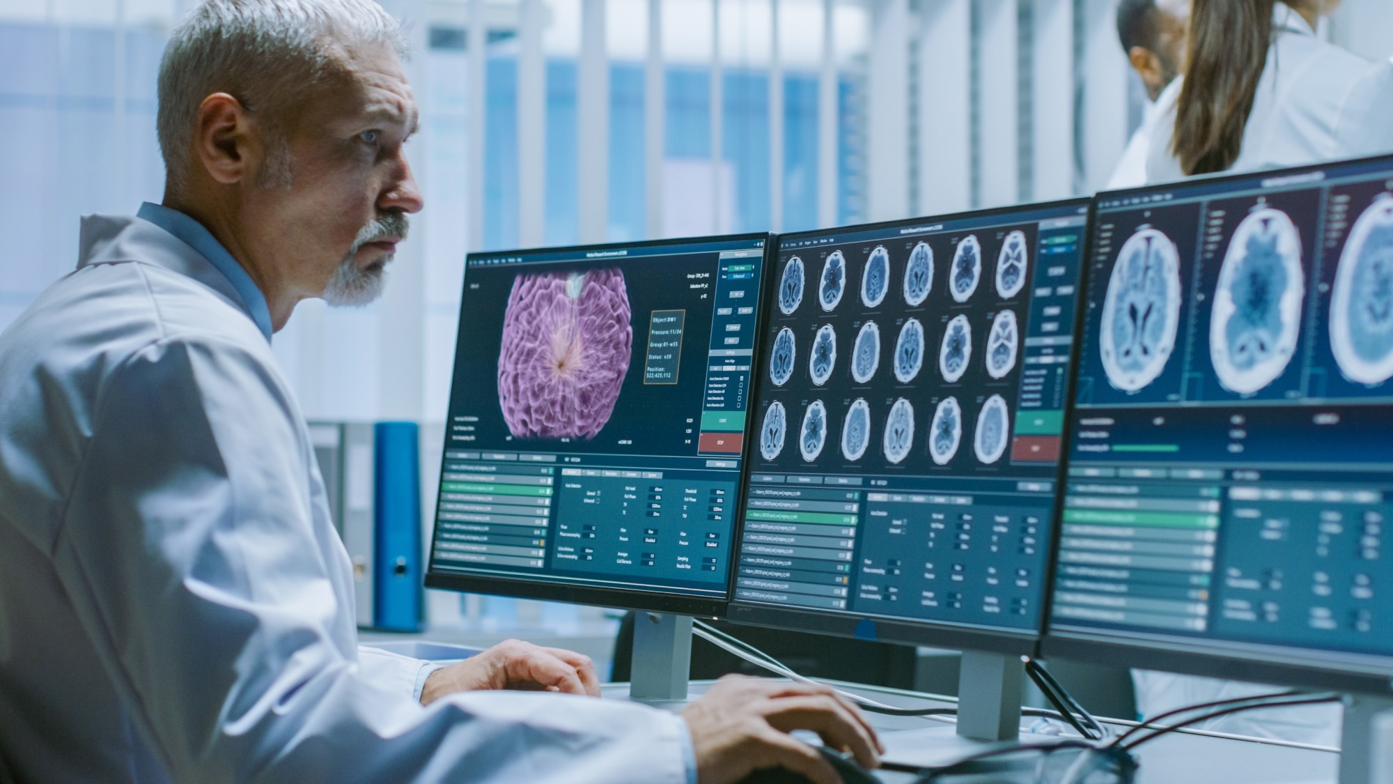
Study: Biomedical application of 2D nanomaterials in neuroscience. Image Credit: Gorodenkoff / Shutterstock.com Study: Biomedical application of 2D nanomaterials in neuroscience. Image Credit: Gorodenkoff / Shutterstock.com

Types of 2D nanomaterials

The thickness of 2D nanomaterials is typically between one or more atomic layers, wherein electrons move freely in the other two dimensions beyond the nanoscale of less than 100 nanometers (nm). These materials exhibit superior biological and physiochemical properties and have been applied in regenerative medicine and tissue engineering.

Some common types of 2D nanomaterials include graphene, layered double hydroxides (LDHs), black phosphorous (BP), transition metal carbides (TMCs), and transition metal dichalcogenides (TMDCs). Many of these nanomaterials are used in neuroscience, particularly for neuroinflammatory reduction, injured neural cell/tissue repairment, synaptic modulation, and stem cell fate regulation.

Graphene, the first discovered 2D nanomaterial, comprises carbon atoms covalently bonded in a hexagonal lattice. Several functionalized forms of graphene-based nanomaterials (GBNs) have been developed, of which include carboxyl graphene, graphene oxide (GO), and reduced GO (RGO).

Another 2D nanomaterial that has recently gained much attention is TMDC. The generalization formula of TMDCs is MX2, where M is the transition metal atoms sandwiched between two layers of chalcogen atoms (X). In 2014, scientists discovered 2D BP, which consists of stable allotropes of phosphorus that are collectively between one and two nm thick.

LDHs interact with organic molecules through the anionic exchange, which is characterized by a phenomenon known as intercalation. This nanomaterial is chemically stable with pH-dependent biodegradability.

Applications of 2D nanomaterials in neuroscience

Different types of 2D nanomaterials have been applied in various aspects of neuroscience, such as neural repair, synaptic stimulation, neurodegenerative diseases (NDs), and glioma.

Neural repair and regeneration

The nervous system has a poor ability to regenerate axonal connections after injury or disease. In addition, it is difficult for drugs to reach damaged areas through the blood-brain barrier (BBB).

Recently, 2D nanomaterials have been extensively explored for their therapeutic potential, particularly in neural repair and regeneration. Since GBNs exhibit significant electrical conductivity and good biocompatibility, they are excellent candidates for neural tissue engineering. 

Graphene substrate has been used as cell scaffolds with optimal electrical stimulations. Furthermore, graphene substrates have been shown to support cell adhesion and promote cell proliferation. Poly (lactic-co-glycolic acid) (PLGA) nanofiber pads coated with GO and methylene blue (MB) improve the ability of neural progenitor cells (NPCs) to counter disease stressors.

Synaptic simulation

Moreover, 2D nanomaterials have been used in the development of artificial synaptic devices that possess optoelectronic, electronic, electrochemical, and mechanical properties.

Graphene, coupled with other materials, forms heterogeneous structures that function as artificial synapses. For example, graphene/tantalum pentoxide/graphene phototransistor exhibits synapse characteristics visible under electromagnetic radiation.

NDs

NDs are chronic diseases characterized by neuronal degeneration and myelin damage, which cause cognitive impairment and morbidity in older adults. Three major types of NDs include Parkinson’s disease (PD), Alzheimer’s disease (AD), and Huntington’s disease (HD), all of which are associated with protein aggregation or misfolding.

Previously, 2D nanomaterials have been used for the early diagnosis and subsequent treatment of NDs. Nickle aluminide LDHs combined with graphene monolayers are used to track dopamine (DA) in neuronal cells in real-time to support DA-based PD diagnosis.

Prussian blue nanoparticle-supported Molybdenum disulfide nanocomposites are also considered a potential DA probe. BP tagged with brain-targeting ligand lactoferrin and loaded with Paeoniflorin has also been used in PD treatment.

LHD scaffolds with short interfering ribonucleic acid (siRNA) promote the ability of siRNAs to target and destroy specific messenger RNAs (mRNAs), which can be used for the treatment of HD. In addition, GO has the capacity to enhance the clearance of mutant huntingtin (Htt) that contributes to HD pathogenesis.

Traumatic diseases

Two leading causes of mortality and morbidity linked to trauma in the central nervous system (CNS) are traumatic brain injury (TBI) and spinal cord injury (SCI).

TBI is associated with brain injury caused by an external mechanical force that might cause temporary or permanent brain impairment. Biodegradable mesoporous silicon nanoparticles encapsulated with GO nanosheets have been developed to transport therapeutic siRNA and effectively silence targeted genes.

SCI has been associated with damage in the spinal cord that could result in varying degrees of quadriplegia or paraplegia. PLGA/GO has been designed to carry brain-derived neurotrophic factor (BDNF) and insulin-like growth factor 1 (IGF-1), which could provide protection to NSCs from oxidative stress. In addition, GO has been deemed the primary nanomaterial for SCI treatment.

Glioma

Glioma, a common craniocerebral tumor caused by the malignant transformation of glial cells in the spinal cord and brain, accounts for about 80% of all malignant brain tumors. Recently, 2D nanomaterials have been proposed for treating and diagnosing gliomas. BP, for example, has superior light absorption characteristics and has been used in photothermal therapy (PTT) of cancers, including glioma.

Journal reference:
  • Li, K., Ji, Q., Liang, H., et al. (2023) Biomedical application of 2D nanomaterials in neuroscience. Journal of Nanobiotechnology 21(181). doi:10.1186/s12951-023-01920-4