Changing stroke rehab and research worldwide now.Time is Brain! trillions and trillions of neurons that DIE each day because there are NO effective hyperacute therapies besides tPA(only 12% effective). I have 523 posts on hyperacute therapy, enough for researchers to spend decades proving them out. These are my personal ideas and blog on stroke rehabilitation and stroke research. Do not attempt any of these without checking with your medical provider. Unless you join me in agitating, when you need these therapies they won't be there.

What this blog is for:

My blog is not to help survivors recover, it is to have the 10 million yearly stroke survivors light fires underneath their doctors, stroke hospitals and stroke researchers to get stroke solved. 100% recovery. The stroke medical world is completely failing at that goal, they don't even have it as a goal. Shortly after getting out of the hospital and getting NO information on the process or protocols of stroke rehabilitation and recovery I started searching on the internet and found that no other survivor received useful information. This is an attempt to cover all stroke rehabilitation information that should be readily available to survivors so they can talk with informed knowledge to their medical staff. It lays out what needs to be done to get stroke survivors closer to 100% recovery. It's quite disgusting that this information is not available from every stroke association and doctors group.

Showing posts with label brain scans. Show all posts

Thursday, September 29, 2022

How Magnetic Brain Scans Could Reveal Brain Age

We could use this to prove that cognitive improvement protocols can reverse the

5 lost years of brain cognition due to your stroke.

Assuming that your doctor has such protocols, that would be a bad assumption.

The latest here:

How Magnetic Brain Scans Could Reveal Brain Age

The scans reveal self-organizing behaviour in the brain that changes as we get older, say scientists.

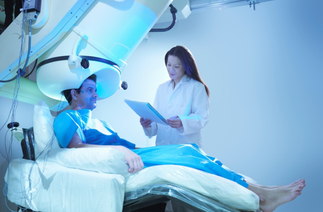
Doctor and patient using magnetoencephalography (MEG) scanner
(Credit:Image Source Trading Ltd/Shutterstock)

Newsletter

Sign up for our email newsletter for the latest science news
 

One of the curious properties of brain activity — the firing of neurons — is that it follows certain patterns. One of these is that brain activity tends to be maintained rather than dampened or amplified.

This turns out to be a special phenomenon of self-organization. Active neurons tend to trigger other neurons. If each active neuron triggers more than one other, any activity is rapidly amplified in a chain reaction. If each neuron triggers less than one other, the activity tends to fizzle out, like a damp firework.

But to maintain activity, each active neuron must trigger about one other neuron. Neuroscientists call this criticality and believe that it maximizes the flow of information through a neural network.

Most healthy brain activity seems to occur in this special critical state.

And that raises an interesting question. Brain function changes substantially as we age. Older people tend to be more forgetful, less focused and more easily distracted. But how does this change the nature of criticality in the brain and can this be observed?

Self-Organized Behaviour

Today, we get an answer of sorts, thanks to the work of Leandro Fosque at Indiana University in Bloomington, and colleagues, who say they have found a correlation between age and critical brain activity that could one day be used for diagnostic monitoring.

The team analyzed brain activity measured over 8 minutes in more than 600 people aged between 18 and 88. The data was gathered using magnetoencephalography (MEG), which measures the magnetic fields generated by the electrical activity of neurons.

This is a dataset known as the Cambridge Centre for Ageing and Neuroscience (Cam-CAN) resting state magnetoencephalography dataset and it is the largest available that spans the full range of adult life.

Of course, it is not possible to measure how many neurons a single active neuron triggers(Acually it can be measured. Use nanowires to listen in on single neurons.)

. But this triggering causes an avalanche of neural activity and it is this avalanche—its size and duration — that MEG reveals.

The relationship between the size of the avalanches versus their duration also follows a clear pattern that is a direct result of critical brain organization.

In fact, when plotted on a log-log graph, this relationship is a straight line. And most healthy brain activity turns out to lie close to this line, a phenomenon that physicists call quasicriticality.

However, an important feature is that this activity is spread along the line. The question Fosque and colleagues ask is whether human factors such as age and gender determine where on this line brain activity sits.

And in crunching the data, they discovered exactly the correlation they were looking for. “We found that there is a small but significant negative correlation between age and the position on the line,” they say.

Critical Avalanches

It turns out that the brain activity of older people tends to be lower on the line suggesting that their brain avalanches are smaller and shorter in duration. The activity is also more susceptible to small changes. This may explain why older people are more easily distracted. The team found other correlations too, such as a statistically significant gender bias. That means it ought to be possible to tell the gender of a brain by measuring its quasicritical behavior.

The results from Fosque and co contradict some earlier papers but are based on a larger data set than has been available before now. The results will need to be validated by others working on different data sets but they have significant potential.

Fosque and co’s findings suggest it may be possible to use MEG brains scans as a diagnostic tool for certain age-related brain conditions and perhaps for measuring the ordinary process of aging in the brain and determining brain age.

And they say that in future work they will look at datasets of the brain activity of people suffering various neurological conditions, such as epilepsy, dementia and depression. Obviously, it is early days for this kind of work. But if it turns out to be fruitful, a new era of neurological diagnostics could be upon us.


Ref: Quasicriticality explains variability of human neural dynamics across life span : arxiv.org/abs/2209.02592

Thursday, April 30, 2020

ASX-listed company show successful results from first clinical trial - portable medical imaging technology

But you never say how fast you are. Can you even compete with these other fast diagnosis tools? It still seems to require a neurologist which I think needs to be removed from the equation. But since I'm not medically trained I will just shut up and be quiet, your stroke medical professionals will explain how fast you need to be treated to get 100% recovery and EXACTLY how this technology will do that. 

Hats off to Helmet of Hope - stroke diagnosis in 30 seconds   February 2017


Microwave Imaging for Brain Stroke Detection and Monitoring using High Performance Computing in 94 seconds March 2017


New Device Quickly Assesses Brain Bleeding in Head Injuries - 5-10 minutes April 2017

The latest here:

ASX-listed company show successful results from first clinical trial


29 April 2020
facebook sharing button
twitter sharing button
sharethis sharing button
EMVision Medical Devices Ltd (EMV), a company that took part in Switzer’s Small and Micro Cap Investor Day on 3 March 2020, has released new information on the ASX, releasing the preliminary images from their clinical trial.
READ MORE: https://switzer.com.au/the-experts/sophia-katsinas/a-taste-of-switzers-small-and-micro-cap-investor-day-event/
EMVision aims to revolutionise the imaging of strokes and traumatic brain injuries through the development and commercialisation of portable medical imaging technology.
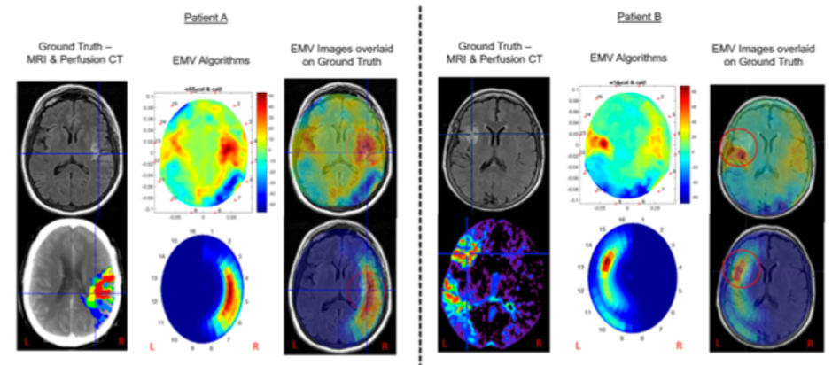
Its first brain scanner is a commercial product at a manageable price that allows for quick, efficient and scalable brain scanning. EMVision’s scanners display strong correlation with CT and MRI scanners. The clinical assessment results show these similarities that produce the detection and localisation of abnormal brain tissue. The advantage of the EMV images, as shown in the images below, is that the EMV images distinguish abnormal brain tissue from healthy brain tissue, which apparently is often less clear in CT scans.


The technology is currently in the clinical trial stage, and the company has its hat in the ring for a share in a $50 million+ Medical Research Future Fund Grant pledged with the Australian Stroke Alliance.
Co-chairs of the Australian Stroke Alliance and past presidents of the World Stroke Organization, Professors Stephen Davis AM and Geoffrey Donnan AO said the results of the trial were “promising”. Professor Davis commented “These early images are clinically promising, clearly showing the effects of ischemic stroke in the same region as the gold standard imaging methods”. While Professor Donnan said “the lightweight portability of the device makes it a potential candidate for emergency stroke imaging in the prehospital setting.”
The machine will be accessible in the prehospital triage stage, meaning parademics and on-the-scene medical professionals will be able to identify the issue earlier, allowing for more targeted treatment options.
EMVision CEO, Dr Ron Weinberger was pleased with the result, saying “We are confident that as we continue to process further stroke patient data, we will demonstrate our unique value proposition to meet a major unmet clinical need in rapid and portable stroke diagnosis and monitoring.”

Wednesday, January 22, 2020

Thousands of lives ‘at risk’ because nearly HALF of NHS hospitals are low of stroke specialists

If your doctors and stroke hospital can't immediately see the obvious solution maybe you want to replace them.  You replace the need for doctors to look at scans by these. And they would be much much faster.

Hats off to Helmet of Hope - stroke diagnosis in 30 seconds   February 2017


Microwave Imaging for Brain Stroke Detection and Monitoring using High Performance Computing in 94 seconds March 2017


New Device Quickly Assesses Brain Bleeding in Head Injuries - 5-10 minutes April 2017

Thousands of lives ‘at risk’ because nearly HALF of NHS hospitals are low of stroke specialists 

Thousands of lives are potentially at risk because half of NHS hospitals are running low of specialist stroke doctors, experts warn. 
Stroke – caused by a block of blood flow to the brain, normally through a blood clot – is the fourth biggest killer in the UK. 
But stroke consultant positions are at a ‘worryingly low level’, a scathing report from a leading charity has warned.
The Stroke Association said Britain is ‘hurtling’ towards a major stroke crisis, unless the NHS can recruit specialist medics.
A lack of specialists on the ward means decisions may not be made quickly enough. Brain scans must be assessed by stroke consultants who also decide what urgent care is needed.
Data analysed by the charity shows there is a large variation in access to the services that stroke patients need across England, Wales and Northern Ireland. 
Ninety-three per cent of NHS hospitals do not employ enough clinical psychologists to support patients following their traumatic ordeal. 
Three quarters of stroke survivors battle depression, anxiety, lack of confidence or mood swings, charities say. 
Stroke Association chief executive Juliet Bouverie said: ‘Unless these workforce issues are urgently addressed, we are hurtling our way to a major stroke crisis in the next few years.  
‘The lack of senior doctors and also of trainees to fill these gaps is worsening and is a ticking time-bomb for an already stretched health service. 
‘The stroke skills gap threatens the sustainability of many services and puts increased pressure on local hospitals. 
‘There are over 100,000 strokes every year in the UK and this is estimated to rise to 150,000 over the next five years which will increase the pressure on stroke wards further.’
The charity highlighted the new findings from the Sentinel Stroke National Audit Programme’s (SSNAP) Acute Organisational Audit Report.
According to these figures, 48 per cent of all hospitals – 81 of 169 hospitals –  have had vacant stroke consultant posts unfilled for at least one year.
It’s an increase on the 40 per cent in 2016 and 26 per cent in 2014. Data suggests there is a similar outlook in Scotland, the BBC reports. 
In London, eight of 22 trusts had at least one vacant position. Almost every trust in East Midlands – seven out of eight – had a vacancy.
Half of hospitals in North of England, Yorkshire and Humber, South West, Wessex and Northern Ireland reported at least one unfilled position. 
‘Gaping holes’ in staffing levels of experienced stroke professionals may jeopardize the recovery of patients, the Stroke Association says. 
Effective treatment of stroke as soon as possible can prevent long-term disability and save lives. 
Specialist doctors are needed to look over brain scans and implement the best treatment as soon as possible to prevent death and lasting muscle weakness, paralysis, stiffness, or changes in sensation. 
Ms Bouverie said: ‘It really matters. Time loss is brain loss.’
Stroke does not just have the potential to affect cognitive function, but the trauma and suddenness of a stroke can also be really difficult to deal with.
One in six people have suicidal thoughts after their stroke, research by the charity has previously found.  
Psychological support is vital for those that need it straight after a stroke as well as during recoveries – but only seven per cent of hospitals are reaching recommended staffing levels. 
Overall, just 16 per cent of hospitals achieved seven out of ten criteria, which include the presence of a stroke specialist and enough nurses at weekends.
Ms Bouverie said: ‘Stroke happens in the brain, the control centre for who we are and what we can do, which is why it is vital that hospitals have the right amount of the right staff ready to support both the mental health and physical effects your stroke can have on you.
‘We are deeply concerned by the rate at which highly qualified stroke doctors are leaving the profession and the slow uptake of stroke medicine by new doctors. 
‘The highest standards of stroke treatment and rehabilitation must be available to all. The progress in stroke treatment and care over the past 10 years run the risk of being wasted without experienced doctors to deliver world class stroke services.’
Professor Tom Robinson, outgoing president of the British Association of Stroke Physicians, said: ‘We must urgently address the lack of professional stroke staff to ensure that patients have access to the best treatment as quickly as possible.’

Wednesday, August 15, 2018

Two simple tests could help to pinpoint cause of stroke

So write up a protocol and get it distributed to all stroke hospitals.  Do you really think every stroke hospital has staff to read research and implement in their hospital? You as a survivor are screwed because your hospital is incompetent in that regard.

Two simple tests could help to pinpoint cause of stroke

Detecting the cause of the deadliest form of stroke could be improved by a simple blood test added alongside a routine brain scan, research suggests.
Combining the with a brain scan could provide key genetic information that may help identify those most at risk from a second , doctors say.
Experts say the new approach could revolutionise the way doctors manage strokes caused by bleeding in the brain, known as intracerebral haemorrhage (ICH).
ICH accounts for up to 50 per cent of all strokes worldwide.(Does no one vet your information? 80-85% are clot based.) Around half of those affected die within one year.
Researchers used the and brain scan images to detect a condition known as cerebral amyloid angiopathy (CAA), which can cause ICH and is linked to a higher risk of further strokes and dementia.
CAA is caused by a build-up of a protein known as amyloid in the walls of vessels in the brain.
University of Edinburgh researchers used computed tomography (CT) scans in more than 100 patients who died following their first ICH. They collected blood samples to test a gene called APOE, which is linked to CAA.
By combining simple CT scan images with a genetic blood test, researchers could accurately spot if an ICH had been caused by CAA.
This new approach could help identify people who are at higher risk after their ICH, scientists say.
It could also improve ICH diagnosis in developing countries as CT scanning and blood testing is available worldwide.
Dr Mark Rodrigues, Wellcome Trust Clinical PhD Programme Fellow at the University of Edinburgh, said: "Identifying the cause of a haemorrhage is important to planning patient care. Our findings suggest that the combination of routine CT scanning with APOE gene testing can identify those whose ICH has been caused by CAA - a group who may be more at risk of another ICH or dementia."
The study is published in Lancet Neurology and was funded by the Medical Research Council, Stroke Association and Wellcome Trust.
More information: Lancet Neurology (2018). DOI: 10.1016/S1474-4422(18)30006-1

Journal reference: Lancet Neurology search and more info website
Provided by: University of Edinburgh search and more info website

Brain scan checklist set to boost care for stroke survivors

Survivors don't care about 'care', they want RESULTS, what the fuck is your stroke hospital doing to deliver results? 
https://medicalxpress.com/news/2018-08-brain-scan-checklist-boost-survivors.html

People who suffer a stroke caused by bleeding in the brain could be helped by four simple checks of their brain scans, research suggests.
The checks could help spot people at risk of further bleeding so they can be monitored more closely.
Experts say this could help improve outcomes for the millions of people around the world who experience a brain bleed each year.
Bleeding in the brain—known as an or ICH—is the most deadly form of stroke.
Only one in five patients survives without . Of the remainder, half are likely to die within a month and half will be left with a long-term disability.
Cases of ICH are diagnosed by , but until now it has been difficult to predict which patients will continue bleeding. Those who do are expected to have worse outcomes.
Research led by the University of Edinburgh analysed data from studies around the world involving more than 5,000 patients.
The team identified four factors that helped doctors predict whether patients were likely to experience further bleeding.
These include the size of the bleed and whether or not the patient was taking medication, such as aspirin or warfarin, to thin their blood or prevent clotting.
Experts say the checks can be applied during routine care to help medical staff decide the best way to continue monitoring each patient. (Wrong focus;which intervention will resolve the damage from the stroke? Survivors don't care about monitoring or prediction, they want 100% recovery. GET THERE!)
Researchers also looked at the benefit of an advanced brain scanning technique—called CT angiography—for predicting a person's risk of ongoing bleeding.
The scan involves injecting a coloured dye into the patient's bloodstream and checking if it can be seen leaking into the brain.
For patients who showed leakage of the dye, the test was of little value in addition to the four simple checks for predicting their risk of ongoing bleeding, researchers found.
Incorporating the four checks into patient care could help to improve survival, especially in low or middle-income countries, where patients may not have access to CT angiography.
Experts from dozens of research centres worldwide contributed to the study, which is the largest of its kind to date.
The research, published in The Lancet Neurology, was funded by the UK's Medical Research Council and the British Heart Foundation.
Professor Rustam Al-Shahi Salman, of the University of Edinburgh's Centre for Clinical Brain Sciences, said: "We have found that four simple measures help doctors to make accurate predictions about the risk of a haemorrhage growing. These can be used anywhere in the world. Better prediction can help us identify which might benefit from close monitoring and treatment. We hope that an app could help doctors to do this. The next step is to find an effective treatment to stop the bleeding."(Great, more followup needed, never occur since we have NO stroke leadership or strategy.)
More information: The Lancet Neurology, (2018). DOI: 10.1016/S1474-4422(18)30253-9

Journal reference: Lancet Neurology search and more info website
Provided by: University of Edinburgh search and more info website

Friday, June 29, 2018

Scientists can predict intelligence from brain scans

So our researchers should come up with objective testing to determine if this loss of cognition via stroke is correct.

brain injury patients were estimated to be around five years older on average than their real age


https://medicalxpress.com/news/2018-06-scientists-intelligence-brain-scans.html
If you've ever lied about your IQ to seem more intelligent, it's time to fess up. Scientists can now tell how smart you are just by looking at a scan of your brain.
Actually, to be more precise, the scientists themselves aren't looking at your brain scan; a machine-learning algorithm they've developed is.
In a new study, researchers from Caltech, Cedars-Sinai Medical Center, and the University of Salerno show that their new computing tool can predict a person's from imaging (fMRI) scans of their resting state brain activity. Functional MRI develops a map of brain activity by detecting changes in blood flow to specific brain regions. In other words, an individual's intelligence can be gleaned from patterns of activity in their brain when they're not doing or thinking anything in particular—no math problems, no vocabulary quizzes, no puzzles.
"We found if we just have people lie in the scanner and do nothing while we measure the pattern of activity in their brain, we can use the data to predict their intelligence," says Ralph Adolphs (Ph.D. '92), Bren Professor of Psychology, Neuroscience, and Biology, and director and Allen V. C. Davis and Lenabelle Davis Leadership Chair of the Caltech Brain Imaging Center.
To train their algorithm on the complex patterns of activity in the human brain, Adolphs and his team used data collected by the Human Connectome Project (HCP), a scientific endeavor funded by the National Institutes of Health (NIH) that seeks to improve understanding of the many connections in the . Adolphs and his colleagues downloaded the and intelligence scores from almost 900 individuals who had participated in the HCP, fed these into their algorithm, and set it to work.
After processing the data, the team's algorithm was able to predict intelligence at statistically significant levels across these 900 subjects, says Julien Dubois (Ph.D. '13), a postdoctoral fellow at Cedars-Sinai Medical Center. But there is a lot of room for improvement, he adds. The scans are coarse and noisy measures of what is actually happening in the brain, and a lot of potentially useful information is still being discarded.
"The information that we derive from the brain measurements can be used to account for about 20 percent of the variance in intelligence we observed in our subjects," Dubois says. "We are doing very well, but we are still quite far from being able to match the results of hour-long intelligence tests, like the Wechsler Adult Intelligence Scale,"
Dubois also points out a sort of philosophical conundrum inherent in the work. "Since the algorithm is trained on intelligence scores to begin with, how do we know that the are correct?" The researchers addressed this issue by extracting a more precise estimate of intelligence across 10 different cognitive tasks that the subjects had taken, not only from an IQ test.
In predicting intelligence from brain scans, the algorithm is doing something that humans cannot, because even an experienced neuroscientist cannot look at a brain scan and tell how intelligent a person is.
"If trained properly, these algorithms can answer questions as complex as the one we are trying to answer here. They are very powerful, but if you actually ask, 'How do they learn? How do they do these things?' These are difficult questions to answer," says co-author Paola Galdi, previously a Ph.D. student at the University of Salerno and now a at the University of Edinburgh.
The study was conducted as part of an ongoing quest to build a diagnostic tool that can tell a great deal about a person's mind from their brain scans. Adolphs and his colleagues say that they would like to one day see MRIs work as well for diagnosing conditions like autism, schizophrenia, and anxiety as they currently do for finding tumors, aneurisms, or liver disease.
"Functional MRI has not yet delivered on its promise as a diagnostic tool. We, and many others, are actively working to change this," says Dubois. "The availability of large data sets that can be mined by scientists around the world is making this possible."
Intelligence was chosen as one of the first test beds for the technology because research has shown that it's very stable over time. That is, a person's IQ score will not vary much over a period of weeks, months, or years.
The researchers also conducted a parallel study, using the same test population and approach, that attempted to predict personality traits from fMRI brain scans. An individual's personality, Adolphs says, is at least as stable as intelligence over a long period of time. The personality test they used divides personality into five scales:
  1. Openness to experience: Preference for new experiences and ideas vs. preference for routine and predictability
  2. Conscientiousness: Self-discipline and thoughtfulness vs. spontaneity and flexibility
  3. Extraversion: Sociability and talkativeness vs. shyness and reservation
  4. Agreeableness: Friendliness and helpfulness vs. antagonism and argumentativeness
  5. Neuroticism: Confidence and predisposition to positive emotions vs. nervousness and predisposition to negative emotions
However, it has turned out to be much more difficult to predict personality using the method the team used for predicting intelligence. But this is not surprising, says Dubois.
"The personality scores in the database are just from short, self-report questionnaires," he says. "That's not going to be a very accurate measure of personality to begin with, so it is no wonder we cannot predict it well from the MRI data."
Adolphs and Dubois say they are now teaming up with colleagues from different fields, including Caltech philosophy professor Frederick Eberhardt, to follow up on their findings.
Papers describing the two studies, titled "Resting-state functional brain connectivity best predicts the personality dimension of openness to experience," and "A distributed network predicts general intelligence from resting-state human neuroimaging data," are available online through bioRχiv; their publication in, respectively, Personality Neuroscience and Philosophical Transactions of the Royal Society, is pending.
More information: Julien Dubois et al. Resting-state functional brain connectivity best predicts the personality dimension of openness to experience, (2017). DOI: 10.1101/215129
Julien C Dubois et al. A distributed brain network predicts general intelligence from resting-state human neuroimaging data, (2018). DOI: 10.1101/257865

Journal reference: Philosophical Transactions of the Royal Society search and more info website
Provided by: California Institute of Technology search and more info

Thursday, May 17, 2018

AI detects stroke, dementia from brain scans

So we could eliminate the neurologist and their inaccuracy in detecting strokes.

Pediatric Stroke Often Misdiagnosed, Treatment Delayed


Doctors tell boy, 15, he had a migraine after rugby tackle - but he was actually suffering a paralyzing stroke which nearly killed him


Factors Associated With Misdiagnosis of Acute Stroke in Young Adults


 The neurologist replacement here:

AI detects stroke, dementia from brain scans

May 16 (UPI) -- Artificial intelligence has been used to detect the most common causes of dementia and stroke -- small vessel damage, according to a study.
Scientists at Imperial College London and the University of Edinburgh in Britain have created machine-learning software to identify and measure the severity of small vessel disease more accurately than some current methods. Their findings were published in the journal Radiology.
The researchers said the tests at Charing Cross Hospital, part of Imperial College Healthcare National Health Service Trust, could pave the way for more personalized medicine and quicker diagnosis in an emergency setting.
"This is the first time that machine learning methods have been able to accurately measure a marker of small vessel disease in patients presenting with stroke or memory impairment who undergo CT scanning," lead author Dr. Paul Bentley, a clinical lecturer at Imperial College London, said. "Our technique is consistent and achieves high accuracy relative to an MRI scan -- the current gold standard technique for diagnosis."
Doctors now diagnose small vessel disease by looking for changes to white matter in the brain during MRI or CT scans. But Bentley said it is often difficult to detect the edges of the SVD, making it difficult to estimate the severity of the disease from CT scans. MRIs are more sensitive, but the scanner might not be available and suited for emergency or older patients.
"Current methods to diagnose the disease through CT or MRI scans can be effective, but it can be difficult for doctors to diagnose the severity of the disease by the human eye," Bentley said. "The importance of our new method is that it allows for precise and automated measurement of the disease."
Studied were historical data of 1,082 CT scans of stroke patients across 70 hospitals in Britain between 2000 and 2014, including cases from the Third International Stroke Trial.
The software, identifying and measuring a marker of SVD, gave a score of how severe the disease was, ranging from mild to severe. These results were compared with information from panel of expert doctors who estimated SVD severity from the same scans. The software was as good as the experts.
In addition, 60 MRI and CT scans were checked in the same subjects. The software was 85 percent accurate at predicting the severity of SVD.
"This is a first step in making a scan reading tool that could be useful in mining large routine scan datasets and, after more testing, might aid patient assessment at hospital admission with stroke," Dr. Joanna Wardlaw, head of neuroimaging sciences at the University of Edinburgh, said.
Bentley said the software can estimate the likely risk of hemorrhage in patients, including whether to treat with clot busters. And he suggested the software can statistically indicate the likelihood of patients developing dementia or immobility because of slowly progressive SVD.

Thursday, May 10, 2018

Advances in Brain Assessment May Improve Care for Stroke Victims

Nothing here is going to actually help survivors until we get exact protocols that address the deficits that this scan points out.
http://washingtondc.legalexaminer.com/miscellaneous/advances-in-brain-assessment-may-improve-care-for-stroke-victims/
Currently, brain scans are used to show doctors if a stroke has occurred in a patient. However, a new computer program in development will be able to tell doctors just how healthy a brain is after a stroke. The predictive software is a joint effort by British researchers at the University of Glasgow’s Institute of Cardiovascular and Medical Sciences and the Stroke Association in the UK, and it may expand a doctor’s ability to accurately treat and identify problems after a stroke—allowing for better long-term rehabilitation and results.
Up to ten times more accurate than current methods, the program translates bits of information stored in brain scans into a single measure called the “brain health index.” The electronic pictures provided by the software also contain information on whether the person is more likely to suffer from dementia or cognitive problems in the future. The program brings these two pieces together, allowing for a full picture of the brain’s state of health and atrophy.
Although this software is not yet in clinicians’ hands, it offers a welcomed breakthrough in post stroke care—until now, suffering a stroke meant an untimely dive into numerous unknowns as the patient progressed down the long road of recovery.
A stroke occurs when a vessel in the brain is blocked by a blood clot or ruptures. This short-term loss of oxygen to the brain is catastrophic to its functioning and can, in the most severe cases, lead to permanent disability or death. In the US, strokes affect people from every walk of life and kill more than 133,000 people annually according to the American Heart Association.
While current medical opinion states there is only a window of six hours before the patient may incur irreversible brain damage that could lead to paralysis and/or impaired physical and mental functioning, there has been recent research and a reliance on advanced brain imaging technology that extends the timeframe to 16 hours when a clot can be removed and thus avoid future complications.
Yet, time is still of the essence and there is often little to no indication of trouble leading up to a stroke. They can occur anywhere, anytime—making it doubly critical that everyone recognize the warning signs. According to the National Institute of Neurological Disorders and Stroke, you should seek immediate medical care if you or a family member experiences the following:
  • Sudden numbness or weakness of the face, arms or legs
  • Sudden confusion or trouble speaking or understanding others
  • Sudden trouble seeing in one or both eyes
  • Sudden trouble walking, dizziness, or loss of balance or coordination
  • Sudden severe headache with no known cause

Remember too that most stroke victims do not die. Millions live with debilitating impairments and up to 30 percent are permanently disabled. These individuals will need care for the rest of their lives—much of which falls on their loved ones at home.

Tuesday, February 27, 2018

USC-led researchers release dataset of brain scans from stroke patients

Unless this contains the physical damage descriptions and protocols used to try to treat such damage this is going to be worthless for stroke recovery.  Does your doctor even know about ATLAS?
https://www.news-medical.net/news/20180221/USC-led-researchers-release-dataset-of-brain-scans-from-stroke-patients.aspx
A USC-led team has now compiled, archived and shared one of the largest open-source datasets of brain scans from stroke patients via a study published Feb. 20 in Scientific Data, a Nature journal.
The data set, known as Anatomical Tracings of Lesion After Stroke (ATLAS), is now available for download; researchers around the world are already using the scans to develop and test algorithms that can automatically process MRI images from stroke patients. In the long run, scientists hope to identify biological markers that forecast which patients will respond to various rehabilitation therapies and personalize treatment plans accordingly.
Stroke is the leading cause of disability in adults, affecting more than 15 million people worldwide each year, according to the World Health Organization. During a stroke, blood flow to part of the brain is cut off. Without oxygen, brain cells die and cease to function. The damaged area, known as a lesion, is what researchers and clinicians study as they design, test and implement recovery programs. Typically, neuroanatomy experts manually draw boundaries around the lesions - in a process called segmentation - but researchers hope to automate this practice so they can examine more images.
"One of our goals is to meta-analyze thousands of stroke MRIs from around the world to understand how the lesions impact recovery," said Sook-Lei Liew, lead author of the study and assistant professor with joint appointments at the Mark and Mary Stevens Neuroimaging and Informatics Institute (INI) within the Keck School of Medicine of USC, the Chan Division of Occupational Science and Occupational Therapy, the Division of Biokinesiology and Physical Therapy and the USC Viterbi School of Engineering.
"We can't do it by hand at the scale of thousands, so we are really interested in helping find better automated ways, using machine learning and computer vision, to identify the lesions and have machines draw those boundaries."
"Dr. Liew's team is making great strides toward improving patient outcomes following stroke," said Provost Professor Arthur Toga, the INI director. "Several other faculty from the institute and across the university have applied their expertise in machine learning, data visualization, informatics and neuroradiology to deliver a valuable set of open-source MR images."
A collaborative effort
The ATLAS team represents a collaborative effort both within USC and beyond. Hosung Kim, assistant professor of neurology at INI, used a neuroimaging analysis pipeline he developed to help standardize the images in the data set. The institute's Tyler Ard, assistant professor of research, created custom software for advanced visualization of the lesioned data set, rendering it into several extremely high-resolution videos and images. Seventeen other co-authors across the university assisted with analysis, clinical characterization, and the collection and storage of data.
Data from the project are stored by the International Neuroimaging Data-Sharing Initiative (INDI), housed at the Child Mind Institute, and by the Inter-University Consortium for Political and Social Research (ICPSR), housed at the University of Michigan. So far, 33 research groups around the world, including from Finland, Iran and Australia, have downloaded the ATLAS data set, which contains 304 manually-segmented MRI scans.
Liew and Kim, along with PhD student Kaori Ito, have already started putting the data set to work. They're testing all of the existing algorithms that attempt to automate the lesion segmentation process to determine which perform the task with greatest accuracy. They presented their work at the annual meeting of the American Society for Neurorehabilitation in November and currently have a paper under review.
The long-term goal
As predictive algorithms improve, a long-term goal is for clinicians to use MRI to inform decisions about stroke patients' treatment and recovery.
"Ultimately, we would run their data through an automated pipeline that would give us some measures of their likelihood of recovery, or more importantly, their likelihood of responding to different types of therapies," Liew said. "We could then personalize their rehabilitation therapy based on their MRI results and, hopefully, improve their recovery."
Stroke researchers who wish to access the data can download a normalized subset (n=229) from INDI or the full dataset (n=304) from ICPSR.

Tuesday, November 21, 2017

Machine learning delivers insight into medication’s effects on stroke patients

Using this we could finally have accurate damage diagnoses which would mean stroke research trials could finally be repeated.  And rehab protocols could be rated for efficacy by  scanning the brain again to see where repair has occurred.

Machine learning delivers insight into medication’s effects on stroke patients

Researchers have used machine learning to identify how individual stroke patients might respond to different medications, based on the unique structure of their brain.
An experiment by University College London (UCL) found that applying computer intelligence to data from from people who had suffered a stroke allowed researchers to see what effect drugs had on brains with varying patterns of damage.
For the study, a machine learning algorithm was applied to CT and MRI scans of 1172 stroke patients and mapped the anatomical pattern of damage throughout the brain of each individual.
The researchers then simulated the effects of certain hypothetical drugs, to see if any reactions that would have been missed by conventional methods could be identified.
They found that the algorithm was particularly advantageous when looking at medication effects that reduced the size of lesions in patients’ brains.
Dr Parashkev Nachev, UCL Institute of Neurology and the study’s lead author, said: “Conventional statistical models will miss an effect even if the drug typically reduces the size of the lesion by half, or more, simply because the complexity of the brain’s functional anatomy – when left unaccounted for – introduces so much individual variability in measured clinical outcomes.”
“Yet saving 50% of the affected brain area is meaningful even if it doesn’t have a clear impact on behaviour. There’s no such thing as redundant brain.”
The researchers explained that the machine learning technique was so effective as it treated the stoke as a unique “fingerprint” in each patient, taking into account the presence or absence of damage throughout the brain accordingly.
Fellow author Tianbo Xu, of the UCL Institute of Neurology explained that stroke trials tend to use relatively few, crude variables, such as the size of the lesion, ignoring whether the lesion is centred on a critical area or at the edge of it.
He added, however, their algorithm learned the entire pattern of damage across the brain, “employing thousands of variables at high anatomical resolution.”
“We used well-established methods of machine learning, teaching the algorithm on subsets of data and then testing its performance on other subsets it had not seen.”
While the study only used data from stoke patients, the researchers aim to apply the technique more widely to clinical trials concerning the brain. They added that it could eventually be applied to other medical fields in order to offer patients more effective, tailored treatment.
Speaking to Digital Health News, Nachev said that machine learning will enable them to better predict the course of illness in each individual patient by taking into account not just one or two factors, “but the wide multiplicity that defines our individuality.”
He said such predictions will be extended to optimal treatment, optimal dose, and so on.
“Second, it will allow us to cast light on biological processes too complex to be modelled with the simple inferential techniques in current use,” Nachev added.
“Since in biology simplicity is the exception, not the rule, this will potentially unlock many new areas of understanding that were previously inaccessible.”
The exact role of machine learning and artificial intelligence in healthcare is yet to be determined, however its implications are widespread.
As well as improving the success of clinical trials, it is hoped that the technology will be capable of analysing large sets of human data and spot healthcare trends that could lead to better preventative healthcare.
“I think in time, machine learning will be seen not as an exotic luxury, but the principal way of making sense of biology, for it is far better equipped to deal with it than the simpler techniques of the past,” Nachev said.

Saturday, November 4, 2017

The Future of the Neurologic Examination

I would put my trust in a CT or MRI scan, way too much human error has occurred in stroke diagnosis.
https://jamanetwork.com/journals/jamaneurology/article-abstract/2656326?
JAMA Neurol. Published online October 2, 2017. doi:10.1001/jamaneurol.2017.2500
The development of precision medicine, gene therapies, advanced imaging techniques, novel monitoring systems, ingestible or injectable sensors, and remote medical care (telemedicine) is leading to remarkable changes in health care. But the increasing ability to deliver care remotely will also reduce physical interactions between physicians and patients, with implications that have barely been explored.
There is no doubt that the art of the neurologic examination is already being lost, as some of these advances come to supplant rather than complement the clinical examination. Indeed, the modern trainee neurologist can perhaps be pardoned for wondering about the place of the clinical examination when, for example, magnetic resonance imaging or computed tomography can detect, localize, and provide prognostic information about a central lesion in just a few minutes and genetic studies can diagnose certain disorders regardless of the clinical findings. The neurologic examination requires time, patience, effort, and expertise and may have to be performed in difficult or unpleasant circumstances, whereas an imaging or laboratory study simply requires completion of a request form and the responsibility is passed to a colleague. Why, then, examine the patient?

First Page Preview at link. 

Sunday, July 16, 2017

Brain scanning could improve dementia diagnosis for two thirds of patients, study finds

You'll likely need this so ask your doctor for a baseline while you can still remember.
1. A documented 33% dementia chance post-stroke from an Australian study?   May 2012.
2. Then this study came out and seems to have a range from 17-66%. December 2013.
3. A 20% chance in this research.   July 2013.

Brain scanning could improve dementia diagnosis for two thirds of patients, study finds

Sarah Knapton
,

Routine brain scanning could improve dementia diagnosis for two thirds of patients, ending years of misdiagnosis, a study has found. 
Currently the only way to determine whether Alzheimer’s is present is to look at the brain of a patient after death.
For patients who are still alive, doctors usually use special cognitive tests which monitor memory and everyday skills such as washing and dressing, but the results are often be misleading or inaccurate. 
Now new findings presented at the Alzheimer’s Association International Conference in London show that Positron Emission Tomography (PET) scans altered the diagnoses for more than two thirds people.
Currently people with early stage Alzheimer’s can wait up to four years to receive a correct diagnosis because PET scans are rarely carried out on the NHS as they cost up to £3,000 a time.
But PET scans show the build-up of sticky amyloid plaques in the brain which prevent neurons from communicating and eventually kill areas, wiping out memories and can help with a definitive diagnosis.


Thousands of people are misdiagnosed because the NHS does not carry out routine brain scanning for people with suspected dementia  Credit: Paula Solloway/Alamy 

Not only do scans pick up problems early, when drugs or lifestyle changes could make a difference, but they could also help reassure people who are suffering mild memory problems that they do not have the disease.
Dr David Reynolds, Chief Scientific Officer at Alzheimer’s Research UK said: “Diagnosing dementia is a complex challenge, and doctors have to gather a range of clues to create a picture of what is going on in the brain.
“This new research highlights that value that amyloid brain scans can bring in helping doctors make a more informed diagnosis, either by indicating or ruling out Alzheimer’s as the possible cause of someone’s dementia symptoms.
“The current drive for life-changing dementia treatments means that in the future, the use of amyloid PET scans or other innovative diagnostic methods will be important to ensure that new medicines reach the right people at the right time.”
Positron Emission Tomography (PET) scans work by picking up how good parts of the brain are at sucking up glucose, which is injected into the body bound to a radioactive tracer which can be seen on screen. Parts of the brain that are clogged up and not functioning will not light up. 
The new study by the Karolinska Institute in Sweden involving 135 people who had been referred for memory problems found that 68 per cent had a change in diagnosis, following the scans.
A separate study led by GE Healthcare in the UK analysed data from four previous studies looking at the use of brain amyloid PET scans in the process of dementia diagnosis, combining information from 1106 people, found the use of brain amyloid PET scans led to a change in diagnosis in 20 per cent of people.
“A negative brain PET scan indicating sparse to no amyloid plaques rules out Alzheimer’s disease as the cause of dementia symptoms,” said Dr James Hendrix, Alzheimer’s Association Director of Global Science Initiatives.
“This makes it a valuable tool to clarify an uncertain or difficult diagnosis. Misdiagnosis is costly to health systems, and expensive and distressing to persons with dementia and their families.”