Showing posts with label protocols. Show all posts
Showing posts with label protocols. Show all posts
Sunday, January 25, 2026


Wednesday, October 20, 2021
Early warning signs of dementia could be lurking in our blood
You'll want the research analyst at your hospital to be following this so if tests reveal you'll be getting dementia you can implement the EXACT DEMENTIA PREVENTION PROTOCOLS that your doctor has competently set up already.
Early warning signs of dementia could be lurking in our blood
ScienceAlert Latest|October 18, 2021
The earlier we can spot dementia, the better we can treat it and prepare for its effects. Through a combination of experiments with humans, mice, and lab samples, researchers recently identified specific microRNAs – those regulatory molecules that control protein production and the body's metabolism – that could be predictors of dementia.
While there's currently no cure for dementia, work continues to find one. Until that happens, giving people the chance to prepare for the loss of memory and cognitive function way before the symptoms start could make a huge difference.
"We need tests that ideally respond before the onset of dementia and reliably estimate the risk of later disease," says neurologist André Fischer from the DZNE (the Deutsches Zentrum für Neurodegenerative Erkrankungen, or the German Center for Neurodegenerative Diseases).
"In other words, tests that give an early warning. We are confident that our current study results pave the way for such tests."
Through an analysis of 132 healthy human volunteers and 53 older people with mild cognitive impairment (MCI), tests on mice looking for signs of neural degeneration, and experiments with cell cultures, the scientists were able to identify three microRNAs linked to mental performance.
Higher levels of the microRNAs were linked to mental decline in mice, as well as in the onset of dementia in the MCI group; of those in that group with elevated biomarker levels, 90% went on to develop Alzheimer's within two years.
The mice and cell cultures studies revealed that the same three microRNAs were associated with inflammatory processes in the brain and neuroplasticity, or how well neurons can form connections.
"In our view, they are not only markers, but also have an active impact on pathological processes," says Fischer. "This makes them potential targets for therapy."
"Indeed, we see in mice that learning ability improves when these microRNAs are blocked with drugs. We've observed this in mice with age-related mental deficits, as well as in mice with brain damage similar to that occurring in Alzheimer's disease."
In other words, the researchers think that the microRNA signatures they've found could also indicate how dementia takes hold of the brain and estimate the future risk. That's potentially very helpful in terms of the development of treatments. For instance, when the microRNAs were blocked in mice, their learning ability was improved.
As of yet, there's no actual blood test that can be used to look for these microRNA biomarkers in a sample. The next step for researchers is a simple, non-invasive screening process, like a blood test, that would be relatively easy to set up and could be used at regular check-ups.
Having more warning time for dementia not only prepares the people affected by it but also gives scientists more time to experiment with treatments before the disease takes hold and to see how effective different approaches might be at the earliest stages.
"When symptoms of dementia manifest, the brain has already been massively damaged," says Fischer. "Presently, diagnosis happens far too late even to have a chance for effective treatment. If dementia is detected early, the odds of positively influencing the course of the disease increase."
The research has been published in EMBO Molecular Medicine.
To read more, click here
Wednesday, June 9, 2021
Cognitive Clock Predicts Brain Health
What is your doctor's protocol to prevent cognitive impairment?
Cognitive Clock Predicts Brain Health
Brain age forecasts outcomes better than chronological age

Cognitive age -- assessed by a novel tool known as a "cognitive clock" -- predicted adverse health outcomes better than chronological age, two independent datasets showed.
A measure of cognitive performance, cognitive age was a strong prognostic indicator of dementia, mild cognitive impairment, and mortality, and was associated more strongly with neuropathology and brain atrophy than chronological age, reported Patricia Boyle, PhD, of Rush University Medical Center in Chicago, and colleagues in Alzheimer's & Dementia.
"Alzheimer's and other diseases of the brain accumulate slowly over time as people get older," Boyle said in a statement. "Age is widely recognized as the main risk factor for Alzheimer's disease, but it's a very imperfect predictor, since not everyone develops dementia as they age."
The cognitive clock provides a specific estimate of cognitive age and can help detect who's at highest risk of developing cognitive impairment in the coming years, Boyle noted.
"For some people, cognition remains fairly stable as they age," she said. "But, for others, cognition declines slowly over time, and still others show steep declines."
To construct the cognitive clock, Boyle and colleagues used long-term cognitive testing data from three studies: the Rush Memory and Aging Project of people who had lived in greater Chicago, the Religious Orders Study of older Catholic clergy from across the U.S., and the Chicago Health and Aging Project, a biracial population-based study.
A total of 1,057 deceased participants in the Memory and Aging Project and the Religious Orders Study with no cognitive impairment at baseline had yearly cognitive assessments including the Mini-Mental State Exam (MMSE) for up to 24 years. Participants also had medical history assessments, neurologic examinations, neurocognitive tests, and brain autopsy.
At baseline, participants were age 79 on average and had a mean 16 years of education, as well as a MMSE of 28.4. Most participants (69%) were female. Participants died at a mean age of 89 with mean MMSE proximate to death of 23.5. At death, 443 people had no cognitive impairment, 232 had mild cognitive impairment, and 316 had Alzheimer's dementia.
The researchers modeled patterns of cognitive decline to produce estimates of cognitive age. After aligning cognitive trajectories to the cognitive clock, they determined an individual's position on the clock at a given time point -- their cognitive age -- to make inferences about brain health. They found clear distinctions among people who had no cognitive impairment, mild cognitive impairment, and Alzheimer's dementia when they modeled trajectories using cognitive age, but no such distinctions when they used chronological age.
Compared with chronological age at death, an older cognitive age was associated with lower cognition, higher frequency of and more rapid time to dementia diagnosis, more rapid time to death, and greater burden of neuropathology. An independent validation sample of 2,592 participants from the Chicago Health and Aging Project confirmed the predictive utility of cognitive age.
The cognitive clock may aid aging research and offer a new tool to identify at-risk individuals, Boyle noted.
"It is very difficult to develop a test or biomarker that accurately predicts health outcomes on an individual level," she said. "This has been a longstanding challenge in aging research. However, we are hoping that with additional research and validation, we may be able to extend the approach applied here to clinical settings."
Disclosures
This study was funded by the NIH and Illinois Department of Public Health.
The researchers reported no conflicts.
Primary Source
Alzheimer's & Dementia
Source Reference: Boyle PA, et al "The cognitive clock: A novel indicator of brain health" Alzheimers Dement 2021; DOI: 10.1002/alz.12351.
Friday, May 14, 2021
Neurological Manifestations Are Prevalent Among Patients Hospitalised With COVID-19
You're already brain damaged, you don't need more.
You need to contact your hospital and doctor RIGHT NOW to ensure they have the proper protocols in place to prevent your catching of COVID-19 from turning into a hospital stay.
Neurological Manifestations Are Prevalent Among Patients Hospitalised With COVID-19
Neurological
manifestations are prevalent among patients hospitalised with
coronavirus disease 2019 (COVID-19), and are associated with higher
in-hospital mortality, according to a study published in JAMA Network Open.
Patients with clinically diagnosed neurological symptoms associated with COVID-19 were 6 times more likely to die in the hospital than those without the neurological complications, according to an interim analysis from the Global Consortium Study of Neurologic Dysfunction in COVID-19 (GCS-NeuroCOVID).
“Very early on in the pandemic, it became apparent that a good number of people who were sick enough to be hospitalised also developed neurological problems,” said lead author Sherry Chou, MD, University of Pittsburgh School of Medicine, Pittsburgh, Pennsylvania. “A year later, we are still fighting an unknown invisible enemy and, like in any battle, we need intel -- we have to learn as much as we can about neurological impacts of COVID-19 in patients who are actively sick and in survivors.”
The GCS-NeuroCOVID is the largest cohort study of neurological manifestations of COVID-19 to date, spanning 133 adult patient sites in all continents except Antarctica.
Among one group of 3,744 hospitalised adult patients with COVID-19, 82% had self-reported or clinically captured neurological symptoms. Nearly 4 out of 10 patients reported having headaches, and approximately 3 out of 10 said they lost their sense of smell or taste. Of the clinically diagnosed syndromes, acute encephalopathy was most common, affecting nearly half of the patients, followed by coma (17%) and strokes (6%).
Despite early concerns about the virus’ ability to directly attack the brain and cause brain swelling and inflammation (meningitis and encephalitis), those events were very rare, occurring in less than 1% of hospitalised COVID-19 patients.
“Acute encephalopathy is by far the most common symptom that we see in the clinic,” said Dr. Chou. “Those patients may be in an altered sensory state or have impaired consciousness, or they don’t feel like themselves and act confused, delirious or agitated.”
The researchers analyzed data from three different types of patient cohorts: the “all COVID-19” cohort, which included all 3,055 hospitalised patients with COVID-19, irrespective of their neurological status; the “neurological” cohort, which included 475 hospitalised patients with clinically confirmed neurological symptoms compiled by the GCS-NeuroCOVID Consortium; and the “ENERGY” cohort, which included 214 hospitalised patients who required evaluation by a consulting neurologist and provided consent to participate in the European Academy of Neurology Neuro-COVID Registry (ENERGY), a formal partner of the GCS-NeuroCOVID Consortium.
The study found that having a pre-existing neurological condition of any kind was the strongest predictor of developing COVID-19-related neurological complications,(that is us) increasing the risk by 2-fold. In addition, having any neurological symptoms related to COVID-19 was associated with a 6-fold higher risk of dying in the hospital.
“Even if the pandemic is completely eradicated, we are still talking about millions of survivors who need our help,” said Dr. Chou. “It is important to find out what symptoms and health problems those patients are facing, and there is still plenty of work for years to come.”
Reference: https://jamanetwork.com/journals/jamanetworkopen/fullarticle/2779759
SOURCE: University of Pittsburgh
Patients with clinically diagnosed neurological symptoms associated with COVID-19 were 6 times more likely to die in the hospital than those without the neurological complications, according to an interim analysis from the Global Consortium Study of Neurologic Dysfunction in COVID-19 (GCS-NeuroCOVID).
“Very early on in the pandemic, it became apparent that a good number of people who were sick enough to be hospitalised also developed neurological problems,” said lead author Sherry Chou, MD, University of Pittsburgh School of Medicine, Pittsburgh, Pennsylvania. “A year later, we are still fighting an unknown invisible enemy and, like in any battle, we need intel -- we have to learn as much as we can about neurological impacts of COVID-19 in patients who are actively sick and in survivors.”
The GCS-NeuroCOVID is the largest cohort study of neurological manifestations of COVID-19 to date, spanning 133 adult patient sites in all continents except Antarctica.
Among one group of 3,744 hospitalised adult patients with COVID-19, 82% had self-reported or clinically captured neurological symptoms. Nearly 4 out of 10 patients reported having headaches, and approximately 3 out of 10 said they lost their sense of smell or taste. Of the clinically diagnosed syndromes, acute encephalopathy was most common, affecting nearly half of the patients, followed by coma (17%) and strokes (6%).
Despite early concerns about the virus’ ability to directly attack the brain and cause brain swelling and inflammation (meningitis and encephalitis), those events were very rare, occurring in less than 1% of hospitalised COVID-19 patients.
“Acute encephalopathy is by far the most common symptom that we see in the clinic,” said Dr. Chou. “Those patients may be in an altered sensory state or have impaired consciousness, or they don’t feel like themselves and act confused, delirious or agitated.”
The researchers analyzed data from three different types of patient cohorts: the “all COVID-19” cohort, which included all 3,055 hospitalised patients with COVID-19, irrespective of their neurological status; the “neurological” cohort, which included 475 hospitalised patients with clinically confirmed neurological symptoms compiled by the GCS-NeuroCOVID Consortium; and the “ENERGY” cohort, which included 214 hospitalised patients who required evaluation by a consulting neurologist and provided consent to participate in the European Academy of Neurology Neuro-COVID Registry (ENERGY), a formal partner of the GCS-NeuroCOVID Consortium.
The study found that having a pre-existing neurological condition of any kind was the strongest predictor of developing COVID-19-related neurological complications,(that is us) increasing the risk by 2-fold. In addition, having any neurological symptoms related to COVID-19 was associated with a 6-fold higher risk of dying in the hospital.
“Even if the pandemic is completely eradicated, we are still talking about millions of survivors who need our help,” said Dr. Chou. “It is important to find out what symptoms and health problems those patients are facing, and there is still plenty of work for years to come.”
Reference: https://jamanetwork.com/journals/jamanetworkopen/fullarticle/2779759
SOURCE: University of Pittsburgh
Sunday, August 2, 2020
New Chest Imaging Guidance on COVID-19 from the World Health Organization
This is after the diagnosis of COVID-19, way too late in the game. What is needed is EXACT PROTOCOLS THAT PREVENT COVID-19 SEQUELAE AS SOON AS YOU ARE DIAGNOSED.
Maybe these? You can't use them because I'm not medically trained.
I'm going to be asking for heparin as a blood thinner because of this:
Common FDA-approved drug may effectively neutralize virus that causes COVID-19
Or this?
CBD may help avert lung destruction in COVID-19
Or this?
Potential therapeutic use of ebselen for COVID-19 and other respiratory viral infections
This following problem needs to be solved by EXACT PROTOCOLS FROM YOUR DOCTOR.
High cortisol levels associated with greater risk of death from COVID-19, levels are high after stroke
Are these in your doctors protocol and are they fast enough?
13 proven natural ways to lower cortisol - Medical News Today
And this from autopsy findings;
Another takeaway is that the findings underscore the importance of getting people on supplementary oxygen quickly to prevent irreversible brain damage.
Oxygen uptake, maybe these?
Sesquiterpenes, a natural compound found in essential oils of Vetiver, Patchouli, Cedarwood, Sandalwood and Frankincense, can increase levels of oxygen in the brain by up to 28 percent
Or this?
University of Glasgow Study Demonstrates the Ability of Oxycyte® to Supply Oxygen to Critical Penumbral Tissue in Acute Ischemic Stroke
Or this? having red blood cells release more oxygen.
Methylene blue shows promise for improving short-term memory
The latest here:
New Chest Imaging Guidance on COVID-19 from the World Health Organization
July 31, 2020
The
World Health Organization (WHO) has published new rapid guidance for
using chest imaging for diagnosing and managing patients who test
positive for COVID-19.
For two months, international experts shared their knowledge and experience via online meetings and reviews. The result is a concise list of recommendations on how providers can best evaluate the how acceptable, feasible, and effective chest X-ray, chest CT, and lung ultrasound will be in addressing COVID-19.
The team published their guidance in the journal Radiology on July 30.
For diagnosis, the team made three recommendations:
For two months, international experts shared their knowledge and experience via online meetings and reviews. The result is a concise list of recommendations on how providers can best evaluate the how acceptable, feasible, and effective chest X-ray, chest CT, and lung ultrasound will be in addressing COVID-19.
The team published their guidance in the journal Radiology on July 30.
For diagnosis, the team made three recommendations:
- Chest imaging does not offer diagnostic accuracy for asymptomatic COVID-19 patients, and use is not suggested.
- Choose RT-PCR, when available and time-effective, over chest imaging in symptomatic COVID-19 patients.
- Chest imaging can be used with symptomatic patients in two instances: when the RT-PCR test is either unavailable or results are delayed and when initial RT-PCR results are negative, but there is a high clinical suspicion of COVID-19.
- For patients with suspected or confirmed COVID-19 who have mild symptoms, use chest imaging – alongside lab assessments – to decide between a hospital admission or home recovery.
- For patients with confirmed infection and moderate-to-severe symptoms, pair imaging with lab assessments to decide between regular ward or intensive care unit admission.
- For hospitalized patients with suspected or confirmed infection and moderate-to-severe symptoms, use chest imaging with lab assessments to inform therapeutic management.
- Do not use chest imaging in hospitalized patients whose symptoms have resolved to make a decision regarding discharge.
Tuesday, April 14, 2020
Alzheimer's: the 'switch-on moment' discovered
Your doctor absolutely needs to understand this so your EXACT PREVENTION PROTOCOLS can be started soon enough. Hopefully your doctor hasn't thrown in up hands in defeat because of this:
Alzheimer's prevention: Does it exist?
Your chances of getting dementia.
1. A documented 33% dementia chance post-stroke from an Australian study? May 2012.
2. Then this study came out and seems to have a range from 17-66%. December 2013.
3. A 20% chance in this research. July 2013.
4. Dementia Risk Doubled in Patients Following Stroke September 2018
5. Parkinson’s Disease May Have Link to Stroke March 2017
You can't use mine, I'm not medically trained, your doctors' better be EXACT.
Dementia prevention 19 ways per Dean
The latest here:
Alzheimer's: the 'switch-on moment' discovered

All studies in dementia patients have failed to show improvements, even if amyloid itself was affected. In a prominent case, a vaccine given to patients was shown to have cleared the brain of amyloid of people who nonetheless died of profound dementia.
Over the same period, studies in people destined to develop the condition because of a genetic mutation reported that the changes leading to dementia begin up to 25 years before any symptoms. One logical interpretation is that attempts to find a cure for dementia may have failed because the patients in drug trials were treated too late in the disease process.
This new thinking led to new treatments being tested as early as possible, for example, at the stage of having widespread amyloid in the brain but no other signs of dementia – so-called preclinical dementia. These studies use methods such as spinal tap or positron emission tomography (a type of brain scan) to confirm that a person has a critical level of amyloid. But there is evidence that already at this very early stage, potentially irreversible damage, such as loss of brain tissue, is occurring.
Researchers have gone further and shown that people who are yet to reach the critical level of amyloid but are accumulating the protein at an accelerated rate, show early signs of dementia-related brain changes, such as changes to mental ability.
Finding fast protein accumulators
Our team wanted to know if such a group of “fast protein accumulators” can be identified among healthy ageing adults. The implication is that these people would be the ones who would benefit most from a drug that interferes with the dementia process, before any damage has set in.To do this, we accessed two US studies that collected repeated spinal tap and amyloid brain scans for decades. We were able to demonstrate that some people are on a particularly aggressive course of build-up of either amyloid or tau, or both. Importantly, there seemed to be a “switch on” moment in the participants’ late 50s when the accumulation suddenly ramped up.
Having a genetic variant that is well known to predispose people to dementia (the e4 version of the APOE gene) made it more likely that the person would be on the aggressive protein-accumulation path and have their “switch on” moment five years earlier, compared with those without an APOE e4 gene version.
We found that the “switch on” moment happens at roughly the same age for both the amyloid and tau proteins. This contradicts the theory that “a brain full of amyloid” is needed to start the cascade leading to dementia. Instead, the processes that lead to dementia run concurrently.
Also, as our studies ran for decades, a number of people eventually developed memory problems. We found that a person that was accumulating both tau and amyloid fast was most likely to be diagnosed with dementia in the decades to follow.

Wearable devices
Our paper shows that we now have the technology to identify people who are on a fast track to developing dementia. Still, it would not be practical to screen for these people by doing repeated spinal taps. Instead, we need to find cheap and easy-to-tolerate methods to predict who belongs to this fast track group of people.We found that tests usually deployed in dementia studies (brain scans, clinic memory tests) were not useful in this respect. It is possible that so early on we need a whole different set of tests that show very minor changes in the way our brains function day to day. Examples of these could be wearable devices that show subtle changes in gait or disturbances in the quality of sleep. Apps that track how well we go about our use of digital technology over time (for example, how fast we are in finding the right word when texting) may also point to people whose brains are under strain.
A number of such digital technologies are being developed and, hopefully, in the not-too-distant future, we will have access to such solutions in both routine clinical practice as well as studies testing new treatments that delay or even prevent dementia.
This article is republished from The Conversation under a Creative Commons license. Read the original article.

Monday, May 20, 2019
New recommendations for stroke systems of care to improve patient outcomes
You goddamn lazy fuckers. we don't need 'care', we need protocols that will provide 100% recovery results. RESULTS, NOT ANYTHING LESS.
New recommendations for stroke systems of care to improve patient outcomes
The policy statement, released during National Emergency Medical Services (EMS) Week, comes as stroke systems of care have seen vast improvements in availability of endovascular therapy, neurocritical care and stroke center certification over the past decade. In addition, innovations such as telestroke and mobile stroke units have increased access for stroke patients to alteplase, a lifesaving, clot-busting drug.
"We have seen monumental advancements in acute stroke care over the past 14 years, and our concept of a comprehensive stroke system of care has evolved as a result," said Opeolu Adeoye, M.D., the chair of the writing group for the statement and associate professor of emergency medicine and neurosurgery at the University of Cincinnati. "These recommendations reflect how far we have progressed and what still needs to be accomplished to maximize patient outcomes in acute stroke care."
The statement recommends that when more than one intravenous alteplase-capable hospital is within reach, Emergency Medical Services (EMS) should consider additional travel time of up to 15 minutes to reach a hospital capable of performing endovascular thrombectomy (also called stent retrievers) for patients suspected of having a severe stroke. Both intravenous alteplase, a clot-dissolving therapy, and endovascular thrombectomy, a procedure to remove a clot mechanically, must be administered quickly to be effective, but not every hospital is able to deliver these services.
"While it is vitally important for patients suspected of having a large vessel blockage to get to the hospital quickly, getting to the right hospital is equally important," Adeoye said.
The statement also addresses disparities in care among racial and ethnic minorities, who are less likely to use EMS and have the lowest awareness of the causes and symptoms of stroke. Among Hispanic and black populations in particular, lack of knowledge of the risk factors and symptoms of stroke can hamper timely stroke care.
The statement also includes the following recommendations:
Education: Stroke systems of care should support local and regional public education initiatives to increase awareness of stroke symptoms with an emphasis on at-risk populations.
Triage: EMS leaders, governmental agencies, medical authorities and local experts should work together to adopt consistent, standardized triage protocols to rapidly identify patients with a known or suspected stroke.
Secondary Prevention: Certified stroke centers should help stroke survivors reduce the of risk of subsequent strokes, consistent with the national guidelines for secondary prevention.
Rehabilitation and Support: A stroke system should provide comprehensive post-stroke care including ongoing primary care and specialized stroke services such as physical, occupational, speech or other therapies on discharge.
Federal and State Policies: Policies should be enacted to standardize the organization of stroke care, lower barriers to seeking emergency care for stroke, ensure stroke patients receive care at appropriate hospitals in a timely manner, and facilitate access to secondary prevention and rehabilitation and recovery resources after stroke.
A stroke occurs every 40 seconds in the U.S., and someone dies of a stroke every four minutes. An estimated 7.2 million Americans aged 20 years or older have had a stroke, and approximately 800,000 people in the U.S. have a new or recurrent stroke each year.
Optimized stroke systems of care that span health care delivery from primordial prevention to rehabilitation and recovery help to ensure patients, caregivers and providers have the tools needed for prevention, treatment and recovery.
Implementation of the American Heart Association's Get With The Guidelines—Stroke at U.S. hospitals has been associated with an 8 percent reduction in mortality at one year and improved functional outcome at hospital discharge.
Explore further
More information:
Stroke (2019). DOI: 10.1161/STR.0000000000000173
Journal information:
Stroke
Provided by
American Heart Association
Thursday, April 4, 2019
Using Both Hands:Tangibles for Stroke Rehabilitation in the Home
The real reason survivors rarely exercise enough is because therapists don't have EXACT rehab protocols specifying what to do and repetitions needed. If the protocol said, 'Do 1,301,000 repetitions and you will get this result', then the survivor will do that. Stop blaming the survivor for your failure to create protocols.
Using Both Hands:Tangibles for Stroke Rehabilitation in the Home
ABSTRACT
Stroke is one of the most common causes of long-term dis-ability in the world, significantly reducing quality of life through impairing motor functions and cognitive abilities.Whilst rehabilitation exercises can help in the recovery of motor function impairments, stroke survivors rarely exercise enough, leading to far from optimal recovery. In this paper, we investigate how upper limb stroke rehabilitation can be supported using interactive tangible bimanual de-vices in the home. We customise the rehabilitation activities based on individual rehabilitation requirements and motivation of stroke survivors. Through evaluation with five stroke survivors, we uncovered insight into how tangible stroke re-habilitation systems for the home should be designed. These revealed the special importance of tailorable form factors as well as supporting self-awareness and grip exercises in order to increase the independence of stroke survivors to carry out activities of daily living.CCS CONCEPTS•Human-centered computing→Field studies;Empir-ical studies in HCI;•Applied computing→Consumerhealth.KEYWORDS
stroke, rehabilitation, bimanual, bilateral, tangible interac-tion, homeACM Reference Format:Mikko Kytö, Laura Maye, and David McGookin. 2019. Using BothHands: Tangibles for Stroke Rehabilitation in the Home. InCHIConference on Human Factors in Computing Systems Proceedings(CHI 2019), May 4–9, 2019, Glasgow, Scotland UK.ACM, New York,NY, USA, 14 pages. https://doi.org/10.1145/3290605.3300612
14 total pages if you want to read this.
Tuesday, April 2, 2019
Guidelines for the Management of Pediatric Severe Traumatic Brain Injury, Third Edition
Does no one working on brain injuries understand that protocols are needed, NOT guidelines. Guidelines are lazy and an effective way not to take responsibility for patient recovery.
Guidelines for the Management of Pediatric Severe Traumatic Brain Injury, Third Edition
Update of the Brain Trauma Foundation Guidelines
Pediatric Critical Care Medicine:
March 2019 - Volume 20 - Issue 3S -
p S1–S82
doi: 10.1097/PCC.0000000000001735
Supplement
Free
Severe Traumatic Brain Injury in Infants, Children, and Adolescents in 2019: Some Overdue Progress, Many Remaining Questions, and Exciting Ongoing Work in the Field of Traumatic Brain Injury Research
In this Supplement to Pediatric Critical Care Medicine, we are pleased to present the Third Edition of the Guidelines for the Management of Pediatric Severe Traumatic Brain Injury (TBI). This body of work updates the Second Edition of the guidelines that was published in 2012 (1).
It represents a substantial effort by a multidisciplinary group of
individuals assembled to reflect the team approach to the treatment of
these complex, critically ill patients that is essential to optimizing critical care
and improving outcomes. This work also represents the strong and
always-evolving partnership between investigators from the medical and
research communities, forged in Chicago in 2000, from which the first
pediatric TBI guidelines
were developed. The mutual trust and respect we share have been the
foundation of our commitment to bringing evidence-based care to children
with TBI.
Updating these guidelines
was particularly exciting to the individuals who have participated in
the previous two editions because several new studies have been
published which begin to address a number of major gaps in the pediatric
TBI literature—gaps that were specifically identified as targets for
future research in earlier editions. For example, we are now able to
include reports on the effects of commonly used sedatives and analgesics
on intracranial pressure (ICP). Similarly, initial head-to-head
comparisons of the influence of agents in routine “real world” use such
as hypertonic saline (HTS), fentanyl, and others now inform these guidelines (2 , 3).
A total of 48 new studies were included in this Third Edition. Although
some progress has been made and should be celebrated, overall the level
of evidence informing these guidelines
remains low. High-quality randomized studies that could support level I
recommendations remain absent; the available evidence produced only
three level II recommendations, whereas most recommendations are level
III, supported by low-quality evidence.
Based in part on a number of requests from the
readership to individual clinical investigators, we have included a
companion article in the regular pages of Pediatric Critical Care Medicine
that presents a “Critical Pathway” algorithm of care for both
first-tier and second-tier (refractory intracranial hypertension)
approaches. The algorithm reflects both the evidence-based
recommendations from these guidelines
and consensus-based expert opinion, vetted by the clinical
investigators, where evidence was not available. An algorithm was
provided in the First but not Second Editions of the guidelines,
and we believe that given the new reports available, along with the
existing gaps in evidence, a combination of evidence-based and
consensus-based recommendations provides additional and much-needed
guidance for clinicians at the bedside. The algorithm also addresses a
number of issues that are important but were not previously covered in
the guidelines,
given the lack of research and the focus on evidence-based
recommendations. This includes addressing issues such as a stepwise
approach to elevated ICP, differences in tempo of therapy in different
types of patients, scenarios with a rapidly escalating need for
ICP-directed therapy in the setting of impending herniation, integration
of multiple monitoring targets, and other complex issues such as
minimal versus optimal therapeutic targets and approaches to weaning
therapies. We hope that the readership finds the algorithm document
helpful, recognizing that it represents a challenging albeit important
step.
Designing and developing this pediatric TBI evidence-based guidelines
document required an expert administrative management team, and to that
end, we are extremely grateful to the staff of the Pacific Northwest
Evidence-based Practice Center, Oregon Health & Science University,
for their vital contribution to this work. We are also grateful to the
Brain Trauma Foundation and the Department of Defense for supporting the
development and publication of these guidelines
documents. We are grateful to the endorsing societies for recognizing
the importance of this work and for the considerable work of the
clinical investigators in constructing the final document. We are also
pleased to have collaborated with the Congress of Neurological Surgeons
and the journal Neurosurgery that is copublishing the Executive Summary document of these guidelines
for its readership. We are also grateful to Hector Wong for serving as
Guest Editor, along with the external reviewers of this final document.
Finally, we thank each of the clinical investigators and coauthors on
this project. We believe that the considerable uncompensated time and
effort devoted to this important project will help to educate clinicians
worldwide and enhance the outcomes of children with severe TBI.
Clinical investigators provided Conflict of Interest Disclosures at the
beginning of the process, which were re-reviewed at the time of
publication. No clinical investigator made inclusion decisions or
provided assessments on publications for which they were an author.
Looking forward, it is important to recognize that these guidelines were written as the Approaches and Decisions in Acute Pediatric TBI Trial (ADAPT) (4–6),
one of the most important in the field of pediatric TBI, was coming to a
close. The ADAPT completed enrollment of 1,000 cases of severe
pediatric TBI and is one example of the recent heightened general
interest in TBI as a disease. This new interest in the importance of TBI
has emerged in part from the recognition of the high prevalence of TBI
across the injury severity spectrum, particularly concussion, and from
the need for new classification systems and new trial design for TBI in
both children and adults (7 , 8).
In addition, the emerging links between TBI and a number of
neurodegenerative diseases have broadened the interest in TBI, have led
to additional support of TBI research, and have produced an
unprecedented level of research in TBI and a quest for new therapies (9–11).
We expect that the results of ADAPT, along with those of other ongoing
and recently completed research in the field, will help provide new
insight and clarity into the acute medical management (MM) of infants,
children, and adolescents with severe TBI, and mandate further
refinement of the recommendations in these documents. We know that we
speak for the entire team of clinical investigators in welcoming the
opportunity to incorporate additional high-level evidence into future
updates of these guidelines.
Saturday, December 22, 2018
Longitudinal Functional Brain Mapping in Supernormals
There is no one in the world that I can go to to give me protocols to assure I become a super ager. At age 90+ I still want to have all the social connections and drinking I do right now. Along with much international travel. For me, 8 days in Ecuador in Jan., 16 days in Italy in Feb.-Mar. 6 days in London in March. I'm sure there will be more for the rest of the year.
Longitudinal Functional Brain Mapping in Supernormals
Cerebral Cortex, Volume 29, Issue 1, 1 January 2019, Pages 242–252, https://doi.org/10.1093/cercor/bhx322
Published:
23 November 2017
Article history
Sunday, December 9, 2018
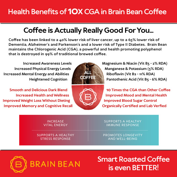
Brain Bean - The Smart Roasted Coffee!
An ad I got, since there is nothing here but fluff, ask your doctor if this is accurate and what the exact diet protocol for it is.
Brain Bean - The Smart Roasted Coffee!
Small
Batched, Smart Roasted Coffee that Maximizes and Keeps 5-10X the Health
Promoting Chlorogenic Acid (CGA) than Traditionally Roasted Coffee
|

|

|
Friday, October 26, 2018
Mumbai MRI scanner back online after tragedy
You'll have to ask what the safety protocol is before you get your MRI scan. Hopefully you are conscious and trust the staff to do everything correctly.
October 25, 2018 -- Nine months after a visitor to Nair Hospital in Mumbai was killed in an MRI accident, the scanner is back in operation on a limited basis.
According to an October 17 report in the Asian Age, the facility is scanning 10 to 12 patients per day until officials are satisfied the system is working properly.
The scanner has been out of service since January, when 32-year-old Rajesh Maruti Maru was killed as he escorted a family member into the MRI room for an examination. Maru was carrying a metal oxygen tank at the time and died when he and the tank were sucked into the magnet bore. According to a previous report, a metal detector that would have detected the tank allegedly was not functioning.
After the accident, technicians from Philips Healthcare inspected the 1.5-tesla Achieva Nova scanner. Hospital authorities claimed that the 9-year-old machine had been functioning normally before the accident, according to the report.
Mumbai MRI scanner back online after tragedy
By AuntMinnie.com staff writersOctober 25, 2018 -- Nine months after a visitor to Nair Hospital in Mumbai was killed in an MRI accident, the scanner is back in operation on a limited basis.
The scanner has been out of service since January, when 32-year-old Rajesh Maruti Maru was killed as he escorted a family member into the MRI room for an examination. Maru was carrying a metal oxygen tank at the time and died when he and the tank were sucked into the magnet bore. According to a previous report, a metal detector that would have detected the tank allegedly was not functioning.
After the accident, technicians from Philips Healthcare inspected the 1.5-tesla Achieva Nova scanner. Hospital authorities claimed that the 9-year-old machine had been functioning normally before the accident, according to the report.
Tuesday, October 23, 2018
Managing LDL-C to Reduce Cardiovascular Disease Risk: Defining Best Clinical Practice
I don't want best clinical practice, that is just laziness. I want a protocol. For this objective starting point ; these exact interventions need to occur. Nothing should be left to chance and interpretation. But that is just my non-medical understanding. Prove me wrong and I can change my position.
Registration and Dinner: 6:30-7 p.m.
Symposium: 7-9 p.m., Sunday, Nov. 11
InterContinental Chicago Grand Ballroom, 7th Floor
Sponsor: Voxmedia - Supporter: Amgen
Program Overview: Nonstatin drugs (ezetimibe, PCSK9 inhibitors) further lower LDL-C and further reduce cardiovascular risk when added to statins, while demonstrating safety at low LDL-C levels.
Symposium: 7-9 p.m., Sunday, Nov. 11
InterContinental Chicago Grand Ballroom, 7th Floor
Sponsor: Voxmedia - Supporter: Amgen
Program Overview: Nonstatin drugs (ezetimibe, PCSK9 inhibitors) further lower LDL-C and further reduce cardiovascular risk when added to statins, while demonstrating safety at low LDL-C levels.
Monday, September 17, 2018
Eating fiber can delay brain aging
But you can't do this on your own until your incompetent doctor and stroke hospital get off their asses and write up diet protocols. How is your doctor making sure you are recovering your lost 5 cognitive years from your stroke?
Eating fiber can delay brain aging

Eating fiber-rich foods — such as broccoli, nuts, oats, beans, and whole-grain bread — might help delay brain aging by triggering the production of a short-chain fatty acid that has anti-inflammatory properties.
This the main takeaway of a new study that was recently published in the journal Frontiers in Immunology.
Rodney Johnson, a professor and the head of the Department of Animal Sciences at the University of Illinois at Urbana-Champaign, is the corresponding author of the study, and Stephanie M. Matt is the first author of the paper.
How fiber lowers inflammation
As Matt and colleagues explain in their study paper, microglia — a major type of immune cell in the brain — tend to become hyperactive and chronically inflamed with age. This inflammation of the microglia is one of the main causes of memory and cognitive decline in old age.Previous research has shown that a drug form of butyrate, which is a short-chain fatty acid that is produced in the colon when bacteria ferment fiber in the gut, can improve memory and reduce inflammation in mice.
However, the precise mechanisms behind this weren't entirely understood. Also, previous research had not shown whether simply increasing the dietary content of fiber would achieve the same results as the drug.

So, Matt and colleagues fed young and aging mice
diets high and low in fiber. Then, the scientists measured the mice's
blood levels of butyrate and their levels of pro-inflammatory substances
in their intestines.
"But," he goes on, "only the old mice showed intestinal inflammation on the low-fiber diet [...] It's interesting that young adults didn't have that inflammatory response on the same diet. It clearly highlights the vulnerability of being old."
Also, consuming a high-fiber diet reduced the intestinal inflammation in aging mice so much that it was indistinguishable from that of young mice.
"Dietary fiber can really manipulate the inflammatory environment in the gut," says Prof. Johnson. What about the brain, however?
Why fiber is good for your brain
A genetic analysis of inflammatory markers
conducted by the scientists found that a high-fiber diet reduced
inflammation in the brain's microglia. The researchers suspect that this
was achieved by diminishing the production of a pro-inflammatory
chemical known as interleukin-1β, which some studies have linked with
Alzheimer's.
Study co-author Jeff Woods, a professor in the Department of
Kinesiology and Community Health at the University of Illinois at
Urbana-Champaign, comments on the findings."We know that diet has a major influence on the composition and function of microbes in the gut and that diets high in fiber benefit good microbes," he points out, "while diets high in fat and protein can have a negative influence on microbial composition and function."
Altering gut microbes, explains Prof. Woods, "is one way in which [diet] affects disease."
Prof. Johnson explains that the findings are relevant to humans, saying, "People are not likely to consume sodium butyrate directly, due to its noxious odor," he says, but, "A practical way to get elevated butyrate is to consume a diet high in soluble fiber."
"What you eat matters. We know that older adults consume 40 percent less dietary fiber than is recommended. Not getting enough fiber could have negative consequences for things you don't even think about, such as connections to brain health and inflammation in general."
Prof. Rodney Johnson
Subscribe to:
Posts (Atom)