Changing stroke rehab and research worldwide now.Time is Brain! trillions and trillions of neurons that DIE each day because there are NO effective hyperacute therapies besides tPA(only 12% effective). I have 523 posts on hyperacute therapy, enough for researchers to spend decades proving them out. These are my personal ideas and blog on stroke rehabilitation and stroke research. Do not attempt any of these without checking with your medical provider. Unless you join me in agitating, when you need these therapies they won't be there.

What this blog is for:

My blog is not to help survivors recover, it is to have the 10 million yearly stroke survivors light fires underneath their doctors, stroke hospitals and stroke researchers to get stroke solved. 100% recovery. The stroke medical world is completely failing at that goal, they don't even have it as a goal. Shortly after getting out of the hospital and getting NO information on the process or protocols of stroke rehabilitation and recovery I started searching on the internet and found that no other survivor received useful information. This is an attempt to cover all stroke rehabilitation information that should be readily available to survivors so they can talk with informed knowledge to their medical staff. It lays out what needs to be done to get stroke survivors closer to 100% recovery. It's quite disgusting that this information is not available from every stroke association and doctors group.

Showing posts with label GPCR. Show all posts

Thursday, April 25, 2024

New Tool Maps Brain Signals with Unprecedented Clarity


This is where stroke leaders would look at this and mutter to themselves; 'We could use this to detect brain signals occurring during stroke recovery; neurogenesis and neuroplasticity, and thus figure out how to make those signals repeatable'. But we have to do human testing first.

But the leaders would already have started listening to brain signals using one of these already. 

1. Use nanowires to listen in on single neurons

2. Or lay a grid across the cortex to listen in.

But we have NO stroke leaders, nothing will get done until we get survivors in charge.

Leaders solve problems, they don't run away from them.

The latest here:

New Tool Maps Brain Signals with Unprecedented Clarity

Summary: Researchers developed an innovative chemical tool to explore how signals like dopamine and epinephrine interact with neurons via G protein-coupled receptors (GPCRs).

This new tool allows for precise detection of neuromodulators across various brain regions with high spatial resolution. It marks cells with a permanent fluorescent signal, facilitating the study of signal pathways and interactions at a cellular level across the entire brain.

This advancement could significantly enhance our understanding of neuronal signaling and improve targeting GPCRs in drug development.

Key Facts:

  1. The tool enables detailed visualization of GPCR-related signals across the entire brain with high spatial resolution, a balance previously unachievable in neuroscience.
  2. It has been tested on opioids and epinephrine, utilizing both green and red fluorescence to track multiple molecules simultaneously.
  3. While the fluorescence takes several hours to manifest and is not suitable for real-time tracking, the tool provides valuable postmortem insights into neuronal pathways and drug targeting.

Source: University of Michigan

University of Michigan researchers have developed a new tool to better understand how chemicals like dopamine and epinephrine interact with neurons.

These chemicals are among a wide variety of signals that get processed in the brain through G protein-coupled receptors (GPCRs), proteins that sit on the surface of neurons to receive messages—in the forms of proteins, sugars, fats, even light—that inform cellular behavior. 
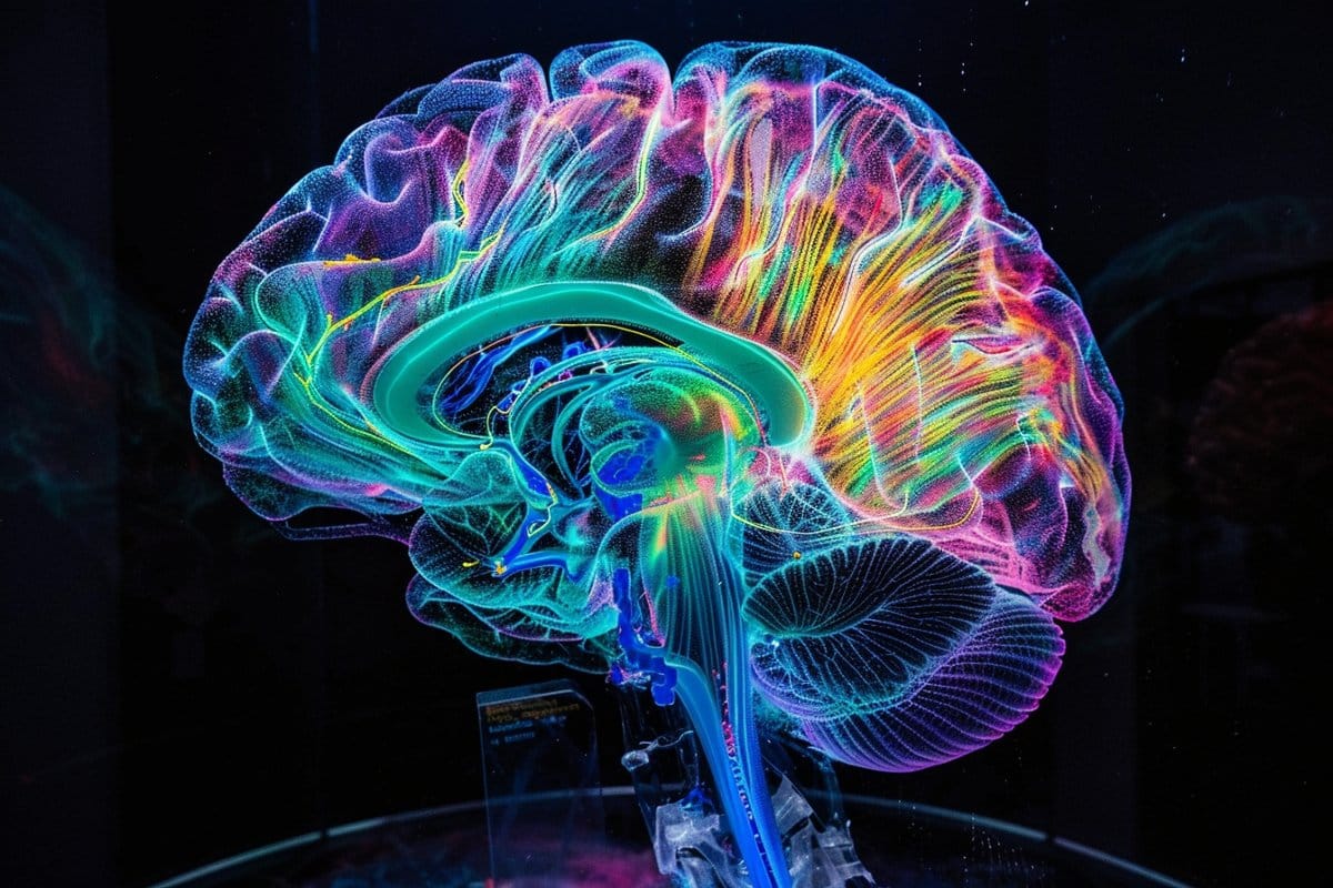
This shows a brain.
Wang’s lab at LSI uses protein engineering to develop technologies that can detect how signaling molecules travel within the brain to reach and interact with specific neurons. Credit: Neuroscience News

GPCRs are involved in an enormous number of biological functions, making them a prime target for treating diseases; more than one-third of FDA-approved drugs target GPCRs. But to fully understand how various molecules interact with GPCRs, researchers need to be able to detect those molecules across the whole brain with high spatial resolution.

“The challenge in our field has been achieving the right balance between a detailed view and the whole picture across the brain,” said Wenjing Wang, a neuroscientist at the U-M Life Sciences Institute. 

LSI faculty member Peng Li said most existing tools can detect a neural modulator either in a small part of the brain with high spatial resolution or in the whole brain with very low resolution.

“But we need to identify the cells that respond to the neuromodulators across various brain regions, in high resolution,” he said.

In a study published in the Proceedings of the National Academy of Sciences, Wang, Li and colleagues unveiled a new chemical tool that achieves both goals for three chemicals that all target GPCRs.

Wang’s lab at LSI uses protein engineering to develop technologies that can detect how signaling molecules travel within the brain to reach and interact with specific neurons. They previously created a tool to reveal the presence of opioids, another GPCR binding partner, at a cellular level.

When the molecule is detected, the tool creates a permanent fluorescent mark in the cells. Thus, researchers can see the specific cells that are highlighted, as well as the whole picture of cells across the brain.

This latest work broadens the utility of that sensor to detect multiple types of GPCR activators, beyond just opioids. So far, the team has tested the tool with opioids and epinephrine in cultured neurons and in mouse models. The team also expanded the tool to use both green and red fluorescence, enabling the tracking of multiple molecules at once.

“Coming from detecting just opioids, we now have a tool that we can begin to easily modulate for various signals that interact with GPCRs,” said Wang, who also is an assistant professor of chemistry at the U-M College of Literature, Science, and the Arts.

“The goal is eventually to even study the interplays of different signaling pathways simultaneously.”

The team cautions that while the tool provides important visualizations of how signals travel across neurons for analysis postmortem, it cannot be used to track chemicals in real time, as it takes several hours for the fluorescence to appear. But it does offer a new path forward for improving understanding of neuronal signaling and the role of GPCRs as drug targets.

“Ideally, we aim to be able to create a brain map for multiple neuromodulators concurrently, offering a comprehensive understanding of the sites of neuromodulation,” said Li, who also is an assistant professor at the U-M School of Dentistry.

About this brain mapping and neurotech research news

Author: Morgan Sherburne
Source: University of Michigan
Contact: Morgan Sherburne – University of Michigan
Image: The image is credited to Neuroscience News

Original Research: Closed access.
“Single-chain fluorescent integrators for mapping G-protein-coupled receptor agonists” by Wenjing Wang et al. PNAS

Tuesday, March 17, 2020

The Protective Effects and Mechanisms of Apelin/APJ System on Ischemic Stroke: A Promising Therapeutic Target

Now we just need whomever is running the stroke strategy to see what followup research needs to be done. Oops, that will never occur. WE HAVE NO STROKE LEADERSHIP AND NO STROKE STRATEGY. 

Until we get stroke survivors in charge nothing useful is going to happen to get to 100% recovery. No one in stroke is even thinking about that goal. 

The Protective Effects and Mechanisms of Apelin/APJ System on Ischemic Stroke: A Promising Therapeutic Target

Yanjun Tian1†, Ruijiao Chen1†, Yunlu Jiang2,3, Bo Bai3, Tongju Yang4* and Haiqing Liu5*
  • 1Collaborative Innovation Center for Birth Defect Research and Transformation of Shandong Province, Jining Medical University, Jining, China
  • 2School of Mental Health, Jining Medical University, Jining, China
  • 3Institute of Neurobiology, Jining Medical University, Jining, China
  • 4Department of Pharmacy, People's Hospital of Zoucheng City, Jining, China
  • 5Department of Physiology, Shandong First Medical University (Shandong Academy of Medical Sciences), Taian, China
The orphan receptor APJ and its endogenous ligand apelin, which are expressed in the brain, are the major components of the apelin/APJ system. Growing evidence shows that the apelin/APJ system plays a vital role in the pathophysiology of cerebral ischemic injury. Targeting the apelin/APJ system may have protective effects on cerebral ischemic injury. In this review, we sum up the latest research progress relating to the actions and therapeutic potential of the apelin/APJ system in ischemic stroke. An in-depth knowledge of the pathophysiological effects of the apelin/APJ system and the underlying mechanisms will help to develop novel therapeutic interventions for ischemic stroke.

Introduction

In 1993, O'Dowd discovered the orphan G protein–coupled receptor (GPCR) APJ while searching for vasopressin receptor subtypes (1). APJ is encoded by a gene located on chromosome 11q12. Although APJ shares 54% homology with angiotensin II receptor type-1 (AT1R) in the hydrophobic transmembrane region, there's no binding site for angiotensin II (1). Tatemoto et al. isolated apelin, the cognate ligand for APJ receptor, from bovine stomach tissue extracts in 1998 (2). The apelin gene, which is located on chromosome Xq25-26.1, encodes the preproapelin of 77 amino acids (apelin-77). Various bioactive isoforms of apelin are derived from apelin-77, including apelin-55, apelin-36, apelin-17, apelin-13, and apelin-12 (3, 4). Recently Chng et al. discovered apela, another endogenous ligand for APJ, which is encoded by a gene located on chromosome 11 and is critical in embryonic development (5, 6). However, in humans, the apela is only expressed in pluripotent cells and kidney (7). The apelin/APJ system mainly refers to APJ and its endogenous ligand, apelin.
Stroke, which is mainly caused by cerebral vascular occlusion and cerebral blood supply disorder, is one of the leading causes of death and disability worldwide, and 87% of cases are ischemic stroke (8). The cerebral infarction area is composed of the ischemic core and penumbra; apoptosis is the main cause of neuronal damage in the penumbra region, which also provides an opportunity for the treatment of ischemic stroke and makes it possible to use drugs to alleviate neuronal injury since the apoptosis is delayed and reversible (9, 10). Neuronal apoptosis in ischemic penumbra is triggered by diffusion of toxic substances released by the dead neurons of the ischemic core in the acute stage of ischemia, while ischemia injury is aggravated after reperfusion, namely ischemia/reperfusion (I/R) injury, which contributes to the neuron apoptosis in the penumbra via numerous biological mechanisms, including excitotoxicity, oxidative and nitrative stress, inflammatory responses, endoplasmic reticulum stress (ERS), and so on (11–14).
The apelin/APJ system is widely expressed in the central nervous system, especially in neurons and oligodendrocytes (15, 16). Growing evidence indicates that the apelin/APJ system is involved in the pathophysiology of ischemic stroke (17, 18). Targeting the apelin/APJ system may have protective effects on cerebral ischemic injury. In this review, we mainly focus on the latest research progress related to the biological functions and therapeutic potential of the apelin/APJ system in ischemic stroke.

Friday, August 9, 2013

Microglial P2Y12 Deficiency/Inhibition Protects against Brain Ischemia

I'm sure your doctor can explain this(snort, snort).
http://www.plosone.org/article/info%3Adoi%2F10.1371%2Fjournal.pone.0070927

Abstract

Objective

Microglia are among the first immune cells to respond to ischemic insults. Triggering of this inflammatory response may involve the microglial purinergic GPCR, P2Y12, activation via extracellular release of nucleotides from injured cells. It is also the inhibitory target of the widely used antiplatelet drug, clopidogrel. Thus, inhibiting this GPCR in microglia should inhibit microglial mediated neurotoxicity following ischemic brain injury.

Methods

Experimental cerebral ischemia was induced, in vitro with oxygen-glucose deprivation (OGD), or in vivo via bilateral common carotid artery occlusion (BCCAO). Genetic knock-down in vitro via siRNA, or in vivo P2Y12 transgenic mice (P2Y12−/− or P2Y12+/−), or in vivo treatment with clopidogrel, were used to manipulate the receptor. Neuron death, microglial activation, and microglial migration were assessed.

Results

The addition of microglia to neuron-astrocyte cultures increases neurotoxicity following OGD, which is mitigated by microglial P2Y12 deficiency (P<0.05). Wildtype microglia form clusters around these neurons following injury, which is also prevented in P2Y12 deficient microglia (P<0.01). P2Y12 knock-out microglia migrated less than WT controls in response to OGD-conditioned neuronal supernatant. P2Y12 (+/−) or clopidogrel treated mice subjected to global cerebral ischemia suffered less neuronal injury (P<0.01, P<0.001) compared to wild-type littermates or placebo treated controls. There were also fewer microglia surrounding areas of injury, and less activation of the pro-inflammatory transcription factor, nuclear factor Kappa B (NFkB).

Interpretation

P2Y12 participates in ischemia related inflammation by mediating microglial migration and potentiation of neurotoxicity. These data also suggest an additional anti-inflammatory, neuroprotective benefit of clopidogrel.

Monday, May 21, 2012

G-Protein-Coupled Receptors in Adult Neurogenesis

The abstract only hints at possibilities. Sign the petition for access to research.
G-Protein-Coupled Receptors in Adult Neurogenesis

Abstract

The importance of adult neurogenesis has only recently been accepted, resulting in a completely new field of investigation within stem cell biology. The regulation and functional significance of adult neurogenesis is currently an area of highly active research. G-protein-coupled receptors (GPCRs) have emerged as potential modulators of adult neurogenesis. GPCRs represent a class of proteins with significant clinical importance, because approximately 30% of all modern therapeutic treatments target these receptors. GPCRs bind to a large class of neurotransmitters and neuromodulators such as norepinephrine, dopamine, and serotonin. Besides their typical role in cellular communication, GPCRs are expressed on adult neural stem cells and their progenitors that relay specific signals to regulate the neurogenic process. This review summarizes the field of adult neurogenesis and its methods and specifies the roles of various GPCRs and their signal transduction pathways that are involved in the regulation of adult neural stem cells and their progenitors. Current evidence supporting adult neurogenesis as a model for self-repair in neuropathologic conditions, adult neural stem cell therapeutic strategies, and potential avenues for GPCR-based therapeutics are also discussed