Changing stroke rehab and research worldwide now.Time is Brain! trillions and trillions of neurons that DIE each day because there are NO effective hyperacute therapies besides tPA(only 12% effective). I have 523 posts on hyperacute therapy, enough for researchers to spend decades proving them out. These are my personal ideas and blog on stroke rehabilitation and stroke research. Do not attempt any of these without checking with your medical provider. Unless you join me in agitating, when you need these therapies they won't be there.

What this blog is for:

My blog is not to help survivors recover, it is to have the 10 million yearly stroke survivors light fires underneath their doctors, stroke hospitals and stroke researchers to get stroke solved. 100% recovery. The stroke medical world is completely failing at that goal, they don't even have it as a goal. Shortly after getting out of the hospital and getting NO information on the process or protocols of stroke rehabilitation and recovery I started searching on the internet and found that no other survivor received useful information. This is an attempt to cover all stroke rehabilitation information that should be readily available to survivors so they can talk with informed knowledge to their medical staff. It lays out what needs to be done to get stroke survivors closer to 100% recovery. It's quite disgusting that this information is not available from every stroke association and doctors group.

Showing posts with label dopamine. Show all posts

Monday, May 16, 2022

A Study Reveals One of the Reasons Why Neurons Die in Parkinson’s Patients

 You'll want your doctor following this carefully so when  protocols are created stopping this neuronal death s/he is ready for your childrens and grandchildren's strokes.

Your risk of Parkinsons here:

Parkinson’s Disease May Have Link to Stroke March 2017 

The latest here:

A Study Reveals One of the Reasons Why Neurons Die in Parkinson’s Patients

Summary: Study implicates the gene DJ1 in neuronal death associated with Parkinson’s disease.

Source: University of Córdoba

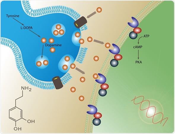
Parkinson’s disease affects about 7 million people worldwide, according to data provided by the World Health Organization (WHO). This neurodegenerative disorder affects the central nervous system and, although its causes are not yet fully understood, it is known that many of its symptoms are due to the loss of neurons that produce dopamine.

A study carried out by a research team at the University of Cordoba has revealed, in mice, one of the reasons for this neuronal loss: the key lies in the protein called DJ1, whose relationship with Parkinson’s disease had already been demonstrated, although until now its exact function was unclear.

This study went a step further in this area, revealing one of this protein’s action mechanisms. To do this, the research team carried out a comparative study of neurons in the brains of mice that have this active gene, and others that lack it, with the aim of “comparing the differences between the two protein patterns and, thus, studying the mechanisms that may be altered,” explains Raquel Requejo, principal investigator of the study and a member of the BIO126 group at the University of Cordoba. 

When dividing is not an option

According to the results of the study, the absence or dysfunction of the gene expressing the DJ1 protein causes the activation of what is known as the cell cycle, the process by which cells divide; in other words, the machinery used by cells to replace others that have died, as occurs, for example, when a wound heals. 

This shows a head and a question mark
Parkinson’s disease affects about 7 million people worldwide, according to data provided by the World Health Organization (WHO). Image is in the public domain


Saturday, April 17, 2021

Nitric oxide and dopamine metabolism converge via mitochondrial dysfunction in the mechanisms of neurodegeneration in Parkinson's disease


Because of the usefulness of nitric oxide in your recovery, your doctor is responsible for adjusting your nitric oxide protocol with this in mind.  Or does your doctor incompetently not even have a nitric oxide protocol?


Your risk of Parkinsons here:

Parkinson’s Disease May Have Link to Stroke March 2017

The latest here:

Nitric oxide and dopamine metabolism converge via mitochondrial dysfunction in the mechanisms of neurodegeneration in Parkinson's disease


Abstract

The molecular mechanisms underlying the degeneration and neuronal death associated with Parkinson's disease (PD) are not clearly understood. Several pathways and models have been explored in an overwhelming number of studies. Overall, from these studies, mitochondrial dysfunction and nitroxidative stress have emerged as major contributors to degeneration of dopaminergic neurons in PD. In addition, an excessive or inappropriate production of nitric oxide (NO) and an abnormal metabolism of dopamine have been independently implicated in both processes. However, the participation of NO in reactions with dopamine relevant to neurotoxicity strongly suggests that dopamine or its metabolites may be potential targets for NO, affecting the physiological chemistry of both, NO and dopamine. In this short review, we provide a critical and integrative appraisal of the nitric oxide-dopamine pathway we have previously suggested and that might be operative in PD. This pathway emphasizes a connection between abnormal dopamine and NO metabolism, which may potentially converge in an integrated mechanism with toxic cellular outcomes. In particular, it encompasses the synergistic interaction of NO with 3,4-dihydroxyphenylacetic acid (DOPAC), a major dopamine metabolite, leading to dopaminergic cell death via mechanisms that involve mitochondrial dysfunction, gluthathione depletion and nitroxidative stress.

Keywords

Parkinson's disease
Nitric oxide
DOPAC
Mitochondrial dysfunction
Nitroxidative stress
Glutathione
View full text

Monday, June 17, 2019

Chronic inflammation removes motivation by reducing dopamine in the brain

Is this one of the reasons survivors lack motivation? I would say no, the motivation lack comes directly from your doctor having NO EXACT PROTOCOLS FOR RECOVERY. If your doctor said do 15 million repetitions of this and you will get this result, you would do 15 million repetitions. I've walked 13,955,868 steps in the last 5 years and still no relief from my ankle, toes and leg spasticity. 

Chronic inflammation removes motivation by reducing dopamine in the brain

Why do we feel listless when we are recovering from an illness? The answer is, apparently, that low-grade chronic inflammation interferes with the dopaminergic signaling system in the brain that motivates us to do things.
This was reported in a new paper published in the journal Trends in Cognitive Sciences.
The research carried out at Emory University explains the links between the reduced release of dopamine in the brain, the motivation to do things, and the presence of an inflammatory reaction in the body. It also presents the possibility that this is part of the body’s effort to optimize its energy expenditure during such inflammatory episodes, citing evidence gathered during their study.
The authors also published an experimental framework based on computational tools, devised to test the theory.
The underlying hypothesis is that the body needs more energy to heal a wound or overcome an infection, for instance, both of which are associated with low-grade inflammation. To ensure that energy is available, the brain uses an adaptive technique to reduce the natural drive to perform other tasks which could potentially drain away the energy needed for healing. This is essentially a recalibration of the specialized reward neurons in the motivation center of the brain, so that ordinary tasks no longer feel like they’re worth doing.
According to the new study, the mechanism of this recalibration is immune-mediated disruption of the dopamine pathway, reducing dopamine release.
The computational technique published by the scientists is designed to allow experimental measurements of the extent to which low-grade inflammation affects the amount of energy available, and the decision to do something based on the effort needed. This could allow us to better understand why and how chronic inflammatory states cause a lack of motivation in other disease conditions as well, including schizophrenia and depression.
Andrew Miller, co-author of the study, says, “If our theory is correct, then it could have a tremendous impact on treating cases of depression and other behavioral disorders that may be driven by inflammation. It would open up opportunities for the development of therapies that target energy utilization by immune cells, which would be something completely new in our field.”
It is already known that immune cells release cellular signaling molecules called cytokines, which affect the functioning of the dopamine-releasing neurons in the area of the brain called the mesolimbic system. This area enhances our willingness to work hard for the sake of a reward.
Dopamine
Image Copyright: Meletios, Image ID: 71648629 via shutterstock.com
Recently, it was discovered that immune cells also enjoy a unique capability to shift between various metabolic states, unlike other cells. This could affect cytokine release patterns in such a way as to signal the brain to conserve available energy for the use of the immune system.
These facts were the foundation of the new hypothesis, which explains it in terms of evolutionary adaptation. In the hypothetical early environment, the immune system, faced with abundant microbial and predatory challenges, needed tremendous amounts of energy. It therefore had its own mechanism to signal other body systems, via the mesolimbic dopamine system, to control the use of energy resources during periods when the organism was undergoing severe or sudden stress.
Modern life is relatively soft and less challenging. With less physical activity, low-grade inflammation is chiefly due to factors such as obesity, chronic stress, metabolic syndrome, aging and other lifestyle illnesses. This could mistakenly cause the mesolimbic dopamine neurons to produce less dopamine. Lower dopamine levels in turn decrease the motivation for work, by reducing the perception of reward while increasing the perception of effort involved. This ultimately conserves energy for use by the immune system.
Previous studies by Miller as well as other scientists have shown that a high level of immune functioning in association with low levels of dopamine and reduced motivation characterizes some cases of schizophrenia, depression and certain other mental health conditions.
The scientists do not think these disorders are caused by the low-grade inflammation, but that some people who have these illnesses are hypersensitive to immune cytokines. This could in turn cause them to lose motivation for daily living.
The scientists are currently performing a clinical trial on people with depression, to test the theory using the computational framework.
Source:
Treadway M. T. et al., (2019). Can’t or Won’t? Immunometabolic Constraints on Dopaminergic Drive. Trends in Cognitive Sciences. https://doi.org/10.1016/j.tics.2019.03.003

Wednesday, April 3, 2019

Effects of Dopamine on Motor Recovery and Training in Adults and Children With Nonprogressive Neurological Injuries: A Systematic Review

Shit, asking for followup because you didn't want to do the research that would prove efficacy one way or the other.  LAZY.  Systematic reviews are lazy. 

But this from Sept. 2001 shows you that nothing is ever done in stroke.

Early Promise For Stroke Patients Given - levodopa  Sept. 2001

And the latest useless stuff here:

Effects of Dopamine on Motor Recovery and Training in Adults and Children With Nonprogressive Neurological Injuries: A Systematic Review 

First Published March 27, 2019 Review Article


Background. The strong link between dopamine and motor learning has been well-established in the animal literature with similar findings reported in healthy adults and the elderly.  
Objective. We aimed to conduct the first, to our knowledge, systematic review of the literature on the evidence for the effects of dopaminergic medications or genetic variations in dopamine transmission on motor recovery or learning after a nonprogressive neurological injury.  
Methods. A PubMed search was conducted up until April 2018 for all English articles including participants with nonprogressive neurological injury such as cerebral palsy, stroke, spinal cord injury, and traumatic brain injury; quantitative motor outcomes; and assessments of the dopaminergic system or medications.
Results. The search yielded 237 articles, from which we identified 26 articles meeting all inclusion/exclusion criteria. The vast majority of articles were related to the use of levodopa poststroke; however, several studies assessed the effects of different medications and/or were on individuals with traumatic brain injury, spinal cord injury or cerebral palsy.  
Conclusions. The evidence suggests that a brain injury can decrease dopamine transmission and that levodopa may have a positive effect on motor outcomes poststroke, although evidence is not conclusive or consistent. Individual variations in genes related to dopamine transmission may also influence the response to motor skill training during neurorehabilitation and the extent to which dopaminergic medications or interventions can augment that response. More rigorous safety and efficacy studies of levodopa and dopaminergic medications in stroke and particularly other neurological injuries including genetic analyses are warranted.

Thursday, August 31, 2017

Injecting stem cells into the brain reverses Parkinson’s symptoms in monkeys

You might very well need this so you better hope that this is followed up better than any stroke research is.

Parkinson’s Disease May Have Link to Stroke

Injecting stem cells into the brain reverses Parkinson’s symptoms in monkeys 


Sunday, July 30, 2017

Vegetative stroke patient, 36, was able to speak and move just 16 DAYS after being given a Parkinson's disease drug

4 posts back to Feb. 2012 and I bet absolutely fucking nothing  has been done to get clinical research done and a stroke protocol written up.

Vegetative stroke patient, 36, was able to speak and move just 16 DAYS after being given a Parkinson's disease drug

  • The unnamed patient was only being kept alive through medical intervention
  • Doctors thought the patient's only option was to be admitted to a nursing home
  • She was able to move and speak in short sentences due to the drug amantadine
  • The patient can now breathe independently, eat precut food and stand with help
  • Experts believe the drug may have pushed her 'above the threshold' for recovery
A vegetative stroke patient who was completely unresponsive to what was going on around her, regained complete consciousness just 16 days after being given a Parkinson's disease drug, a case report reveals. 
The unnamed woman, 36, who was only being kept alive by medical intervention, was able to speak in short sentences after being given the dopamine-boosting drug, known as amantadine.
Unable to move, doctors thought her only option was to be admitted to a nursing home, yet the woman, believed to be from Berlin, can now eat and stand.
Experts believe the drug may have pushed the woman, who was diagnosed with unresponsive wakefulness syndrome, 'above the threshold' for recovery.
Amantadine is used to treat Parkinson's disease and 'flu. It is thought to increase levels of the 'feel-good' hormone dopamine in the brain. Dopamine is involved in regulating movement.

Monday, March 13, 2017

How a Simple Smile Benefits Your Brain and Body

Can your doctor and stroke hospital even manage to implement this costless intervention for your stroke therapy? Or are they that incompetent that this will slide right past them because it is too much work? Thanks to Debbie Hampton for this.

How a Simple Smile Benefits Your Brain and Body 


Tuesday, November 29, 2016

Experts say playing trivia games can provide a dopamine rush much like gambling, without the negative effects

I bet your doctor and stroke hospital  will never provide this basically free intervention.  When I wasn't traveling for work I would play Team Trivia twice a week at bars. Great for your social connections and with drinking some alcohol great for your balance training. Triple your stroke rehab at one time/place. I bet your doctor never thinks of this.
http://www.healthline.com/health-news/obscure-facts-is-good-for-mental-health#2
It can be quite satisfying and doesn’t have many downsides.
That’s how psychologists describe the mental health benefits of trivia.
The way people play trivia games continues to evolve whether it’s folks enjoying Trivial Pursuit at home or attending a pub trivia night.
But the basic premise remains the same: People enjoy the thrill of providing correct answers to questions about lesser-known facts.
Trivia Question #1
Who was the first U.S. president born in a hospital?
“You get a rush or a neuroreward signal or a dopamine burst from winning,” John Kounios, Ph.D., professor of psychology and director of the doctoral program in applied cognitive and brain sciences at Drexel University in Pennsylvania, told Healthline. “I think whenever you’re challenged with a trivia question and you happen to know it, you get a rush. It’s sort of like gambling.”
He said the benefits can also be similar to those of playing a video game.
However, unlike gambling and even video games, Kounios says trivia is generally not a habit that’s a problem.
“I don’t think there are any pitfalls,” he said. “Like anything else that’s fun, it takes up time.”

Monday, June 20, 2016

Want to Remember Something? Workout 4 Hours After Learning to Help Retention

How will your doctors and therapists use this to create SPECIFIC stroke rehab protocols? I'm guessing never.
http://www.biosciencetechnology.com/news/2016/06/want-remember-something-workout-4-hours-after-learning-help-retention?

It’s known that good sleep is important for memory formation, but a new study suggests that delayed exercise can also help boost retention of newly learned memories.
The waiting game is key to the improvement, as hitting the gym immediately after learning doesn’t result in the same benefits, according to the findings published June 16 in Current Biology
For the study researchers brought in 72 people of average fitness and taught them 90 picture-location associations, where they learned where certain objects had appeared on the screen.  The participants were then split into three groups: one-third performed 35 minutes of interval training on an exercise bike immediately after learning, one-third waited four hours and then exercised, and the last group did not perform any exercise.  The biking exercise was at an intensity of up to 80 percent of the participants’ maximum heart rate.
Two days later the participants returned and underwent brain MRI’s as they were tested on how much they remembered from the pictures.  There was no difference in retention between the group that exercised immediately and those who abstained from working out.  Interestingly, those that exercised four hours after learning remembered almost 10 percent more material than the other groups.
“Our results suggest that appropriately timed physical exercise can improve long-term memory and highlight the potential of exercise as an intervention in educational and clinical settings,” the researchers concluded.
However, what the exact optimal time for exercise after learning is not yet known.  A longer or shorter delay may have better, or worse results.
The researchers also say that the cause of this effect on memory is not entirely clear yet, however they suggest that it may be the result of naturally occurring chemical compounds known as catecholamines, such as dopamine and norepinephrine.  Animal studies previously suggested that catecholamines can improve memory consolidation and that exercise can boost these hormones.
“These proteins help stabilizing new memory traces, which would otherwise be lost,” Dr. Guillen Fernadez, a professor of cognitive neuroscience at Radboud University Medical Center told The Huffington Post.  “Physical exercise is at the start of this sequence, because it is accompanied by the release of dopamine and norepinephrine.”
Up next the researchers plan to set up a similar experiment to understand the molecular mechanism of exercise and its influence on learning and memory, and further investigate the ideal timing.

Wednesday, March 16, 2016

Posttraumatic Stress Disorder: Does the Gut Microbiome Hold the Key?

Since we have a 23% chance of survivors getting PTSD, your doctor should already have stroke protocols to prevent that and be following this with great interest.

Posttraumatic Stress Disorder: Does the Gut Microbiome Hold the Key?


  1. Sophie Leclercq, PhD1,2
  2. Paul Forsythe, PhD1,3
  3. John Bienenstock, MD1,2
  1. 1McMaster Brain-Body Institute at St Joseph’s Healthcare Hamilton, Hamilton, Ontario
  2. 2Department of Pathology and Molecular Medicine, McMaster University, Hamilton, Ontario
  3. 3Firestone Institute for Respiratory Health and Department of Medicine, McMaster University, Hamilton, Ontario
  1. John Bienenstock, MD, McMaster Brain-Body Institute at St. Joseph’s Healthcare Hamilton, 50 Charlton Avenue East, Hamilton, ON L8 N 4A6, Canada. Email: bienens@mcmaster.ca

Abstract

Gut bacteria strongly influence our metabolic, endocrine, immune, and both peripheral and central nervous systems. Microbiota do this directly and indirectly through their components, shed and secreted, ranging from fermented and digested dietary and host products to functionally active neurotransmitters including serotonin, dopamine, and γ-aminobutyric acid. Depression has been associated with enhanced levels of proinflammatory biomarkers and abnormal responses to stress. Posttraumatic stress disorder (PTSD) appears to be marked in addition by low cortisol responses, and these factors seem to predict and predispose individuals to develop PTSD after a traumatic event. Dysregulation of the immune system and of the hypothalamic-pituitary-adrenal axis observed in PTSD may reflect prior trauma exposure, especially early in life. Early life, including the prenatal period, is a critical time in rodents, and may well be for humans, for the functional and structural development of the immune and nervous systems. These, in turn, are likely shaped and programmed by gut and possibly other bacteria. Recent experimental and clinical data converge on the hypothesis that imbalanced gut microbiota in early life may have long-lasting immune and other physiologic effects that make individuals more susceptible to develop PTSD after a traumatic event and contribute to the disorder. This suggests that it may be possible to target abnormalities in these systems by manipulation of certain gut bacterial communities directly through supplementation or indirectly by dietary and other novel approaches.

Friday, August 8, 2014

The DARS (Dopamine Augmented Rehabilitation in Stroke) trial: protocol for a randomised controlled trial of Co-careldopa treatment in addition to routine NHS occupational and physical therapy after stroke

It already has these benefits:
Dopamine restores reward prediction errors in old age
Dopamine effect triggered by just a tiny taste of beer
The effects of the dopamine agonist rotigotine on hemispatial neglect following stroke
I'm sure your doctor knows all about this.

The DARS (Dopamine Augmented Rehabilitation in Stroke) trial 
Bipin B Bhakta, Suzanne Hartley, Ivana Holloway, J Alastair Couzens, Gary A Ford, David Meads, Catherine M Sackley, Marion F Walker, Sharon P Ruddock and Amanda J Farrin
For all author emails, please log on.
Trials 2014, 15:316  doi:10.1186/1745-6215-15-316
Published: 8 August 2014

Abstract (provisional)

Background

Stroke has a huge impact, leaving more than a third of affected people with lasting disability and rehabilitation remains a cornerstone treatment in the National Health Service (NHS). Recovery of mobility and arm function post-stroke occurs through re-learning to use the affected body parts and/or learning to compensate with the lesser affected side. Promising evidence suggests that the addition of Co-careldopa to physical therapy and occupational therapy may improve the recovery of arm and leg movement and lead to improved function.

Methods

Dopamine Augmented Rehabilitation in Stroke (DARS) is a multi-centre double-blind, randomised, placebo, controlled clinical trial of Co-careldopa in addition to routine NHS occupational therapy and physical therapy as part of early stroke rehabilitation. Participants will be randomised on a 1:1 basis to either Co-careldopa or placebo. The primary objective of the trial is to determine whether the addition of six weeks of Co-careldopa treatment to rehabilitation therapy can improve the proportion of patients who can walk independently eight weeks post-randomisation.

Discussion

The DARS trial will provide evidence as to whether Co-careldopa, in addition to routine NHS occupational and physical therapy, leads to a greater recovery of motor function, a reduction in carer dependency and advance rehabilitation treatments for people with stroke.
Trial registration: ISRCTN99643613 assigned on 4 December 2009.

The complete article is available as a provisional PDF. The fully formatted PDF and HTML versions are in production.


Wednesday, June 5, 2013

Catechol-O-Methyltransferase Polymorphism Influences Outcome After Ischemic Stroke

No clue here.
http://nnr.sagepub.com/content/27/6/491.abstract?etoc

Abstract

Background. To explore whether a polymorphism in dopamine metabolism influences the effectiveness of neurological rehabilitation and the outcome after ischemic stroke. Methods. The Barthel Index (BI) and the Rivermead Motor Assessment (RMA) were assessed in 78 moderately affected stroke patients (1) after they had entered a neurological inpatient rehabilitation, (2) after 4 weeks of rehabilitation therapy, and (3) 6 months later. Polymorphisms of the gene encoding catechol-O-methyltransferase (COMT) were determined. BI and RMA results were analyzed with respect to the genetic profiles of COMT. Results. Carriers of COMT Val/Val alleles showed better results in BI and RMA than COMT Met/Met carriers at all 3 time points. Val/Met carriers exhibited results in between the homozygotes, suggesting a gene–dose relationship. Altogether, BI and RMA results were highly correlated. Conclusion. Stroke patients with COMT Val/Val alleles had higher motor functions and abilities of activities of daily living even at the beginning of the rehabilitation period. All patient groups improved during the rehabilitation period to a similar degree, suggesting that physical therapy is comparably effective in all polymorphism subtypes.

Monday, April 15, 2013

Sunday, September 16, 2012

The effects of the dopamine agonist rotigotine on hemispatial neglect following stroke

You'll have to ask your doctor about this.
http://www.hubmed.org/display.cgi?uids=22761293
Hemispatial neglect following right-hemisphere stroke is a common and disabling disorder, for which there is currently no effective pharmacological treatment. Dopamine agonists have been shown to play a role in selective attention and working memory, two core cognitive components of neglect. Here, we investigated whether the dopamine agonist rotigotine would have a beneficial effect on hemispatial neglect in stroke patients. A double-blind, randomized, placebo-controlled ABA design was used, in which each patient was assessed for 20 testing sessions, in three phases: pretreatment (Phase A1), on transdermal rotigotine for 7-11 days (Phase B) and post-treatment (Phase A2), with the exact duration of each phase randomized within limits. Outcome measures included performance on cancellation (visual search), line bisection, visual working memory, selective attention and sustained attention tasks, as well as measures of motor control. Sixteen right-hemisphere stroke patients were recruited, all of whom completed the trial. Performance on the Mesulam shape cancellation task improved significantly while on rotigotine, with the number of targets found on the left side increasing by 12.8% (P = 0.012) on treatment and spatial bias reducing by 8.1% (P = 0.016). This improvement in visual search was associated with an enhancement in selective attention but not on our measures of working memory or sustained attention. The positive effect of rotigotine on visual search was not associated with the degree of preservation of prefrontal cortex and occurred even in patients with significant prefrontal involvement. Rotigotine was not associated with any significant improvement in motor performance. This proof-of-concept study suggests a beneficial role of dopaminergic modulation on visual search and selective attention in patients with hemispatial neglect following stroke.

Wednesday, July 4, 2012

Skin patch improves attention span in stroke patients

So I wonder what else stimulates dopamine receptors? maybe Ritalin from this research in 1998. Doesn't anyone follow previous research? Where is the 1998 research in the stroke therapy protocol? Who has the stroke therapy protocol?
http://medicalxpress.com/news/2012-07-skin-patch-attention-span-patients.html
Hemi-spatial neglect, a severe and common form of inattention that can be caused by following a stroke, is one of the most debilitating symptoms, frequently preventing patients from living independently. When the right side of the brain has suffered damage, the patient may have little awareness of their left-hand side and have of objects that they have seen, leaving them inattentive and forgetful. Currently there are few treatment options.
The randomised control trial took 16 patients who had suffered a stroke on the right-hand side of their brain and assessed to see whether giving the drug rotigotine improved their ability to concentrate on their left-hand side. The results showed that even with treatment for just over a week, patients who received the drug performed significantly better on attention tests than when they received the .
Rotigotine acts by stimulating receptors on for dopamine, a chemical normally produced within the brain.
Professor Masud Husain who led the study at the Institute of Neurology at UCL says: “Inattention can have a devastating effect on stroke patients and their families. It impacts on all aspects of their lives. If the results of our clinical trial are replicated in further, larger studies, we will have overcome a major hurdle towards providing a new treatment for this important consequence of stroke.
“Milder forms of inattention occur in other brain disorders, across all ages - from ADHD (attention deficit hyperactivity disorder) to Parkinson’s disease. Our findings show that it is possible to alter attention by using a drug that acts at specific receptors in the brain, and therefore have implications for understanding the mechanisms that might cause in conditions other than stroke.”
The work, published in the journal Brain, was funded by the Medical Research Council (MRC) and Wellcome Trust.

Monday, May 21, 2012

G-Protein-Coupled Receptors in Adult Neurogenesis

The abstract only hints at possibilities. Sign the petition for access to research.
G-Protein-Coupled Receptors in Adult Neurogenesis

Abstract

The importance of adult neurogenesis has only recently been accepted, resulting in a completely new field of investigation within stem cell biology. The regulation and functional significance of adult neurogenesis is currently an area of highly active research. G-protein-coupled receptors (GPCRs) have emerged as potential modulators of adult neurogenesis. GPCRs represent a class of proteins with significant clinical importance, because approximately 30% of all modern therapeutic treatments target these receptors. GPCRs bind to a large class of neurotransmitters and neuromodulators such as norepinephrine, dopamine, and serotonin. Besides their typical role in cellular communication, GPCRs are expressed on adult neural stem cells and their progenitors that relay specific signals to regulate the neurogenic process. This review summarizes the field of adult neurogenesis and its methods and specifies the roles of various GPCRs and their signal transduction pathways that are involved in the regulation of adult neural stem cells and their progenitors. Current evidence supporting adult neurogenesis as a model for self-repair in neuropathologic conditions, adult neural stem cell therapeutic strategies, and potential avenues for GPCR-based therapeutics are also discussed

Sunday, May 13, 2012

New study discovers powerful function of single protein that controls neurotransmission

This news should drive every stroke researcher wild with ideas of how to create uses for stroke rehab.
http://www.eurekalert.org/pub_releases/2012-05/nyph-nsd051112.php

Research findings may lead to new drugs designed to improve communication between brain cells and effectively treat neurological disorders

NEW YORK (May 13, 2012) -- Scientists at Weill Cornell Medical College have discovered that the single protein -- alpha 2 delta -- exerts a spigot-like function, controlling the volume of neurotransmitters and other chemicals that flow between the synapses of brain neurons. The study, published online in Nature, shows how brain cells talk to each other through these signals, relaying thoughts, feelings and action, and this powerful molecule plays a crucial role in regulating effective communication.
In the study, the investigators also suggest how the widely used pain drug Lyrica might work. The alpha 2 delta protein is the target of this drug and the new work suggests an approach to how other drugs could be developed that effectively twist particular neurotransmitter spigots on and off to treat neurological disorders. The research findings surprised the research team, which includes scientists from University College London.
"We are amazed that any single protein has such power," says the study's lead investigator Dr. Timothy A. Ryan, professor of Biochemistry and associate professor of Biochemistry in Anesthesiology at Weill Cornell Medical College. "It is indeed rare to identify a biological molecule's function that is so potent, that seems to be controlling the effectiveness of neurotransmission."
The researchers found that alpha 2 delta determines how many calcium channels will be present at the synaptic junction between neurons. The transmission of chemical signals is triggered at the synapse by the entry of calcium into these channels, so the volume and speed of neurotransmission depends on the availability of these channels.
Researchers discovered that taking away alpha 2 delta from brain cells prevented calcium channels from getting to the synapse. "But if you add more alpha 2 delta, you can triple the number of channels at synapses," Dr. Ryan says. "This change in abundance was tightly linked to how well synapses carry out their function, which is to release neurotransmitters."
Before this study, it was known that Lyrica, which is used for neuropathic pain, seizures and fibromyalgia, binds to alpha 2 delta, but little was understood about how this protein works to control synapses.
Lifting up the Hood Dr. Ryan is building what he calls a "shop manual" of neurological function, much of which centers on synaptic neurotransmission. In 2007 and 2008, he discovered crucial clues to how neurons repackage the chemicals used to signal across synapses. In 2011, Dr. Ryan discovered that distinct neurons differently tune the speed by which they package these chemicals. And in a recent study published April 29 in Nature Neuroscience, he described, for the first time, the molecular mechanisms at the synapse that control the release of dopamine, a crucial neurotransmitter.
"We are looking under the hood of these machines for the first time," he says. "Many neurological diseases are considered to arise from pathologies of synaptic function. The synapse is so complex; at least a few thousand genes control how they work. Repairing them through treatment requires that we understand how they work."
Dr. Ryan and his team often use two tools to conduct these studies -- they pin fluorescent tags on to molecules involved in synaptic function, and use ultra sensitive microscopy technology to watch these molecules up close and in real-time.
The researchers used the same toolkit to examine the function of calcium channels, which triggers neurotransmission. "At all synapses, the secretion of a neurotransmitter is driven by the arrival of an electric impulse, initiated by another neuron," Dr. Ryan says. When this impulse arrives at the nerve terminal it triggers the opening of calcium channels. The calcium that rushes in is the key trigger that drives a synapse to secrete its neurotransmitter.
"We have known for the past half century that calcium is a key controller of neurotransmission," he says. "Any small change in calcium influx has a big impact on neurotransmission."
Protein Acts like a Shipping Label But the number of calcium channels at the synapse is not static. Neurons constantly replace worn out channels, and to do this, they build the channels in the neuron's cell body and then package them up and ship them to the nerve terminal. In some cases, that is a very long journey -- as much as a few feet, such as the distance between the brain and the base of the spinal cord or the length of a leg.
In the study, researchers tagged fluorescent proteins onto a gene that encodes protein that makes a calcium channel and delivered it to neurons. They then watched the progress of the newly formed channels as they made their way, from day four to day seven, from the bodies of neurons to the synapse.
They also manipulated the levels of alpha 2 delta, a suspected calcium channel partner, and discovered that when the protein was increased, more calcium channels were moved to the synapse. Less alpha 2 delta reduced the flow. "We discovered that alpha 2 delta made the decision of how many calcium channels should be shipped the length of the neuron to the synapse," Dr. Ryan says. "It's like the channels couldn't be transported without an alpha 2 delta shipping label."
The research team found however that alpha 2 delta must work in at least two steps. When they impaired a piece of alpha 2 delta that resembles proteins that are involved in how cells bind to each other, they found that this broken alpha 2 delta could still help get calcium channels shipped down to synapses. But once there, they no longer helped drive neurotransmitter release. "This means that not only does alpha 2 delta help to get calcium channels shipped out, but it also implies that something at the synapse has to sign-off on receiving the calcium channels, putting them in the right place for them to do their job," Dr. Ryan says.
The researchers suggest that Lyrica might work by interfering with this final step since the piece of alpha 2 delta they "broke" that prevents the signing-off resembles parts of proteins that allows them to stick to each other in a kind of handshake.
These findings suggest that future therapies designed to manipulate neurotransmission could try to target this handshaking process, Dr. Ryan says. To do this will require that researchers identify the missing partner in the handshake.
"We hope these exciting findings are providing a new direction in how to make better drugs to control communication between brain cells," Dr. Ryan says.
###
The study was funded by the National Institutes of Mental Health and the Welcome Trust. Co-authors of the study include Dr. Michael B. Hoppa from Weill Cornell Medical College, and Dr. Beatrice Lana, Dr. Wojciech Margas, and Dr. Annette C. Dolphin from University College London.