Changing stroke rehab and research worldwide now.Time is Brain! trillions and trillions of neurons that DIE each day because there are NO effective hyperacute therapies besides tPA(only 12% effective). I have 523 posts on hyperacute therapy, enough for researchers to spend decades proving them out. These are my personal ideas and blog on stroke rehabilitation and stroke research. Do not attempt any of these without checking with your medical provider. Unless you join me in agitating, when you need these therapies they won't be there.

What this blog is for:

My blog is not to help survivors recover, it is to have the 10 million yearly stroke survivors light fires underneath their doctors, stroke hospitals and stroke researchers to get stroke solved. 100% recovery. The stroke medical world is completely failing at that goal, they don't even have it as a goal. Shortly after getting out of the hospital and getting NO information on the process or protocols of stroke rehabilitation and recovery I started searching on the internet and found that no other survivor received useful information. This is an attempt to cover all stroke rehabilitation information that should be readily available to survivors so they can talk with informed knowledge to their medical staff. It lays out what needs to be done to get stroke survivors closer to 100% recovery. It's quite disgusting that this information is not available from every stroke association and doctors group.

Showing posts with label Mesenchymal Stem Cells. Show all posts

Saturday, July 25, 2026

Cannabidiol Drives Efficient Neural Differentiation of Human Wharton’s Jelly Mesenchymal Stem Cells: A Small-Molecule Approach for In Vitro Neurogenesis

Will your competent? doctor conclude that this is worth writing a protocol on to get you better recovery? Or, WILL NOTHING BE DONE, LIKE USUAL?

Of course your incompetent? doctor won't get further research going!

 Cannabidiol Drives Efficient Neural Differentiation of Human Wharton’s Jelly Mesenchymal Stem Cells: A Small-Molecule Approach for In Vitro Neurogenesis

  Erfan Motalebzadeh 1 , Akram Tajik 2 , Raheleh Halabian 3 , Nafiseh Abbasabadi 4, Hanieh Ahmadi 5, Ali Salimi 6* 1 Department of Biology, Basic Science Faculty, Science and Research Branch, Islamic Azad University, Tehran, Iran 2 Department of Cellular and Molecular Biology, Faculty of Advanced Science and Technology, Tehran Medical Sciences, Islamic Azad University, Tehran, Iran 3 Applied Microbiology Research Center, Biomedicine Technologies Institute, Baqiyatallah University of Medical Sciences, Tehran, Iran 4 Department of Biology, Science and Research branch, Islamic Azad University, Tehran, Iran 5 Department of Cell and Molecular Biology, Faculty of Biological Sciences, Kharazmi University, Tehran, Iran 6 Tissue Engineering and Regenerative Medicine Research Center, New Health Technologies Institute, Baqiyatallah University of Medical Sciences, Tehran, Iran Corresponding Author: Ali Salimi, PhD, Associate Professor, Tissue Engineering and Tissue Engineering and Regenerative Medicine Research Center, New Health Technologies Institute, Baqiyatallah University of Medical Sciences, Tehran, Iran. Tel: +989128596570, E-mail: salimiali@bmsu.ac.ir Received August 11, 2025; Accepted November 5, 2025; Online Published June 30, 2026 

 Abstract 

 Introduction: 
Over the past decade, research on small molecules such as cannabidiol (CBD) has expanded due to their ability to modulate biological pathways through protein interactions. Understanding how mesenchymal stem cells (MSCs) differentiate into neural-like cells is crucial for developing effective therapies for neurological disorders. This study aimed to investigate the efficacy of cannabidiol, as a cost effective small-molecule inducer, in promoting the neural differentiation of human Wharton’s jelly MSCs (hWJ-MSCs) in vitro and to determine the optimal non-cytotoxic dose for this purpose. 
 Materials and Methods: 
The optimal CBD dose (5 μg/ml) was first determined using the MTT assay and acridine orange/ethidium bromide (AO/EB) staining. To evaluate neural differentiation potential, the expression of neural marker genes, MAP-2, NSE, Oligo-2, β-tubulin III, and GFAP, was assessed by real-time PCR at 7 and 14 days’ post-induction. 
 Results: 
The results demonstrated significant upregulation of neural marker genes at 7 and 14 days after CBD treatment, confirming successful induction of neural differentiation. 
 Conclusions: 
The designed protocol is highly effective and efficient for inducing neural differentiation. Further in vivo studies using biocompatible scaffolds are necessary to elucidate the underlying mechanisms and therapeutic implications of these findings. 
 Keywords: 
Cannabidiol, Small Molecules, Wharton’s Jelly Mesenchymal Stem Cells, Neural-Like Cells Citation: Motalebzadeh E, Tajik A, Halabian R, Abbasabadi N, Ahmadi H, Salimi A. Cannabidiol Drives Efficient Neural Differentiation of Human Wharton’s Jelly Mesenchymal Stem Cells: A Small-Molecule Approach for In Vitro Neurogenesis. J Appl Biotechnol Rep. 2026;13(2):2080-2088. doi:10.30491/jabr.2025.540602.1903

Monday, January 5, 2026

From Bench to Bedside: The Evolving Landscape of Stem Cell Therapies for Stroke Rehabilitation


But why go thru all the trouble of stem cells if exosomes are the reason for the benefits? Which must be why no one seems to be monitoring stem cell survival.

Application of stem cell-derived exosomes in ischemic diseases: opportunity and limitations

The latest here:

From Bench to Bedside: The Evolving Landscape of Stem Cell Therapies for Stroke Rehabilitation

First published: 28 December 2025
Academic Editor: Alain Chapel

Abstract

Globally, stroke stands as a principal cause of death and disability, presenting formidable challenges in rehabilitation. Conventional therapeutic modalities often fail to restore functional capabilities fully, underscoring the need for innovative treatment strategies. Stem cell therapy emerges as a revolutionary approach, capitalizing on the regenerative capabilities of stem cells to improve neurological function poststroke. This review evaluates the roles of various stem cell types—mesenchymal stem cells (MSCs), neural stem cells (NSCs), and induced pluripotent stem cells (iPSCs)—in the realm of stroke recovery. It elucidates their distinct biological mechanisms, evaluates their therapeutic impact based on clinical trial data, and discusses their efficacy in fostering neural repair and recovery. MSCs are particularly noted for their role in immunomodulation and promotion of angiogenesis and neurogenesis, with clinical evidence supporting their safety and effectiveness in stroke recovery. NSCs are lauded for their ability to differentiate into diverse neural lineages. They integrate into neural circuits to enhance synaptic connectivity and neuroplasticity. iPSCs, known for their versatility, can be tailored to patient-specific needs and are shown in preclinical settings to reduce infarct size and promote the survival of neuronal cells. However, the field grapples with challenges, including optimizing stem cell transplantation timing, precision in cell delivery, integration efficiency, and immune system compatibility. These issues call for harmonization of methodologies across ongoing studies to ensure the reliability and consistency of therapeutic outcomes. This review highlights the promising future and challenges of stem cell therapy for treatment of stroke.

Sunday, November 24, 2024

Neuroplasticity-enhancing therapy using glia-like cells derived from human mesenchymal stem cells for the recovery of sequelae of cerebral infarction

Word, words and more words; but NOTHING TELLS ME IN PLAIN ENGLISH if this gets stroke survivors recovered.

Neuroplasticity-enhancing therapy using glia-like cells derived from human
mesenchymal stem cells for the recovery of sequelae of cerebral infarction

Eun Ji Lee, Min-Ju Lee, Ye Jin Ryu, Sang-Hyeon Nam, Rokhyun Kim, Sehyeon Song, Kyunghyuk Park, Young Jun Park, Jong-Il Kim, Seong-Ho Koh, Mi-Sook Chang PII: S1525-0016(24)00749-4 DOI: https://doi.org/10.1016/j.ymthe.2024.11.022 Reference: YMTHE 6643 To appear in: Molecular Therapy Received Date: 23 April 2024 Accepted Date: 15 November 2024 Please cite this article as: Lee EJ, Lee M-J, Ryu YJ, Nam S-H, Kim R, Song S, Park K, Park YJ, Kim J-I, Koh S-H, Chang M-S, Neuroplasticity-enhancing therapy using glia-like cells derived from human mesenchymal stem cells for the recovery of sequelae of cerebral infarction, Molecular Therapy (2024), doi: https://doi.org/10.1016/j.ymthe.2024.11.022. This is a PDF file of an article that has undergone enhancements after acceptance, such as the addition of a cover page and metadata, and formatting for readability, but it is not yet the definitive version of record. This version will undergo additional copyediting, typesetting and review before it is published in its final form, but we are providing this version to give early visibility of the article. Please note that, during the production process, errors may be discovered which could affect the content, and all legal disclaimers that apply to the journal pertain. © 2024 Published by Elsevier Inc. on behalf of The American Society of Gene and Cell Therapy. Abstract Despite a dramatic increase in ischemic stroke incidence worldwide, effective therapies for attenuating sequelae of cerebral infarction are lacking. This study investigates the use of human mesenchymal stem cells (hMSCs) induced toward glia-like cells (ghMSCs) toameliorate chronic sequelae resulting from cerebral infarction. Transcriptome analysis demonstrated that ghMSCs exhibited astrocytic characteristics, and assessments conducted ex vivo using organotypic brain slice cultures demonstrated that ghMSCs exhibited superior neuroregenerative and neuroprotective activity against ischemic damage compared to hMSCs. The observed beneficial effects of ghMSCs were diminished by pre-treatment with a CXCR2 antagonist, indicating a direct role for CXCR2 signaling. Studies conducted in rats subjected to cerebral infarction demonstrated that ghMSCs restored neurobehavioral functions and reduced chronic brain infarction in a dose-dependent manner when transplanted at the subacute-to- chronic phase. These beneficial impacts were also inhibited by a CXCR2 antagonist. Molecular analyses confirmed that increased neuroplasticity contributed to ghMSCs’ neuroregenerative effects. These data indicate that ghMSCs hold promise for treating refractory sequelae resulting from cerebral infarction by enhancing neuroplasticity and identify CXCR2 signaling as an important mediator of ghMSCs’ mechanism of action./ Introduction Stroke ranks as the second leading cause of mortality, following ischemic heart disease. The prevalence of stroke, including instances of young-onset stroke, is anticipated to rise significantly in the coming years.1,2 Cerebral infarction due to ischemia accounts for over 80% of strokes,3 and while thrombolytic therapies exist, approximately 50% of patients who receive such therapy following the onset of ischemic cerebral infarction fail to respond.4 Moreover, while oral antithrombotic drugs may prevent the recurrence of cerebral infarction, they are not treatments for already damaged brain areas. Since 50 to 80% of patients with cerebral infarction suffer from serious neurological aftereffects, which increase the social and economic burden on patients and their guardians, there is a critical need for novel therapies that preserve or restore the function of brain regions damaged by cerebral infarction.5 It is widely acknowledged that the adult brain has a limited capacity for regeneration after injuries, and that this capacity diminishes as humans age.6 Considering that most patients with cerebral infarction are elderly, it is unrealistic to expect that damage induced by cerebral infarction, which includes neuronal loss, synaptic loss, gliosis, and decreased blood supply, will recover spontaneously thereby reducing the incidence of long-lasting sequelae.7 Consequently, establishing approaches to enhance brain regeneration, plasticity, and blood supply in the damaged region would constitute a breakthrough in treating patients with sequelae. Over the past few decades, cell-based therapies have emerged as new therapeutic modalities for treating cerebral infarction.8 While numerous cell types have been evaluated as therapeutics, human mesenchymal stem cells (hMSCs) have gained favor due to their ready availability, amenability to large-scale expansion, and documented ability to secrete various paracrine acting factors with pro-angiogenic, neurotrophic, and anti-inflammatory activity. Recent studies have demonstrated the differentiation of hMSCs into neural phenotypes.9,10 Moreover, mouse bone marrow-derived MSCs have been differentiated into neurons, astrocytes, and oligodendrocytes.11 Our previous work has also demonstrated the potential of late-passage hMSCs, when induced to adopt a glia-like phenotype (ghMSCs), to exhibit neuroprotective effects in pre-clinical stroke models.12 The therapeutic benefits of ghMSCs are attributed to their secretion of neurotrophic factors and their induction into astrocyte-like cells.12-14
Astrocytes can be classified into two major subtypes: A1 and A2. A1 astrocytes, often found in various human neurodegenerative diseases, are neurotoxic as they secrete neurotoxins and upregulate complement cascade genes that damage synapses. Conversely, A2 astrocytes promote neuronal survival and tissue repair, primarily through the upregulation of neurotrophic factors.15 We hypothesize that the A2 astrocyte-like properties of ghMSCs may underlie the neuroprotective effects observed in our previous studies.12,14 Recognizing the significance of A2 markers on ghMSCs, this study sought to clarify the role of A2 astrocyte-like properties in mediating neuroprotection and neuroregeneration. To achieve this, we first induced astrocytic induction in hMSCs and validated the induction efficacy using RNA-sequencing (RNA-seq) and protein analyses to confirm the A2 astrocyte- like phenotype of ghMSCs. We then tested the ability of these induced astrocytes to protect against oxygen-glucose deprivation (OGD)-induced cell death in rodent brain slices, an ex vivo model of cerebral infarction. We further investigated whether the neuroprotective effects might be mediated through the activation of the CXCR2 signaling pathway by exposing neonatal rat brain slices to a CXCR2 antagonist. Finally, we evaluated the therapeutic efficacy of ghMSC transplantation in vivo in an adult rat model of middle cerebral artery occlusion (MCAO). Our findings demonstrate that ghMSCs exhibit superior neuroprotective and neuroregenerative effects in these models compared to hMSCs and highlight the activation of CXCR2 signaling as a key mechanism driving these beneficial effects.  Results Changes in cell morphology and gene expression support induction of an astrocytic phenotype in ghMSCs Consistent with our previous studies on ghMSCs derived from hMSCs,12-14 hMSCs initially exhibited a flattened, fibroblast-like morphology prior to induction (Day 0). However, at the end of the induction period (Day 12), ghMSCs adopted a morphology similar to astrocytes (Figure 1A). To determine if phenotypic differences reflect changes in cell function, we performed RNA-seq on hMSCs obtained from two different donors and their corresponding ghMSCs. Deposited data from three immortalized human astrocyte lines were also used as a reference. Unsupervised hierarchical clustering analysis of the most highly expressed 10,000 genes indicated that ghMSCs were more closely related to human astrocytes than to hMSCs (Figure 1B). This analysis also identified differentially expressed genes (DEGs, adjusted P- value < 0.05 and log2(fold change) > 1) between ghMSCs vs. hMSCs with 313 up- and 63 down-regulated (Figure 1C). Notably, genes encoding the cytokine ligands, such as CXCL1, CXCL3, and CXCL8, which all bind CXCR2,16 were significantly upregulated in ghMSCs vs. hMSCs. Included among the 313 DEGs were the astrocyte specific markers SLC16A6, SNAP25, SOX9, APOE, SLC16A4, DIO2, SLC13A (GLAST), and GJA1 (Table S1). Furthermore, the expression of PTGS2 and SPHK1 in ghMSCs indicates that the cells expressed an A2 astrocyte- like phenotype, which is associated with neuroprotection.15
Gene set enrichment analysis (GSEA) of these DEGs returned the Gene ontology (GO) terms cell communication, regulation of cell population proliferation, cell migration, tissue development, neuron differentiation, and cytokine activity (Figure 1D). We also conducted GSEA using a manually curated gene set comprising 65 astrocyte markers including 63 obtained from PangladDB with the addition of SOX2 and PAX6. GSEA results indicated an enrichment of astrocyte marker genes in ghMSCs with an adjusted P-value < 0.1 (Figure 1E). These results confirm previous studies indicating that ghMSCs exhibit glia-like characteristics.12
ghMSCs exhibit increased expression of astrocyte markers and release of cytokines and trophic factors Immunohistochemistry, flow cytometry and ELISA assays of condition media all show increases proteins associated with A2 astrocytes in ghMSCs, compared to hMSCs. Immunocytochemistry analysis demonstrated that expressed levels of astrocyte-specific markers, SRY-box transcription factor 9 (SOX9) and glutamate aspartate transporter 1 (GLAST), were significantly higher in ghMSCs than in hMSCs17,18 (Figure 1F). Additionally, flow cytometry analysis demonstrated that levels of glial fibrillary acidic protein (GFAP) and GLAST were also significantly higher in ghMSCs than in hMSCs (Figure 1G). Lastly, Western blot analysis demonstrated a substantial increase in expression of SOX9, an astrocyte-specific nuclear marker in adult brain,17 in ghMSCs vs. hMSCs (Figure 1H). Using a human cytokine and trophic factor array, we further showed that ghMSCs secreted significantly higher levels of interleukin-8 (IL-8, CXCL8), growth-related oncogene (GRO- α/β/γ, CXCL1, 2, 3), GRO-α (CXCL1), monocyte chemoattractant protein-1 (MCP-1, CCL2), vascular endothelial growth factor (VEGF), insulin-like growth factor binding protein-4 (IGFBP-4), and hepatocyte growth factor (HGF) compared to hMSCs (Figure 1I). Differences in expressed levels of IL-8, GRO-α/β/γ, and IGFBP-4 between ghMSCs and hMSCs were also confirmed by ELISA (Figure 1J).
Whole-exome sequencing reveals that the induction of ghMSCs from hMSCs does not 18 superior therapeutic effects at lower concentrations than recombinant peptides, potentially reducing side effects. Synergistic effects among factors secreted by ghMSCs may also produce enhanced therapeutic impacts. Notably, results in this study are consistent with previous findings showing significantly higher IGFBP-4 secretion, which exhibited a neuroprotective effect in both in vitro and in vivo models of acute ischemia, by ghMSCs compared to hMSCs.12 IGFBP-4 has been shown to activate the AKT pathway, which promotes cell survival and proliferation.43,44 Activation of CXCR2, triggered by ligands, such as CXCL1/2/3 and CXCL8 also leads to the stimulation of the AKT pathway, promoting cell survival and proliferation.16 Both IGFBP-4 and CXCR2 pathways converge on the AKT pathway, suggesting potential synergistic effects in mediating the neuroprotective properties of ghMSCs, as observed in both our previous and current studies. We propose that the enhanced therapeutic effects of ghMSCs may result from synergistic interactions among multiple secreted factors. Furthermore, in our study, we utilized late-passage hMSCs rather than early-passage hMSCs. While the use of late-passage hMSCs raises concerns due to potential senescence, obtaining sufficient quantities of early-passage hMSCs for clinical applications can be challenging and expensive. Therefore, it is essential to develop efficient methods that allow the use of late-passage hMSCs while maintaining or even enhancing their therapeutic properties. Our approach aims to efficiently convert late-passage hMSCs into cells with A2 astrocyte-like properties. This strategy could potentially offer comparable or even superior efficacy to early- passage hMSCs, making it a more practical and scalable option for clinical use. The results of WES further confirmed that induction of ghMSCs from hMSCs did not elevate the risk of tumor development, ensuring the safety of these cells for potential clinical application. Despite the promising results, our study has several limitations. The in vivo results were conducted exclusively in male rats. In middle-aged humans, the incidence of ischemic stroke is higher in men compared to women, and clinical symptoms are more severe in men.45,46 19 Additionally, it is widely known that female sex hormones have the ability to protect against ischemic damage as evidenced in a previous study.47 Although the symptom severity is greater for male than female rats after MCAO surgery, the therapeutic efficacy of MSC transplantation is the same.48 Consequently, it will be important in future studies to extend our findings to female rats. Considering that cerebral infarction mainly occurs in aged individuals, it will also be important to repeat studies using rats of varying ages.49,50 Numerous investigations have analyzed the susceptibility to ischemic damage across different age groups. In these studies, older rats demonstrated a significantly elevated mortality rate of 43.5%, which is substantially higher compared to the 6.3% observed in younger cohorts. However, there was no statistically significant difference in the extent of cerebral infarction between the age groups up to 28 days post-MCAO surgery.50,51 Therefore, employing older rats in research may necessitate a greater number of animal sacrifices. It is recommended that further investigations be carried out following a more thorough examination of the impact of ischemic injury in older rats. Although our research does not entirely replicate the cerebral environment of an aged rat, we have assessed the efficacy of ghMSCs transplantation during the subacute to chronic phases of the stroke, employing a temporal phase post-surgery in 8-week-old rats, a model frequently utilized in similar studies.52-54 As mentioned above, we conducted the experiments under specific conditions in many aspects. Further studies in less restrictive environments are needed for future clinical applications, and although no adverse effects of treatment were observed during our experimental period, long-term toxicity of transplanted ghMSCs should also be explored. In this study, we employed organotypic brain slice culture to elucidate the underlying mechanisms of action of ghMSCs on cell survival and neuroregeneration in brain tissues. The rationale for selecting organotypic brain slice cultures stems from their extensive use in neuroscience research19,20 and the fact that they preserve cytoarchitecture and physiological features of the brain thereby enabling the study of the microenvironment, including cell-cell 20 interactions, neuronal networks, and synaptic organization, as well as impacts in specific brain regions.19,20 For our experiments, we selected rats at postnatal day 7 (P7) as opposed to 8-week- old rats, which were used in in vivo studies. This decision was based on the fact that slices from adult brains require meticulous optimization for long-term culturing, such as reducing thickness, given the limited cell survival and the lack of retention of cytoarchitectural organization over an extended period.19,20 Therefore, brain slices from early postnatal days are preferred because they show greater resistance to mechanical trauma during preparation, though it is important to note that the brain microenvironment at this developmental stage may differ from that of adults.19,20 Our results clearly indicate a significant increase in cell death in the cortex following OGD/R treatment compared to the normal group, with the cortex being the primary region affected. Importantly, at least 60% of the dead cells in the cortex were identified as neurons (Figure 2B). These findings underscore the vulnerability of the cortex to OGD/R-induced damage. This is consistent with the fact that the cortex is significantly affected in stroke patients and plays a crucial role in essential functions, such as cognition, motor skills, and sensory processing.55 Therefore, the ex vivo organotypic brain slice model used in our study serves as a relevant representation of the pathophysiological state observed in cerebral infarction patients, reinforcing the translational value of our findings. In conclusion, our results demonstrate that ghMSCs exhibit distinct gene expression signatures resembling astrocytes and possess superior therapeutic efficacy compared to hMSCs in treating sequelae resulting from chronic cerebral infarction, particularly motor sequelae. They also implicate activation of CXCR2 signaling in endogenous cells as a mode of action of ghMSCs. Given the superior neuroregenerative effects of ghMSCs over hMSCs, we postulate that ghMSCs represent a promising therapeutic alternative for treating patients with sequelae of cerebral infarction, deserving consideration for testing in clinical trials.458Journal Pre-proof21 Materials & methods Compliance with ethical standards for animal welfare and identification of source of human cells All studies involving animals were approved by the Institutional Animal Care and Use Committee (IACUC) of Seoul National University (SNU-221206-5) and Hanyang University (2022-0034A), Republic of Korea. Sprague-Dawley (SD) rats at postnatal day 7 (P7), used for the culture of organotypic brain slices, were obtained from Orient Bio (Gapyeong, Korea) and sacrificed on the same day for use in experiments. Male SD rats, aged 8-9 weeks and weighing 250–280 g, used for MCAO surgery, were procured from KOATECH (Pyeongtaek, Korea). These animals were housed in a controlled environment with constant humidity (50 ± 10%), temperature (22 ± 2℃), and a 12-hour light/dark cycle (lights on at 8 a.m.). Diet and water were provided ad libitum Adult hMSCs from the bone marrow of healthy donors were purchased from STEMCELL Technologies Inc. (70022, Vancouver, Canada) and Lonza (PT-2501, Basel, Switzerland). The in vivo experiments involving hMSCs and ghMSCs were reviewed and approved by the Institutional Review Board of Hanyang University (HYUIRB-202202-007). Cell culture Adult hMSCs were cultured in low-glucose Dulbecco’s modified Eagle’s medium (DMEM) supplemented with 10% fetal bovine serum (FBS; 16000044, Gibco, MA, USA) and 1% penicillin/streptomycin (P/S; 15140, Gibco). Previously published protocols were used to generate ghMSCs.12-14 Briefly, hMSCs were treated for 24 h with 1 mM β-mercaptoethanol (63689, Sigma-Aldrich, St Louis, MO, USA) followed by 0.28 μg/mL all-trans-retinoic acid (R2625, Sigma-Aldrich) for three days. Thereafter, cells were treated with a cocktail containing 22 10 ng/mL basic fibroblast growth factor (bFGF; Peprotech, Rocky Hill, NJ, USA), 5 ng/mL platelet-derived growth factor-AA (PDGF-AA; 100-13A, Peprotech), 10 μM forskolin (F6886, Sigma-Aldrich), and 200 ng/mL recombinant human heregulin-β1 (HRG-β1; 396-HB, R&D Systems, Minneapolis, MN, USA) for 8 days and then harvested for experiments. RNA extraction, library generation, and RNA Quant-sequencing RNA was extracted from cells using the Qiagen RNeasy kit according to the manufacturer's instructions (74004, Qiagen, Hilden, Germany). RNA quality was assessed with an Agilent 2100 bioanalyzer using an RNA 6000 nano chip (5067-1511, Agilent Technologies, Santa Clara, CA, USA). Libraries were constructed using the QuantSeq 3’-mRNA-Seq Library Prep Kit (113.96, Lexogen Inc., Vienna, Austria) according to the manufacturer’s instructions at the Genomic Medicine Institute Research Service Center. The quality of the libraries was checked with an Agilent 2100 bioanalyzer using a High Sensitivity DNA Chip and sequenced paired-end (2 × 150 bp read length) on the Hiseq platform (Illumina, San Diego, CA, USA) to produce 2 Gb of data per sample. Raw data quality control including per base sequence quality and GC content was performed using FastQC (v0.11.9). Each read’s unique molecular index (UMI) was extracted with the ‘extract’ function from UMI-tools (v1.1.2).56 Using Illumina sequencing adapters and polyA fasta as reference files, sequencing reads were trimmed using the bbduk.sh script from the BBMap software (38.87). After trimming, reads were then aligned to human reference genome (GRCh38/hg38) using STAR aligner (2.7.9a)57 together with Samtools (1.13+htslib-1.13).58 PCR duplicates were removed using UMI-tools dedup function. HTSeq (v0.13.5)59 htseq-count function was used to quantify gene expression. Data analysis of RNA-seq The read counts and counts per million (CPM) values of the generated RNA-seq data 23 (ghMSCs and hMSCs) were imported into R. For human astrocytes, RNA-seq raw data were retrieved from a previously published study.60 Deposited data from three immortalized human astrocyte lines (SRR2557092, SRR2557093 and SRR2557094) were utilized as a reference.61 These fastq files were aligned to the human reference genome (GRCh38/hg38) specifically generated for RSEM (1.3.3), using rsem-calculate-expression function, together with STAR aligner (2.7.9a). Subsequently, the count data from publicly available astrocytes, combined with those from ghMSCs and hMSCs, underwent normalization and batch correction through the limma package within the DESeq2 tool.60 The most variable 10,000 genes across all samples were then selected for unsupervised hierarchical clustering, and a heatmap was generated using the pheatmap package. Differential expression analysis was then performed using DESeq2 package, and volcano plots of DEG were visualized using the Enhanced Volcano package. Base-mean and fold changes were calculated within the DESeq2 algorithm with statistical significance assumed at Padj < 0.05. Then, significant DEG genes were identified with an adjusted P-value < 0.05 and log2(fold change) > 1. In the DEG list, we applied the scoring system of log2(fold change) value multiplied by negative logP value to rank the significant genes. A web-based tool, the g:profiler, was then used to evaluate the ontology to identify the underlying pathways of the genes identified. GO terms were plotted by https://www.bioinformatics.com.cn/en, a free online data analysis and visualization platform. The GO analysis considered P adjust values below 0.05. GSEA was performed using astrocyte markers downloaded from PanglaoDB.62 A total of 18,640 genes were ranked according to the stat value calculated by DESeq2 package, with upregulation observed in ghMSCs compared to hMSCs. The analysis utilized an adjusted P- value of 0.0962. WES and data processing 24 Genomic DNA was extracted from frozen cells with the DNeasy Blood and Tissue Kit (69504, Qiagen). Agilent SureSelectXT Low Input Target Enrichment System (Agilent Technology Inc.) was used for DNA library preparation. All experiments were performed according to the manufacturer’s instructions. Exome libraries for WES were sequenced on an Illumina platform. Most bioinformatic analyses of sequencing data were performed using the computing server at the Genomic Medicine Institute Research Service Center. We aligned the DNA sequence reads to the human reference genome (GRCh38) using Burrows–Wheeler Aligner (BWA).63 Thereafter, we performed preprocessing procedures for BAM files, including local realignment around insertions/deletions (INDELs) and base recalibration, according to the Genome Analysis Toolkit (GATK) best practices document.64,65 Somatic single nucleotide polymorphism (SNPs) and INDELs specific to ghMSCs were called with GATK Mutect2 using hMSCs as matching normal sample and gnomAD project allele frequency information for filtering of likely germline variation. GATK FilterMutectCalls was performed to filter outputs from Mutect2 calling with tumor segmentation file from GetPileupSummaries and contamination table from CalculateContamination on minimum allele fraction of 0.001 and minimum reads per strand 1. All mutations were annotated with OncoKB-Annotator. For SNP and INDELs specific to ghMSCs, we measured the tumorigenicity by mutations annotated as ‘Likely Oncogenic’ or ‘Oncogenic’ by the OncoKB-Annotator. All the analyses used the computing server at Genomic Medicine Institute Research Service Center. Immunocytochemistry staining Immunocytochemistry was performed to investigate expression levels of astrocyte markers, SOX9 and GLAST, in cells. Cells were seeded at 1 × 10⁴ cells per well onto 12-mm cover glass (Marienfeld, Lauda-Königshofen, Germany), fixed with 4% PFA, washed with phosphate- buffered saline (PBS), and permeabilized with 1% bovine serum albumin (BSA) in PBS containing 0.1% Triton X-100 (TX-100) for 20 min. Blocking was performed with 5% normal goat serum (NGS) in PBS containing 0.1% TX-100 for 1 h. Cells were incubated overnight at 4°C with primary antibodies diluted in blocking solution (5% NGS in PBS containing 0.1% TX-100), then washed and treated for 1 h at room temperature (RT) with secondary antibodies diluted in PBS. After washing, cells were stained with 4',6-diamidino-2-phenylindole (DAPI) (P36931, Invitrogen, MA, USA) for 20 min at RT in the dark. The cells were washed again with PBS and mounted with a fluorescence mounting medium (S302380-2, Dako Agilent, Santa Clara, CA, USA). Stained cells were imaged using inverted fluorescence microscopy (DM6B, Leica, Wetzlar, Germany). All DAPI-positive cells were counted as total cells, and the proportion of cells stained with each primary antibody was expressed as a percentage of total cells. Relative fluorescence intensities were quantified using ImageJ software (National Institutes of Health, NIH, Bethesda, MD, USA). Fluorescence-activated cell sorter analysis Cultured ghMSCs and hMSCs were detached with TrypLE Select (Gibco) and centrifuged at 1,200 RPM for 5 min. The pellets were resuspended in 5% FBS/PBS (FACS buffer). Cells were washed with FACS buffer, fixed with 4% paraformaldehyde (PFA) for 20 min, and permeabilized for 15 min with 0.4% TX-100 at RT. Cells were incubated with the fluorescence- conjugated primary antibodies against human anti-GFAP-fluorescein isothiocyanate (FITC, 1:250; 561449, BD Biosciences, Franklin Lakes, NJ, USA) and GLAST-phycoerythrin (PE, 1:200, 130-118-483, Miltenyi Biotec., Bergisch Gladbach, Germany) for 1 h at RT in the dark. The cells without antibody binding were used as controls. Cells were washed with FACS buffer twice and strained with a 40 μm strainer. Then, cells were transferred to a polystyrene round bottom tube. Percentages of cells expressing each marker were calculated based on 10,000 gated cell events. FACS performance was acquired on a BD LSRFortessa™ X-20 flow cytometer and analyzed with BD FACSDiva™ software (BD Biosciences). Assays of human cytokines and growth factors To prepare conditioned medium (CM) from cultured hMSCs and ghMSCs, cells (4 × 10³ cells/cm2) were rinsed four times with PBS and incubated in serum-free Neurobasal-A medium (NB; 10888022, Gibco) for 18 h. The CM from ghMSCs or hMSCs was analyzed using a Human Cytokine Array C1000 (AAH-CYT-1000-2, RayBiotech, Norcross, GA, USA). Briefly, array membranes were blocked with a blocking buffer and incubated with 1 mL of CM at 4℃ overnight. Array membranes were washed three times with wash buffer I and twice with wash buffer II. Membranes were then incubated at 37℃ with 1 mL of biotinylated antibody cocktail for 2 h with horseradish peroxidase (HRP)-streptavidin, and visualized by chemiluminescence (ChemiDoc MP, Bio-Rad, Hercules, CA, USA).
More at link.

Wednesday, April 12, 2023

Mesenchymal stem cell-based treatments for stroke, neural trauma, and heat stroke

 If your doctor doesn't know about this or can't explain it, you don't have a functioning stroke doctor,  they've had 6 years to become well versed in this.

RUN AWAY!

Mesenchymal stem cell-based treatments for stroke, neural trauma, and heat stroke

First published: 03 August 2016
Citations: 69

Abstract

Background

Mesenchymal stem cell (MSC) transplantation has been reported to improve neurological function following neural injury. Many physiological and molecular mechanisms involving MSC therapy-related neuroprotection have been identified.

Methods

A review is presented of articles that pertain to MSC therapy and diverse brain injuries including stroke, neural trauma, and heat stroke, which were identified using an electronic search (e.g., PubMed), emphasize mechanisms of MSC therapy-related neuroprotection. We aim to discuss neuroprotective mechanisms that underlie the beneficial effects of MSCs in treating stroke, neural trauma, and heatstroke.

Results

MSC therapy is promising as a means of augmenting brain repair. Cell incorporation into the injured tissue is not a prerequisite for the beneficial effects exerted by MSCs. Paracrine signaling is believed to be the most important mediator of MSC therapy in brain injury. The multiple mechanisms of action of MSCs include enhanced angiogenesis and neurogenesis, immunomodulation, and anti-inflammatory effects. Microglia are the first source of the inflammatory cascade during brain injury. Cytokines, including tumor necrosis factor-α, interleukin-1β, and interleukin-6, are significantly produced by microglia in the brain after experimental brain injury. The proinflammatory M1 phenotype of microglia is associated with tissue destruction, whereas the anti-inflammatory M2 phenotype of microglia facilitates repair and regeneration. MSC therapy may improve outcomes of ischemic stroke, neural trauma, and heatstroke by inhibiting the activity of M1 phenotype of microglia but augmenting the activity of M2 phenotype of microglia.

Conclusion

This review offers a testable platform for targeting microglial-mediated cytokines in clinical trials based upon the rational design of MSC therapy in the future. MSCs that are derived from the placenta provide a great choice for stem cell therapy. Although targeting the microglial activation is an important approach to reduce the burden of the injury, it is not the only one. This review focuses on this specific aspect.

1 Introduction

Neuroinflammation is a hallmark of brain injury

Inflammation is a hallmark of stroke (Lambertsen, Biber, & Finsen, 2012), traumatic brain injury (TBI) (Mannix & Whalen, 2012), and heatstroke pathology (Chen, Lin, & Chang, 2013). The cytokines that modulate tissue injury in ischemic stroke, TBI, spinal cord injury (SCI), or heatstroke, including tumor necrosis factor-α (TNF-α), interleukin (IL)-1, and IL-6, are potential targets for future therapy. The production of these cytokines in greatly increased by microglia in the brain the first 24 hours after experimental stroke (Clausen, Lambertsen, Meldgaard, & Finsen, 2005; Clausen et al., 2008; Hill et al., 1999; Lambertsen, Meldgaard, Ladeby, & Finsen, 2005). Interleukin-1β and TNF-α are produced by a largely segregated population of microglia and infiltrating macrophages after ischemic stroke in mice (Clausen et al., 2008). This has promoted the hypothesis that inhibiting proinflammatory cytokine production may be a therapeutic approach in treating brain injury (Barone & Parsons, 2000). Indeed, according to an observational study that involved 629 consecutive patients with chronic neurological, neuropsychiatric, and clinical impairment after stroke and TBI, the perispinal administration of etanercept produces clinical improvement (Tobinick, Rodriguez-Romancce, Levine, Ignatowski, & Spengler, 2014). In addition, various drugs or strategies improve outcomes of experimental heatstroke by reducing the overproduction of these proinflammatory cytokines resulting from heat stress (Chen et al., 2013).

Microglial activation is involved in brain injury pathology

In contrast to their well-known deleterious roles, TNF-α and IL-6 have also been shown to exhibit neuroprotective properties. In both TNF-deficient mice (Bruce et al., 1996; Gary, Bruce-Keller, Kindy, & Mattson, 1998; Lambertsen et al., 2009; Taoufik et al., 2007) and IL-6-deficient mice (Herrmann et al., 2003), infarct sizes were significantly increased following cerebral injury. In addition, TNF-α and IL-6 double-receptor knockout mice had higher mortality rates than did their wild-type controls following heatstroke collapse (Leon, Blaha, & DuBose, 2006). Adult IL-6 knockout mice have also shown to compromise neurogenesis (Bowen, Dempsey, & Vemuganti, 2011). A complete lack of TNF-α or IL-6 might be detrimental to neurogenesis in the adult brain (Monje, Toda, & Palmer, 2003; Vallières, Campbell, Gage, & Sawchenko, 2002). This can be concluded by previous studies that show that an appropriate baseline level of TNF-α or IL-6 is necessary and essential for neurogenesis or host defense, whereas higher levels of TNF-α or IL-6 are detrimental to neurogenesis or host defense.

Microglia are activated rapidly in response to central nervous system injury and produce proinflammatory cytokines, growth factors, reactive oxygen species, nitric oxide, and glutamate (Block & Hong, 2005; Jin, Yang, & Li, 2010; Stolp & Dziegielewska, 2009). An appropriate state of microglial activation is necessary and crucial for host normal neurogenesis and defense; however, microglial overactivation results in deleterious and neurotoxic consequences. Proinflammatory cytokines, such as TNF-α, IL-1β, and IL-6, which are increasingly expressed during experimental stroke, have a crucial role in the progression of neuronal loss and brain injury (Banati, Gehrmann, Schubert, & Kreutzberg, 1993; Barone et al., 1997; Rothwell, Allan, & Toulmond, 1997).

Recent developments in magnetic resonance (MR) and positron emission tomography (PET) imaging techniques have demonstrated that increased binding of the peripheral benzodiazepine receptor (PBR) PET ligand 11C-RK11195 is interpreted as a marker of microglial activation and hence neuroinflammation in several brain diseases (Denes et al., 2010). Increases in 11C-PK11195 binding are found 30 days after stroke in patients, which suggests a contribution of microglial activation to ongoing processes in the ischemic brain (Price et al., 2006). In a rat stroke model, evidence supports a role for microglia as a central mediator in the ongoing processes of stroke damage (Gelosa et al., 2014). In addition, microglial activation is involved in other neurodegenerative disease models, such as Alzheimer's and Parkinson's disease (Mosher & Wyss-Coray, 2014; Walker et al., 2014), traumatic brain injury (Chio, Lin, & Chang, 2015), and heatstroke (Chen et al., 2013).

Tumor necrosis factor-alpha levels in both serum and cerebrospinal fluid are found to be significantly elevated in ischemic stroke, traumatic brain injury, and heatstroke (Chen et al., 2013; Chio et al., 2015; Gelosa et al., 2014). Activation of TNF receptor 1 (TNF-R1) is believed to promote proinflammatory and proapoptotic action, astrogliosis, leukocyte extravasation, and disrupted blood–brain barrier (BBB) permeability (McCoy & Tansey, 2008). However, other results have demonstrated that TNF-R1 is required for erythropoietin receptor and vasculoendothelial growth factor expression and protective effects in primary cortical neurons after ischemic and excitotoxic injury (Taoufik et al., 2008).

MSC therapy may improve outcomes of brain injury by modulating microglial activation

Mesenchymal stem cells (MSCs) can be derived from different sources, including bone marrow, adipose tissue, the umbilical cord, and the placenta. Currently, clinical trials are being conducted to investigate the therapeutic effects of human MSCs in many cardiovascular and neurodegenerative disorders (Kalladka & Muir, 2014; Mastri, Lin, & Lee, 2014). In addition to their multilineage differentiation potential, MSCs may exert their regenerative effect via the production of multiple paracrine factors (Kalladka & Muir, 2014; Mastri et al., 2014). Production of IL-6, vascular endothelial growth factor (VEGF), hepatocytes growth factor (HGF), brain-derived neurotrophic factors (BDNF), glial-derived neurotrophic factor (GDNF), neurotrophin-3 (NT3), fibroblast growth factor (FGF), and thrombospondins can be promoted by MSCs. It is well known that neural injury results in BBB breakdown and the infiltration of tissue neutrophils and macrophages into damaged brain tissue, which causes microglial activation. In addition, microglia have been promoted as a compelling target for treating infectious and inflammatory diseases of the brain (Chio et al., 2015; Denes et al., 2010; Rock & Peterson, 2006). It is likely that MSC therapy may improve outcomes of brain injury by modulating microglial activation. Although targeting the microglial activation is an important approach to reduce the burden of the injury, it is not the only one.

MSCs fulfill the criteria that have been established by the international society of cellular therapy

Mesenchymal stem cells are multipotent, self-renewing cells (Friedenstein, Petrakova, Kurolesova, & Frolova, 1968). They fulfill the following criteria that have been established by the International Society of Cellular Therapy (Dominici et al., 2006): (i) adherence to plastic, (ii) expression of CD105, CD73, and CD90; lack of expression of CD45, CD34, CD14, CD116, CD79a, CD19, and HLA11; and (iii) ability to differentiate into osteoblasts, adipocytes, and chondroblasts in vitro. Due to extensive self-renewal capacity, their ease of isolation, and their presence during young and fetal life, MSCs that are derived from the placenta are an appropriate source for stem cell therapy.

In this review, we collected publications that pertain to MSC therapy and cerebral injury that is caused by stroke, neural trauma, and heatstroke. In doing so, we emphasized the mechanisms of MSC therapy-related neuroprotection, which were identified using an electronic search (e.g., using PubMed). It reports the feasibility of MSCs to improve neurological function after injury. It focuses on adult injuries such stroke, TBI, and heatstroke. It summarizes the pathophysiology of the injury briefly and offers an overview of MSCs therapeutic approaches.

2 Therapeutic Effects of MSCs in Ischemic Stroke

Neonatal stroke rats or mice

Neonatal stroke occurs frequently in live birth and presents motor dysfunction, cognitive deficits, and epilepsy (Ferriero, 2004; Kirton & de Veber, 2009). However, treatment options are not currently available. The transplantation of MSCs into neonatal animal models of ischemic stroke promotes functional recovery by stimulating neurogenesis, oligodendrogenesis, and axonal remodeling (van Velthoven, Kavelaars, van Bel, & Feijene, 2010a,b; Yasuhara et al., 2008). The beneficial effect of MSC transplantation might involve the augmentation of the secretion of growth and differentiation factors and the fostering of an environment that stimulates both angiogenesis and neurogenesis (van Velthoven et al., 2010b, 2012, 2013) (Table 1). The secretome that has been obtained from MSCs contains several neurotrophic factors, including insulin-like growth factor-1 and brain-derived neurotrophic factor, which are responsible for the protective effects of MSCs that were observed in studies with in vitro and in vivo neuronal injury models (Wei et al., 2009). When compared with adults, it is believed that newborns benefit more from cell therapy because newborns have an increased brain plasticity as well as a different pathophysiology of the injury. In addition, in newborns the microglial activation is more pronounced as microglial activation is present during physiological brain development as well.

Table 1. Effects of mesenchymal stem cells (MSCs) therapy on ischemic stroke damage
Treatment regimens Main results References no.
1. Neonatal stroke rats or mice received intranasal or intracerebral injection of MSCs Decreasing cerebral damage by reducing both overproduction of IL-6 and TNF-α and microgliosis, but stimulating neurogenesis (e.g., increased production of HGF, VEGF, IGF, EGF, 6FGF, IL-10, GDNF, BDNF, NF3, angiopoietin, TGF, and I-CAM 1 van Velthoven et al. (2010a,b), Yasuhara et al. (2008), van Velthoven et al. (2012, 2013), Wei et al. (2009)
2. Adult stroke rats received intravenous or intracerebral injection of MSCs Decreasing cerebral damage by stimulating synaptogenesis and vessel density, reducing apoptosis in the ischemic boundary zone, and increasing proliferation of progenitor cells in the subventricular zone. Wakabayashi et al. (2010), Xu et al. (2010); Bao et al. (2011), Lin et al. (2011); Walker et al. (2010), Wei et al. (2012), Ma et al. (2013), Tang et al. (2014a,b), Cheng et al. (2015)
3. Adult stroke monkeys received intracerebral injection of MSCs Reducing cerebral damage by stimulating production of IL-10 Li et al. (2010)
4. Adult stroke patients received intravenous injection of MSCs Reducing cerebral damage by promoting nerve cell proliferation Weimann et al. (2003) Bang et al. (2005) Lee et al. (2010)
  • MSCs, mesenchymal stem cells; IL-6, interleukin-6; TNF-α, tumor necrosis factor-α; IL-10, interleukin-10; VEGF, vascular endothelial growth factor; HGF, hepatocytes growth factor; BDNF, brain-derived neurotrophic factor; GDNF, glial-derived neurotrophic factor; NT3, neurotrophin-3; FGF, fibroblast growth factor; IGF-1, insulin-like growth factor; EGF, epidermal growth factor; TGF, transforming growth factor; ICAM-1, intercellular adhesion molecule-1.

Adult ischemic stroke models

2.2.1 MSC therapy improves outcomes of stroke mainly by secreting paracrine factors

Mesenchymal stem cells have the potential to differentiate into osteoblasts, chondrocytes, adipocytes, hepatocytes, and neurons (Sanchez-Ramos et al., 2000). Although, MSCs are able to pass through the BBB (Kopen, Prockop, & Phinney, 1999), MSCs that are transplanted by intracerebral or intravenous routes minimally and selectively migrate to the ischemic boundary sites (Li et al., 2002; Zhao et al., 2002). Considering the small number of MSCs in injured brain tissue, the presence of therapeutic neurotrophic factors that are secreted by MSCs apparently confers neuroprotection. This suggests that providing the therapeutic molecules that are secreted by these cells can be neuroprotective (Borlongan, Hadman, Sanberg, & Sanberg, 2004). Although MSCs have been shown to localize only to the injured brain using immunohistochemistry (Chen et al., 2001; Vendrame et al., 2004), intravenously transplanted human MSCs are functionally involved in repair in ischemic stroke rats, possibly by providing human insulin-like growth factor 1 (IGF-1), vascular endothelial growth factor (VEGF), epidermal growth factor (EGF), basic fibroblast growth factor (FGF), and neurotrophic neurotrophic factors to the host brain (Wakabayashi et al., 2010). Xu and colleagues (Xu et al., 2010) further suggested that the transplantation of neuronal cells induced from human MSCs improves neurological function after stroke without cell fusion.

2.2.2 MSC therapy improves outcomes of stroke by stimulating angiogenesis, neurogenesis, and synapse formation

The mechanisms that underlie the beneficial effects of transplanted MSCs include transdifferentiation into the neural lineage as well as the induction of neurogenesis, angiogenesis, and synapse formation in rodents (Kurozumi et al., 2005; Li et al., 2002; Shen et al., 2006; Wislet-Gendebien et al., 2005). Transplantation of MSCs protects against cerebral injury and upregulates IL10 expression in Macaca fascicularis (Li et al., 2010), thereby suggesting the activation of endogenous neurotrophins. Angiogenesis that is induced by MSC transplantation promotes endogenous neurogenesis, which may produce functional recovery after cerebral injury in rats with ischemic stroke (Bao et al., 2011). Both histology and MRI reveal that human umbilical MSCs promote recovery after ischemic stroke in rats (Lin et al., 2011). The beneficial effects of MSC therapy are associated with improved revascularization in ischemic injured tissues.

2.2.3 MSC therapy attenuates neuronal death by suppressing activated microglia

During the acute phase of cerebral injury, the expression of neuronal and microglial IL-6 is elevated in the injured penumbra (Berti et al., 2002; Block, Peters, & Nolden-Koch, 2000). Direct intrathecal implantation of MSCs results in enhanced neuroprotection. The implantation of MSCs into the injured brain activates resident stem cells niches via an NF kappa B-mediated increase in IL-6 production (Walker et al., 2010). Microglia have also been implicated in the pathogenesis of a number of neurodegenerative diseases, such as stroke, Alzheimer's disease, dementia, and multiple sclerosis (Danton & Dietrich, 2003). Microglia can defend against brain damage, but excessive or sustained microglia activation can contribute to apoptotic cell death (Ohmi et al., 2003). Bone marrow MSCs result in the suppression of activated microglia and to a delay of neuronal death (Ohmi et al., 2003; Wei, Fraser, Lu, Hu, & Yu, 2012). Human MSCs also stimulate angiogenesis in focal cerebral injury by increasing expression of α-tubulin and angiopoietin 1 and 2 (Ma et al., 2013). MSC treatment reduces the expression of inflammatory cytokines in lipopolysaccharide-activated microglia and subsequently reduces aquaporin-4 expression and apoptosis of astrocytes after cerebral injury (Tang, Cai, et al. 2014a, Tang, Liu, et al., 2014b). In addition, the survival and function of transplanted MSCs after focal cerebral injury can be enhanced by melatonin pretreatment (Tang et al., 2014b). Both neurological deficit and brain edema and infarct volume are significantly decreased postischemic stroke with MSC treatment via the tail vein (Tang et al., 2014a). MSCs also protect against brain injury in the mouse by stimulating the production of TGF-β (transforming growth factor), but reduce proinflammatory cytokines (e.g., IL-1, TNF-α) (Cheng et al., 2015). Thus, it appears that MSCs improve outcomes of ischemic stroke in animal models by stimulating neurotrophic factors production and endogenous neurogenesis and modulating neuroinflammation.

Stroke patients

Systemic delivery of MSCs has also been shown to be a feasible and safe therapy for treating ischemic stroke patients (Tang et al., 2014b). Long-term follow-up data further indicate a contribution of transplanted MSCs to Purkinje neurons in human adult brains (Bang, Lee, Lee, & Lee, 2005; Lee et al., 2010; Weimann, Charlton, Brazelton, Hackman, & Blau, 2003). Both clinical (Bang et al., 2005; Cheng et al., 2015; Tang et al., 2014a) and experimental (Lee et al., 2015) studies demonstrate that the outcomes of ischemic stroke in patients and rodents are greatly improved by MSC therapy. Furthermore, earlier administration of MSCs produces an improved functional recovery, survival rate, stroke recurrences, or adverse effects.

A more recent report has shown that CD4+ CD28- T cells (also called CD28 null cells) are increased in the clinical setting of acute ischemic stroke (Tuttolomondo et al., 2015). Among these T cells, CD28 null cells produce high amounts of γ-interferon and TNF-α and thus may have a direct pathogenetic role in neuronal damage. It is not known whether the peripheral frequency of CD28 null cells in acute ischemic stroke can be affected by MSC therapy.

 
More at link.

Thursday, November 24, 2022

The role of mesenchymal stem cell transplantation for ischemic stroke and recent research developments

But why go thru all the trouble of stem cells if exosomes are the reason for the benefits? Which must be why no one seems to be monitoring stem cell survival.

Application of stem cell-derived exosomes in ischemic diseases: opportunity and limitations

The latest here:

The role of mesenchymal stem cell transplantation for ischemic stroke and recent research developments

Li Zhou, Jiani Wang, Jiagui Huang, Xiaosong Song, Youlin Wu, Xia Chen, Yongjun Tan and Qin Yang*
  • Department of Neurology, The First Affiliated Hospital of Chongqing Medical University, Chongqing, China

Ischemic stroke is a common cerebrovascular disease that seriously affects human health. However, most patients do not practice self-care and cannot rely on the current clinical treatment for guaranteed functional recovery. Stem cell transplantation is an emerging treatment studied in various central nervous system diseases. More importantly, animal studies show that transplantation of mesenchymal stem cells (MSCs) can alleviate neurological deficits and bring hope to patients suffering from ischemic stroke. This paper reviews the biological characteristics of MSCs and discusses the mechanism and progression of MSC transplantation to provide new therapeutic directions for ischemic stroke.

Introduction

Stroke is a common neurological disease affecting human survival and health; it is characterized by high morbidity, mortality, disability, and a high recurrence rate (1). Statistically, more than 13.7 million people suffer from strokes worldwide annually, and 5.8 million die (2). More remarkable, ischemic stroke incidence is increasing yearly due to the aging population and other reasons. Therefore, ischemic stroke has received increasing attention as the most common type (accounting for ~70% of strokes) (3).

Ischemic stroke is a pathological process caused by a blood circulatory disorder in the brain that leads to neuronal cell death or softening of the brain tissue. As a terminally differentiated cell, the death of a large number of neuronal cells leads to irreversible damage to brain tissue. Early recovery of blood volume in the ischemic area and reduction of nerve cell death are the key points in the treatment of ischemic stroke. However, treatments such as thrombolysis and mechanical thrombectomy benefit only 5% of patients because of narrow therapeutic windows and severe treatment complications (46). Thus, further research for safer and more effective ways is still warranted (7).

Stem cells are primitive and unspecialized cells that can develop into diverse specialized cells through mitosis and differentiation and have the potential to regenerate a variety of tissues and organs (8). Extensive preclinical evidence suggests that stem cell transplantation therapy can alleviate brain tissue damage by directional proliferation and differentiation of nerve cells and other pathways. A large number of abnormal nerve cell deaths can occur after an ischemic stroke, and stem cell transplantation will be a viable treatment to relieve neurological deficits in the future (9).

Various types of stem cells have been studied in animal models or clinical studies, such as neural progenitor cells (NPC), mesenchymal stem cells (MSC), endothelial progenitor cells (EPC), and human umbilical cord blood cells (HUCBCs) (10). However, these kinds of stem cells all have limitations in therapeutic effects. For example, EPC therapy faces ethical problems (11). NPCs are tricky to harvest and have a low proliferation rate (12). The treatment of engineered cells, such as induced pluripotent stem cells (iPSC), NT2N cells, CTX0E3, and SB623, is hampered by technology (13, 14). Nevertheless, it is worth noting that MSC cells have become the preferred cells for treating ischemic stroke due to their characteristics, such as high availability, efficient isolating and culturing, high immune tolerance, and fewer treatment complications. Furthermore, MSC cell therapy is not contrary to social ethics (15, 16).

In this paper, we analyze the biological characteristics of MSCs and the neuroprotective mechanism in treating ischemic stroke with the hope of providing new therapeutic directions for ischemic stroke.

Overview of MSCs

MSCs were first described as spindle-bone marrow stromal cells adhered to plastic by Friedenstein and his colleagues in 1970 (17). Four years later, they found that MSCs can form colonies outside the body that adhere to the wall like fibroblasts. Hence, MSCs are also known as cluster unit fibroblasts (CFU-Fs) (18). In 1991, Caplan coined the term “mesenchymal stem cells” and predicted that these mesodermally derived cells would represent the main arsenal of autologous therapies for regenerative purposes (19). With their development in recent decades, MSCs have become the most widely studied stem cell population. They are widely used in clinical trials and/or the treatment of various diseases, especially neurological diseases (20, 21).

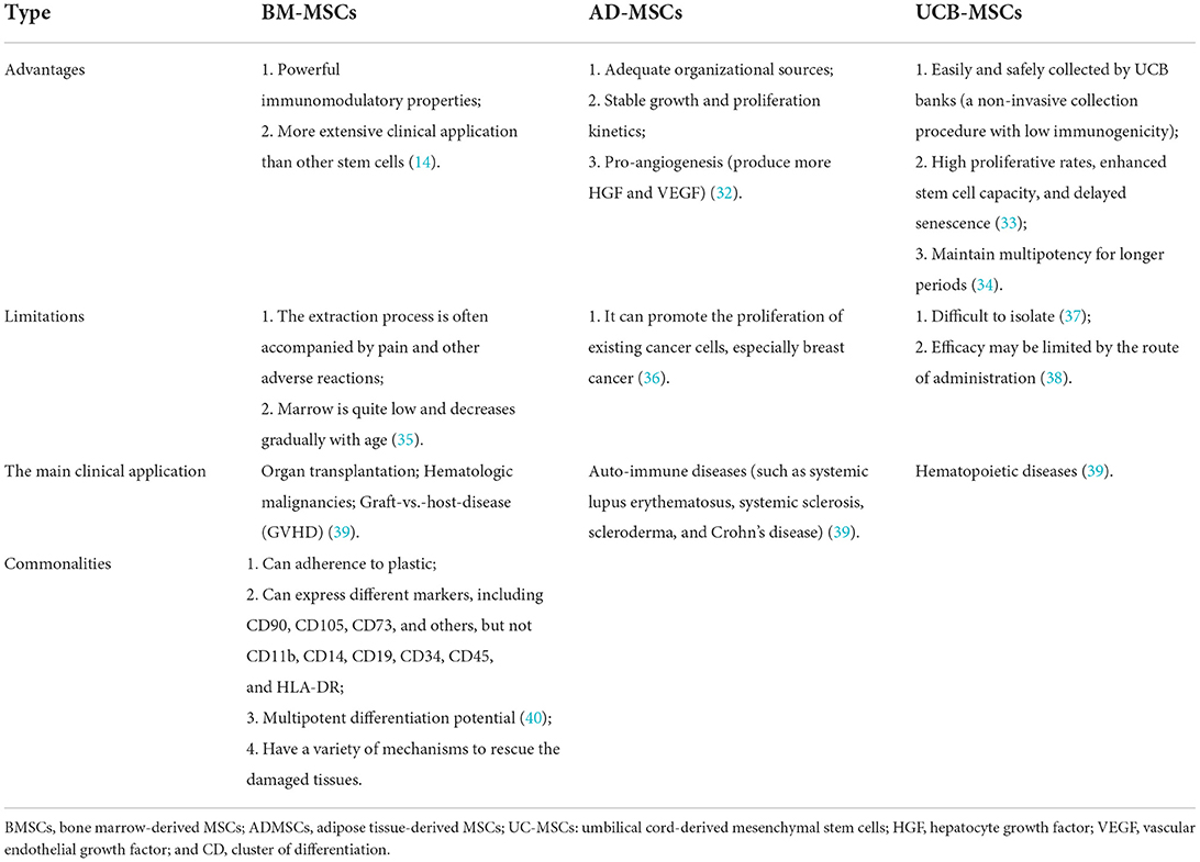
MSCs were isolated from bone marrow for the first time. MSCs have previously been isolated from a variety of tissues, such as the lung, liver, kidney, placenta, fallopian tubes, endometrial polyps, adipose tissue, dental pulp, salivary glands, inferior turbinate, umbilical cord blood, menstrual blood, and other tissues (22, 23). They are plastic-adherent and can express mesenchymal markers, including CD90, CD105, CD73, and others, but cannot express CD11b, CD14, CD19, CD34, CD45, and human leukocyte antigen (HLA)-DR (24). MSCs can be harvested from different tissues, and various donor characteristics restrict the surface markers, quality, and isolated numbers of MSCs. Currently, the most frequently reported sources of MSCs utilized in clinical trials are the bone marrow, adipose tissue, and umbilical cord. MSCs obtained from adipose tissue (AD-MSCs) can express CD49d and produce more HGF and VEGF than bone marrow-derived stem cells (BM-MSCs) (25). Compared with bone marrow-derived stem cells, the number of cells obtained from 1 g of fat tissue may be 500 times greater than that of the same weight of bone marrow (26). However, BM-MSCs are safer than AD-MSCs because they can promote the proliferation of existing cancer cells, especially breast cancer (27). Both BM-MSCs and AD-MSCs have significant neurotrophic potential to stimulate neurite growth in DRG-neurons despite different growth factors, which further supports the feasibility of MSC-based stroke treatment (28). Recent investigations into the transplantation of human umbilical cord mesenchymal stem cells (hUC-MSCs)in stroke models have displayed favorable results, including a reduction in infarct size, improved functional recovery, and increased expression of several neuroprotective factors (including VEGF and BDNF) (29). Yet, their isolation can be difficult (30). MSCs from other sources, such as canine-derived MSCs (cMSCs), have not obtained sufficient clinical evidence and cannot be directly applied (31) (Table 1).

TABLE 1
www.frontiersin.org

Table 1. Advantages and disadvantages of MSCs from different sources.

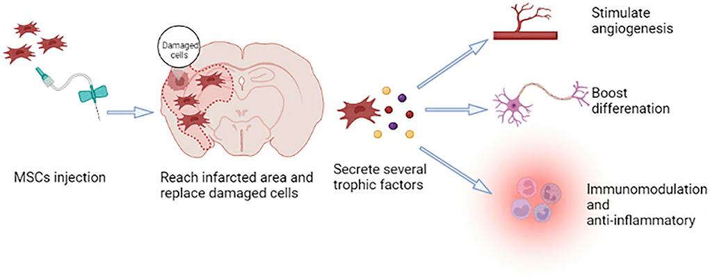
MSCs can self-renew and show polymorphic differentiation (41). They can differentiate into mesoderm cells (described above), endoderm (smooth muscle cells), and ectoderm (neurons) cells under certain conditions (42), which can promote the repair of various damaged tissues (41). Neural regeneration, including neurogenesis, angiogenesis, and synaptic plasticity, is crucial for functional recovery after a stroke. Because MSCs have the characteristics of plasticity, multidirectional differentiation, immunomodulation, and anti-inflammatory, they have the potential to participate in brain regeneration, which can promote tissue repair after ischemic stroke (Figure 1) (43).

FIGURE 1
www.frontiersin.org

Figure 1. Mechanism of MSC therapy.

The role and research progress of MSC transplantation in the treatment of ischemic stroke

Since Azizi et al. published the first report on the transplantation of human BM-MSCs into the rat brain in 1998, an increasing number of studies on treating neurological diseases by MSC transplantation have been conducted. Moreover, MSC transplantation therapy is gradually shifting from laboratory to clinical therapy. Successful clinical studies demonstrate the clinical transformation of MSC transplantation in treating ischemic stroke. Researchers have adopted various methods (intravenous, artery, and intrathecal injection) to administer MSCs to patients with ischemic stroke. They have focused on the safety, feasibility, and short-term effectiveness of MSCs in treating ischemic stroke. This section summarizes several clinical trials to explore the feasibility of MSCs in treating patients with ischemic stroke.

Plentiful preclinical studies of MSC transplantation therapy in ischemic stroke provide a theoretical basis for clinical practice

Medication, rehabilitation training, and physical union therapy have not been effective as experimental treatments for ischemic stroke(So isn't it obvious that you need too get much earlier in the process and stop the 5 causes of the neuronal cascade of death in the first days, saving millions to billions of neurons? And you can't figure that out?)

. Except for thrombolysis and mechanical thrombectomy, no effective medications or procedures have yet been developed. In this situation, new therapeutic strategies using multiple mechanisms are sought, with MSC transplantation being one of them.

In the preclinical studies, researchers explore different sources of MSCs, feasibility, security, and specific mechanisms of MSC-based therapy. First, using in vitro models, they isolate MSCs from various tissues and demonstrate cell differentiation, neuroprotection, neurogenesis, and angiogenesis. In in vivo models, MSCs are injected into animals by different pathways. Researchers demonstrate that MSC transplantation has a potential therapeutic activity that can repair damaged brain tissue, and it seems feasible and secure (summarized in Table 2).

TABLE 2
www.frontiersin.org

Table 2. Preclinical studies of MSCs for the treatment of ischemic stroke.

Model in vitro

The in vitro propagation of MSCs is a three-step process: Extracted from various tissues, MSCs are separated and obtained using density gradient centrifugation digesting culture before being cultured in a plastic cell tissue culture bottle for 3–5 days for further expansion. These steps are then repeated to expand adhered MSCs (63). The following are detailed procedures: To begin, isolate MSCs from multiple tissues such as bone, adipose tissue, tooth tissue, and others using Percoll or Ficoll density gradient centrifugation. Second, rinse MSCs with buffer once to eliminate contaminants before cultivation in 10% fetal bovine serum (FBS) or FBS substitutes, and incubate them in flasks at 37 °C in a humidified 5% CO2 incubator for 2 days. Non-adherent cells are removed by replacing the medium with a fresh one. Subsequently, the attached MSCs proliferate for 2–3 weeks with regular medium change. When the cells have grown to cover about 80% of the flask, it is critical to separate them and allow them to proliferate continually (63).

At present, researchers agree on the multi-lineage differentiation and transplantation potential of MSCs to replace lost tissue after ischemic stroke. When these isolated MSCs are treated with corresponding growth factors or induction medium, they can differentiate into various cell types from different blastoderms (64, 65). These differentiated neuron cells also have functional activity. For example, electrophysiological measurements show that the differentiated cells have voltage-gated sodium and potassium currents, which can be reversibly blocked by tetrodotoxin and tetraethylammonium, respectively (6, 9). However, we need more evidence to prove whether the differentiated cells can fire repetitive action potentials (64).

In addition, MSCs have great neuroprotective effects and promote neurite outgrowth in vitro. Liu et al. claim that BM-MSCs can promote the survival of oxygen-glucose deprivation (OGD) injured neurons, promote axonal outgrowth, and upregulate the expression of GAP-43 when they are cocultured for 48 h with neurons following OGD injury (66). BM-MSCs can also stimulate neurite outgrowth of DRG neurons (67). When hippocampal slices or cortical neurons are cocultured with hMSCs or MSC-derived SB623 cells separated by a semi-porous membrane or with MSC- or SB623 cell-conditioned medium following OGD, neural cell death or damage is decreased. Moreover, 11 neurotrophic factors are identified as secreted by MSCs and/or SB623 cells, and most of them are potentially beneficial to neural tissue following an ischemic insult (68). Furthermore, BM-MSCs from normal healthy and cerebral ischemia rats increase neuronal survival and connectivity in glial-neuron mixed cultures (69). These reports support the fact that MSCs have neuroprotective effects and stimulate neurite development in vitro.

Lastly, the ability of MSCs to stimulate angiogenesis, participate in vascularization, and re-establish a blood supply is evaluated in in vitro models. It is also the fundamental process of tissue repair (70). In in vitro models, MSCs can differentiate into endothelial lineage cells to protect ECs against hypoxia-induced cell death (71), promote the formation of endothelial rings (65), improve the paracrine activity of angiogenic growth factors and EC migration, and form mature vascular tissue (70). Hypoxic preconditioning enhances the pro-angiogenic effects of MSCs by increasing the expression of angiogenic growth factors and boosting the proliferation and migration of ECs (72).

Thus, MSCs show great promise in vitro, including the potential for cell differentiation, neuroprotection, neurogenesis, and pro-angiogenesis. Therefore, several studies have ulteriorly transplanted MSCs into animal models of ischemic stroke and evaluated the outcomes as discussed below.

More at link.