Showing posts with label NeuRA Blog. Show all posts
Showing posts with label NeuRA Blog. Show all posts
Wednesday, April 3, 2019


Monday, September 12, 2016
New imaging technique will help understand muscle contracture after a stroke
How long before this is rolled out to your stroke hospital? Probably won't do one damn bit of good right now since there is no cure for spasticity. And a phone number for your doctor to call.
https://blog.neura.edu.au/2016/09/muscle-contracture-after-stroke/#sthash.G6k2GnGc.dpbs
Cutting-edge imaging technologies are being used to understand what causes muscle contracture after a stroke and how we can improve treatments.

DTI is an MRI-based imaging technique typically used to measure white matter in the brain. Recent advancements in imaging technology have allowed researchers, like ourselves, to more accurately measure muscle fibres as well.
A recent study we conducted revealed that DTI produces more reliable images and minimises the effect of errors typically found in ultrasound images. This is because muscles have complex 3D shapes, so 3D methods such as DTI have many advantages over the more conventional 2D techniques like ultrasound imaging.
We’ll put this information to good use in a new clinical trial aimed at understanding the cause of muscle contracture (stiffening of muscles) in stroke. Contracture is a common cause of disability in people who have had a stroke and, at present, the mechanisms that cause contracture are poorly understood. Additionally, current intervention strategies to treat contracture have been shown to be ineffective. Clearly, there is a need to improve our current understanding of contracture so that it can be treated effectively.
We are seeking volunteers to participate in the study. Participants could be people of any age who have had a stroke and have some physical disability, or healthy people aged over 60. Participation in the study involves coming to NeuRA and having an MRI scan. The visit, including the scan, takes around two hours.
If you are interested in finding out more about the study, please call NeuRA on 02 9399 1832 and ask to speak with Arkiev D’Souza, or send an email to a.dsouza@neura.edu.au with your enquiry.
https://blog.neura.edu.au/2016/09/muscle-contracture-after-stroke/#sthash.G6k2GnGc.dpbs
Cutting-edge imaging technologies are being used to understand what causes muscle contracture after a stroke and how we can improve treatments.
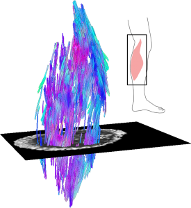
Example of a 3D reconstruction of the muscle fibre
structure of a human calf muscle (medial gastrocnemius) using diffusion tensor
imaging.
We believe a lot of fundamental questions about muscles have yet to be
answered, but that may be about to change with the use of diffusion tensor
imaging (DTI).DTI is an MRI-based imaging technique typically used to measure white matter in the brain. Recent advancements in imaging technology have allowed researchers, like ourselves, to more accurately measure muscle fibres as well.
A recent study we conducted revealed that DTI produces more reliable images and minimises the effect of errors typically found in ultrasound images. This is because muscles have complex 3D shapes, so 3D methods such as DTI have many advantages over the more conventional 2D techniques like ultrasound imaging.
We’ll put this information to good use in a new clinical trial aimed at understanding the cause of muscle contracture (stiffening of muscles) in stroke. Contracture is a common cause of disability in people who have had a stroke and, at present, the mechanisms that cause contracture are poorly understood. Additionally, current intervention strategies to treat contracture have been shown to be ineffective. Clearly, there is a need to improve our current understanding of contracture so that it can be treated effectively.
We are seeking volunteers to participate in the study. Participants could be people of any age who have had a stroke and have some physical disability, or healthy people aged over 60. Participation in the study involves coming to NeuRA and having an MRI scan. The visit, including the scan, takes around two hours.
If you are interested in finding out more about the study, please call NeuRA on 02 9399 1832 and ask to speak with Arkiev D’Souza, or send an email to a.dsouza@neura.edu.au with your enquiry.
Cutting-edge
imaging technologies are being used to understand what causes muscle
contracture after a stroke and how we can improve treatments.
 We believe a lot of fundamental questions about muscles have yet to
be answered, but that may be about to change with the use of diffusion
tensor imaging (DTI).
We believe a lot of fundamental questions about muscles have yet to
be answered, but that may be about to change with the use of diffusion
tensor imaging (DTI).
DTI is an MRI-based imaging technique typically used to measure white matter in the brain. Recent advancements in imaging technology have allowed researchers, like ourselves, to more accurately measure muscle fibres as well.
A recent study we conducted revealed that DTI produces more reliable images and minimises the effect of errors typically found in ultrasound images. This is because muscles have complex 3D shapes, so 3D methods such as DTI have many advantages over the more conventional 2D techniques like ultrasound imaging.
We’ll put this information to good use in a new clinical trial aimed at understanding the cause of muscle contracture (stiffening of muscles) in stroke. Contracture is a common cause of disability in people who have had a stroke and, at present, the mechanisms that cause contracture are poorly understood. Additionally, current intervention strategies to treat contracture have been shown to be ineffective. Clearly, there is a need to improve our current understanding of contracture so that it can be treated effectively.
We are seeking volunteers to participate in the study. Participants could be people of any age who have had a stroke and have some physical disability, or healthy people aged over 60. Participation in the study involves coming to NeuRA and having an MRI scan. The visit, including the scan, takes around two hours.
If you are interested in finding out more about the study, please call NeuRA on 02 9399 1832 and ask to speak with Arkiev D’Souza, or send an email to a.dsouza@neura.edu.au with your enquiry.
- See more at: https://blog.neura.edu.au/2016/09/muscle-contracture-after-stroke/#sthash.G6k2GnGc.dpuf
Example
of a 3D reconstruction of the muscle fibre structure of a human calf
muscle (medial gastrocnemius) using diffusion tensor imaging.
DTI is an MRI-based imaging technique typically used to measure white matter in the brain. Recent advancements in imaging technology have allowed researchers, like ourselves, to more accurately measure muscle fibres as well.
A recent study we conducted revealed that DTI produces more reliable images and minimises the effect of errors typically found in ultrasound images. This is because muscles have complex 3D shapes, so 3D methods such as DTI have many advantages over the more conventional 2D techniques like ultrasound imaging.
We’ll put this information to good use in a new clinical trial aimed at understanding the cause of muscle contracture (stiffening of muscles) in stroke. Contracture is a common cause of disability in people who have had a stroke and, at present, the mechanisms that cause contracture are poorly understood. Additionally, current intervention strategies to treat contracture have been shown to be ineffective. Clearly, there is a need to improve our current understanding of contracture so that it can be treated effectively.
We are seeking volunteers to participate in the study. Participants could be people of any age who have had a stroke and have some physical disability, or healthy people aged over 60. Participation in the study involves coming to NeuRA and having an MRI scan. The visit, including the scan, takes around two hours.
If you are interested in finding out more about the study, please call NeuRA on 02 9399 1832 and ask to speak with Arkiev D’Souza, or send an email to a.dsouza@neura.edu.au with your enquiry.
- See more at: https://blog.neura.edu.au/2016/09/muscle-contracture-after-stroke/#sthash.G6k2GnGc.dpuf
Wednesday, June 29, 2016
Cutting-edge imaging technologies are being used to understand what causes muscle contracture after a stroke and how we can improve treatments.
The first studies I've seen that are looking into objectively describing contractures. This first step is required before we can even attempt to prevent it from happening. This come from Neuroscience Research Australia (NeuRA).
Page 29 for the stroke info:
https://www.neura.edu.au/neura-profile/2016/#28
Page 29 for the stroke info:
https://www.neura.edu.au/neura-profile/2016/#28
Neuroscience Research Australia (NeuRA).
Neuroscience Research Australia (NeuRA).
Sunday, August 17, 2014
Contracture: understanding mechanisms and testing treatments
One more sequelae about stroke we know nothing about. Damn, is there anything that our doctors know about treating stroke problems?
From NeuRA Blog is an initiative of Neuroscience Research Australia
The latest here:
Contracture: understanding mechanisms and testing treatments
NeuRA
Blog is an initiative of Neuroscience Research Australia - See more at:
http://blog.neura.edu.au/about/#sthash.XtTRZ8p5.dpuf
NeuRA
Blog is an initiative of Neuroscience Research Australia - See more at:
http://blog.neura.edu.au/about/#sthash.XtTRZ8p5.dpuf
There
has been surprisingly little research into the mechanisms of
contracture. As a result, the mechanisms are poorly understood. - See
more at:
http://blog.neura.edu.au/2014/08/18/contracture/?utm_source=rss&utm_medium=rss&utm_campaign=contracture#sthash.eOop3nIO.dpuf
There has been surprisingly little research into the mechanisms of contracture. As a result, the mechanisms are poorly understood. Who the hell is tasked with solving these problems? They've only been around since stroke was known about.
One more thing you as a survivor is going to have to solve or prevent on your own.
Subscribe to:
Posts (Atom)