Changing stroke rehab and research worldwide now.Time is Brain! trillions and trillions of neurons that DIE each day because there are NO effective hyperacute therapies besides tPA(only 12% effective). I have 523 posts on hyperacute therapy, enough for researchers to spend decades proving them out. These are my personal ideas and blog on stroke rehabilitation and stroke research. Do not attempt any of these without checking with your medical provider. Unless you join me in agitating, when you need these therapies they won't be there.

What this blog is for:

My blog is not to help survivors recover, it is to have the 10 million yearly stroke survivors light fires underneath their doctors, stroke hospitals and stroke researchers to get stroke solved. 100% recovery. The stroke medical world is completely failing at that goal, they don't even have it as a goal. Shortly after getting out of the hospital and getting NO information on the process or protocols of stroke rehabilitation and recovery I started searching on the internet and found that no other survivor received useful information. This is an attempt to cover all stroke rehabilitation information that should be readily available to survivors so they can talk with informed knowledge to their medical staff. It lays out what needs to be done to get stroke survivors closer to 100% recovery. It's quite disgusting that this information is not available from every stroke association and doctors group.

Showing posts with label arginine. Show all posts

Sunday, November 30, 2025

Arginine slows amyloid buildup and improves brain outcomes in Alzheimer’s disease models

Will your competent? doctor guarantee that the protocol they are writing up on this will 100% prevent Alzheimers?  Why not? Can they at least get human testing going? Or are they that fucking incompetent in not knowing of that need?

All this earlier research which I bet your doctor/hospital knows nothing!

  • arginine (4 posts to December 2016)
  • arginine-rich peptides (1 post to February 2015)
  • Cationic arginine-rich peptides (1 post to March 2020)
  • poly-arginine peptides (1 post to February 2015)
  • Do you prefer your doctor and hospital incompetence NOT KNOWING? OR NOT DOING?

    Arginine slows amyloid buildup and improves brain outcomes in Alzheimer’s disease models

    New preclinical evidence shows that oral arginine reduces toxic amyloid structures and eases behavioral impairments in Alzheimer’s models, highlighting a potential therapeutic path that now requires careful human dose and safety evaluation.
    Study: Oral administration of arginine suppresses Aβ pathology in animal models of Alzheimer

    Study: Oral administration of arginine suppresses Aβ pathology in animal models of Alzheimer's disease. Image Credit: Kateryna Kon / Shutterstock

    A recent study published in the journal Neurochemistry International showed that arginine suppresses amyloid beta () pathology in preclinical Alzheimer’s disease (AD) models. AD is the most common dementia, affecting more than 50 million individuals worldwide. The aggregation of  peptides into fibrils and oligomers is likely the upstream event in the pathogenesis of AD. While various therapies have been developed targeting , they have limited efficacy, adverse effects, and high costs. As such, safe and cost-effective orally administered therapeutic approaches to inhibit  aggregation are needed.

    Rationale for Testing Arginine as an Anti-Aggregation Agent

    Previously, the authors showed that arginine suppresses the aggregation of polyglutamine (PolyQ) proteins, exerting therapeutic effects in preclinical models of PolyQ diseases. Moreover, oral arginine administration was found to improve patients with spinocerebellar ataxia type 6, a PolyQ disease. Given that arginine is a chaperone that inhibits protein misfolding and aggregation, the authors hypothesized that it might suppress the aggregation of .

    In Vitro Assays Demonstrate Strong Inhibition of Aβ Aggregation

    In the present study, researchers investigated whether arginine suppresses the aggregation of  in preclinical models of AD. First, they examined the suppression of  aggregation in vitro and incubated 42 peptides in the presence of varying concentrations of arginine. The aggregation of 42 peptides was determined by Thioflavin T (ThT) fluorescence intensity.

    The team observed that co-incubation of 42 and arginine substantially reduced ThT fluorescence intensity in a dose-dependent manner, decreasing 42 aggregation by 80% at 1 mM arginine. Electron microscopy showed that fibrils were shortened, and biochemical analyses indicated reduced insoluble 42 fractions with unchanged soluble 42 levels, consistent with reduced fibrillar aggregation.
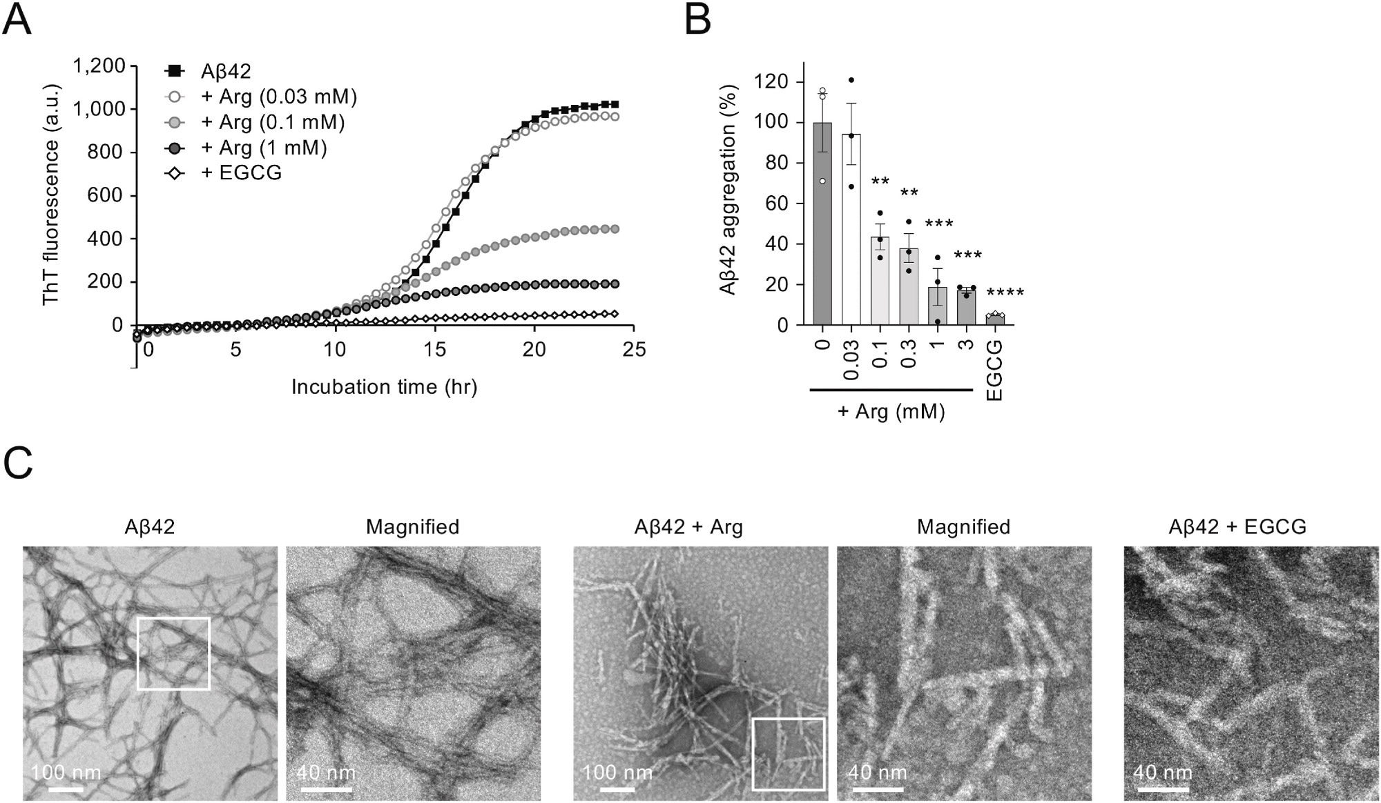
    Arginine suppresses Aβ42 aggregation in vitro. (A) Effect of arginine on Aβ42 aggregation in vitro. The incubation of Aβ42 peptide (5 μM) at 37 °C resulted in an increase in Thioflavin T (ThT) fluorescence after a short lag phase, which eventually reached a plateau. (B) Bar graph showing the ratio of Aβ42 aggregation in the presence of arginine at 24 h. (C) EM images of Aβ42 amyloid fibrils. Scale bars, 100 nm (left images) and 40 nm (right images). The panel on the right is a magnified image of the boxed region in the left panel. Statistical analysis in (B) was performed to assess differences from the control group (Aβ42 without arginine) by one-way ANOVA followed by the Dunnett

    Arginine suppresses Aβ42 aggregation in vitro. (A) Effect of arginine on Aβ42 aggregation in vitro. The incubation of Aβ42 peptide (5 μM) at 37 °C resulted in an increase in Thioflavin T (ThT) fluorescence after a short lag phase, which eventually reached a plateau. (B) Bar graph showing the ratio of Aβ42 aggregation in the presence of arginine at 24 h. (C) EM images of Aβ42 amyloid fibrils. Scale bars, 100 nm (left images) and 40 nm (right images). The panel on the right is a magnified image of the boxed region in the left panel. Statistical analysis in (B) was performed to assess differences from the control group (Aβ42 without arginine) by one-way ANOVA followed by the Dunnett's multiple comparisons test. ∗∗p < 0.01, ∗∗∗p < 0.001, ∗∗∗∗p < 0.0001.

    Arginine Reduces Amyloid Deposition in Drosophila AD Models

    Next, the researchers examined in vivo suppression of  aggregation in a Drosophila AD model carrying the 42 transgene with the E22G Arctic mutation (42arc). 42arc expression in the compound eyes led to abnormal deposition in larval eye discs and shrinkage of adult eyes due to  toxicity. However, oral arginine administration substantially decreased 42 deposition and suppressed eye shrinkage in a dose-dependent manner.

    Mouse Studies Show Reduced Amyloid Deposition and Plaque Burden

    The team further evaluated arginine in App knock-in mice carrying Arctic, Beyreuther, or Iberian, and Swedish mutations (AppNL-G-F mice), which develop age-dependent  deposition and behavioral abnormalities. The mice received 6% arginine orally from 5 weeks of age. Body weight did not differ between treated AppNL-G-F, untreated AppNL-G-F, or wildtype mice.

    Monday, November 24, 2025

    Supplement Shows Promise in Blocking Amyloid in Alzheimer’s

    Your incompetent? doctor and hospital won't get human testing going, will they?

    All this earlier research which I bet your doctor/hospital knows nothing!

  • arginine (2 posts to December 2016)
  • arginine-rich peptides (1 post to February 2015)
  • Cationic arginine-rich peptides (1 post to March 2020)
  • poly-arginine peptides (1 post to February 2015)
  • Do you prefer your doctor and hospital incompetence NOT KNOWING? OR NOT DOING?

    Supplement Shows Promise in Blocking Amyloid in Alzheimer’s

    Summary: A new study shows that oral arginine, a naturally occurring amino acid, can significantly suppress amyloid-β aggregation in Alzheimer’s disease models. Researchers found that arginine not only prevented Aβ42 from clumping but also reduced plaques and inflammation in mouse and fruit fly models carrying Alzheimer’s-related mutations.

    Treated mice showed improved cognitive performance alongside reduced neuroinflammation, indicating broad neuroprotective effects. Because arginine is clinically safe, affordable, and widely available, it holds strong potential as a repurposed therapeutic strategy pending further human studies.

    Key Facts:

    • Aggregation Blocker: Arginine reduced Aβ42 aggregation in vitro and in Alzheimer’s disease models.
    • Neuroprotective Effects: Treatment lowered plaques, decreased inflammatory markers, and improved behavior.
    • Clinical Potential: Arginine’s safety and low cost make it a promising candidate for rapid therapeutic repurposing.

    Source: Kindai University

    Alzheimer’s disease (AD), a progressive neurodegenerative disorder, is one of the leading causes of dementia worldwide, and currently has no definitive cure. Although antibody-based therapies that target amyloid β (Aβ) have recently been developed, their clinical effectiveness remains limited. 

    Sunday, June 15, 2025

    Aspartate in the Brain: A Review

     Ask your competent? doctor what will be done with this knowledge to get you recovered. NOTHING, LIKE USUAL? 

    All this earlier research which I bet your doctor knows nothing!

  • arginine (2 posts to December 2016)
  • arginine-rich peptides (1 post to February 2015)
  • Cationic arginine-rich peptides (1 post to March 2020)
  • poly-arginine peptides (1 post to February 2015)
  • beta-amyloid peptides (2 posts to February 2019)
  • nitric oxide (120 posts to March 2011)
  • endothelial nitric oxide (2 posts to January 2019)
  • Do you prefer your doctor and hospital incompetence NOT KNOWING? OR NOT DOING?

    Aspartate in the Brain: A Review

    Review
  • Open access
  • Published:
  • Volume 50, article number 199, (2025)Cite this articleDownload PDF

    You have full access to this open access article

    Abstract

    l-Aspartate (aspartic acid; C4H7NO4; 2-aminobutanedoic acid) is a non-essential α-amino acid found ubiquitously throughout the body, including in the brain. Aspartate is one of the protein-forming amino acids and the formation of tRNA-aspartate complex is catalysed by aspartyl tRNA synthetase. Free aspartate, which is the main subject of this review, plays key roles in metabolism, as an amino donor and acceptor. It contributes to the synthesis of protein, arginine and nitric oxide, asparagine, N-acetylaspartate and N-methyl-d-aspartate. Its major metabolic role in the brain is recycling reducing equivalents (protons) between the cytoplasm and mitochondrial matrix as part of the malate-aspartate shuttle. l-Aspartate’s actions on synaptic receptors, as well as its possible presence in nerve terminals and synaptic vesicles, are, in principle, consistent with a role as an excitatory neurotransmitter. The evidence is far from conclusive and at times controversial. The role of d-aspartate in brain function is even less certain but, it appears that, rather than being a minor neurotransmitter, d-aspartate is more likely to be involved in fine regulation of endocrine and homeostatic processes. Much research remains to be done in this area. The diversity of its functions and chemistry make aspartate a complex molecule to investigate and measure in vivo. Perturbations of aspartate metabolism have been described in a range of neurological deficits, particularly those of white matter. Here, we examine what is known about the various roles of aspartate in brain, its metabolism, transport and compartmentation, its role as a neurotransmitter or a more general signalling molecule, and what is currently known about its role(s) in disease processes.

    More at link.

    Malate Aspartate Shuttle—Linkage to Phosphorylation State

    The malate-aspartate shuttle (Fig. 1) plays a major role in the movement of reducing equivalents (NADH) into, or out of, the mitochondrion.

    Fig. 1
    figure 1

    Wednesday, March 13, 2024

    Fenugreek seeds: A superfood for health and wellness, says new review

    Ask your doctor if this needs to be added to your diet protocol. If you don't have a diet protocol containing EXACT amounts your doctor is massively incompetent!

    Fenugreek seeds: A superfood for health and wellness, says new review

    The growing interest in adopting a healthier lifestyle has been accompanied by an increased use of healthy food ingredients, which refers to naturally occurring bioactive compounds that have functions in the human body related to human health. These foods are often referred to as functional foods and are crucial for preventing disease, managing chronic conditions, and providing nutritional value.

    A recent study published in the Journal of Food Science discusses the dietary importance and possible applications of fenugreek seeds in foods and beverages.

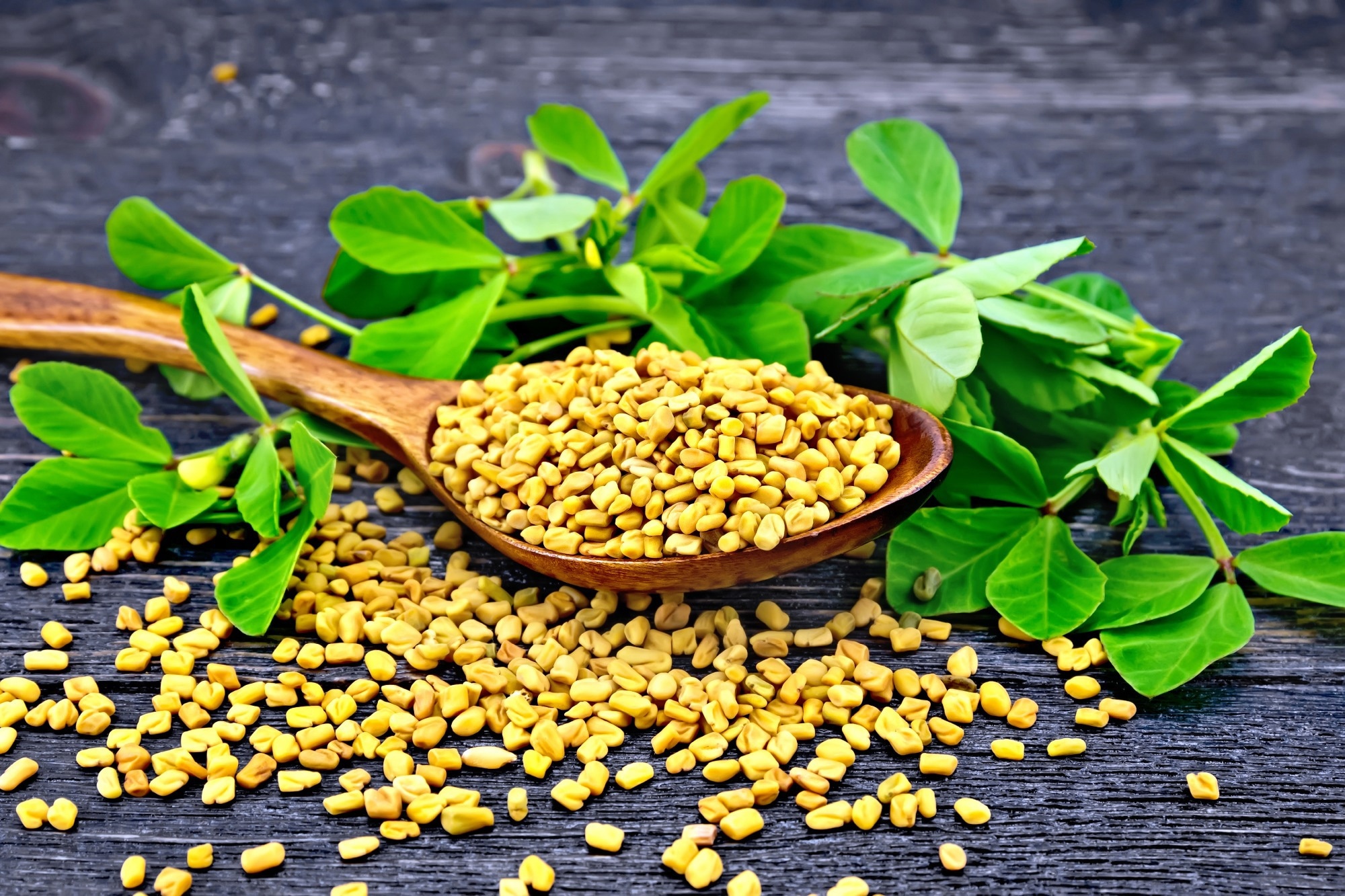
    Study: Current perspectives on fenugreek bioactive compounds and their potential impact on human health: A review of recent insights into functional foods and other high value applications. Image Credit: kostrez / Shutterstock.com

    Nutritional composition of fenugreek seeds

    Each 100 grams (g) of fenugreek seeds comprises 60% carbohydrates, 25% dietary fiber, 23 g protein, 6 g lipids, and 9 g water. Fenugreek is particularly rich in potassium, phosphorus, magnesium, and calcium. Fresh fenugreek leaves contain about 86% water, 6% carbohydrates, 4% protein, and about 1% each of fiber and fat.

    Fenugreek seed carbohydrates have a high glycemic index (GI), thus demonstrating their potential to reduce blood sugar levels, as well as total and low-density lipoprotein (LDL) cholesterol levels. The equimolar galactose:mannose ratio is responsible for a distinctive type of gum that is high in molecular weight and water solubility compared to other plant gums.

    Extruded snack foods fortified with fenugreek have a lower GI, better nutritional and functional profile, as well as a long shelf life. For example, the addition of GM to a chickpea-rice blend reduced the GI of this product from 68% to 43%.

    Germination increases, and roasting reduces dietary fiber, respectively. Roasted fenugreek seed has lower carbohydrate content but higher protein content.

    Between 13-39% of fenugreek seed is protein, which is similar to other legumes used as food, though it differs with the variety. As compared to the husk, the endosperm contains six-fold the protein content.

    Fenugreek seed contains proteins that are resistant to heat denaturation, are very stable, water-soluble, and form stable foams and films. In curries, soups, sauces, bread, meat dishes, cheeses, and desserts, fenugreek seeds provide taste, texture, and thickening, in addition to their non-nutritional benefits.

    Soaking, germinating, and roasting fenugreek seeds increases protein digestibility by 10-15%. Comparatively, certain processes like brief blanching can increase the vitamin content of fenugreek seeds.

    Glutamic acid, aspartic acid, and arginine are the primary amino acids in fenugreek seeds. The main volatile compound is 4-hydroxyisoleucine (4-HIL), which is converted to sotolone, the principal taste-producing molecule. Importantly, 4-HIL, an amino acid not involved in protein synthesis, mediates many of the metabolic actions of fenugreek.

    Fenugreek seed lipids include phospholipids and glycolipids, mostly unsaturated triacylglycerols in the form of polyunsaturated fatty acids (PUFA). The high omega-6:omega-3 ratio of fenugreek seeds is nearly 3:1, which is similar to that of hemp, promotes health, and reduces diet-related chronic disease risk. Phytosterols like campesterol and β-Sitosterol comprise 56% to 72% of the total sterols in fenugreek seeds, respectively.

    Nutraceuticals in fenugreek include saponins like dioscin and diosgenin, alkaloids, phenolics, and volatile essential oils (Eos). Other nutraceutical compounds present in fenugreek include flavonoids like quercetin and ellagic acid, eugenol and linalool, as well as trigonelline.

    Diosgenin is used to synthesize various steroidal drugs, including progesterone and cortisone. Saponin hydrolysis to sapogenins is also capable of producing many compounds that are more bioactive than the parent compounds.

    Trigonelline is the most abundant alkaloid present in fenugreek and is potentially protective against type 2 diabetes and neurodegenerative diseases. Triogenelline has been shown to reduce blood lipids, support kidney and liver function, and prevent cancerous changes. Trigonelline may also prevent bacterial and viral infections and appears to act synergistically with other compounds.

    Health benefits of fenugreek seeds

    Some benefits associated with fenugreek include blood sugar control, lower blood lipids, anticancerous activity, immunomodulation, and pain relief, thereby offering protective effects for the heart and vascular system, gastrointestinal tract, liver, brain, and endocrine system. Fenugreek is also used for preserving reproductive function and relieving skin inflammatory conditions.

    Fenugreek compounds act by restoring beta cell function in the pancreas, reducing hepatic neogluconeogenesis, as well as upregulating antioxidant and hepatoprotective enzymes. Improved insulin signaling and antioxidant activity is also associated with trigonelline. Fenugreek can also restore gut microbiota composition, thereby improving metabolic function and glucose tolerance with secondary beneficial effects on other organ systems.

    Fenugreek seeds may also regulate appetite, prevent cognitive decline, promote wound healing, treat asthma, reduce dysmenorrhea and muscle pain, and modulate menopausal symptoms.

    How is fenugreek used?

    Fenugreek (Trigonella foenum-graecum L.) is an herb bearing small brown seeds with a unique taste and nutritional value. Fenugreek seeds, leaves, and stems are often used for culinary and medicinal purposes.

    Fenugreek seeds provide soups, spice blends, desserts, and teas with a somewhat bittersweet taste. Additionally, biopolymers synthesized from fenugreek are used to stabilize and texturize a wide range of foods.

    Fenugreek is a promising ingredient for functional foods and is a ‘generally recognized as safe (GRAS)’ flavoring agent in the United States and other countries. Multiple forms of fenugreek are available, including seed powder, leaf flour, seed gum, seed husk, Eos, and extracts, as well as edible film.

    Specialty foods like pasta, bread, milk analogs, low-fat dairy cheese, or flavor-enhanced cheeses have been produced using fenugreek. Meat products can also be made more functional by incorporating fenugreek without noticeably altering the taste. Fenugreek leaves have high antioxidant and antimicrobial content, thus preventing the spoiling and rancidity of meat when used in a marinade.

    Fenugreek is tolerable in humans when used at therapeutic doses, except for rare or transient minor side effects like nausea, abdominal pain, or dizziness. In diabetics, fenugreek overuse may lead to hypoglycemia and, if used with drugs that induce low potassium levels, may cause hypokalemia. Fenugreek may also interact with oral anticoagulants to increase bleeding risk and interfere with the absorption of oral drugs.

    Journal reference:
    • Alu’datt, M. H., Rababah, T., Al-ali, S., et al. (2024). Current perspectives on fenugreek bioactive compounds and their potential impact on human health: A review of recent insights into functional foods and other high value applications. Journal of Food Science. doi:10.1111/1750-3841.16970.

    Monday, March 16, 2020

    Cationic Arginine-Rich Peptides (CARPs): A Novel Class of Neuroprotective Agents With a Multimodal Mechanism of Action

    So which one of the 5 causes of the neuronal cascade of death in the first week is this addressing? I wish the president of that great stroke association would enforce the elimination of the useless word neuroprotection and replace it with the neuronal cascade of death. That at least sounds deadly requiring immediate attention.


    Cationic Arginine-Rich Peptides (CARPs): A Novel Class of Neuroprotective Agents With a Multimodal Mechanism of Action

    Bruno P. Meloni1,2,3*, Frank L. Mastaglia2,3 and Neville W. Knuckey1,2,3
    • 1Department of Neurosurgery, QEII Medical Centre, Sir Charles Gairdner Hospital, Nedlands, WA, Australia
    • 2Perron Institute for Neurological and Translational Science, Nedlands, WA, Australia
    • 3Centre for Neuromuscular and Neurological Disorders, The University of Western Australia, Nedlands, WA, Australia
    There are virtually no clinically available neuroprotective drugs for the treatment of acute and chronic neurological disorders, hence there is an urgent need for the development of new neuroprotective molecules. Cationic arginine-rich peptides (CARPs) are an expanding and relatively novel class of compounds, which possess intrinsic neuroprotective properties. Intriguingly, CARPs possess a combination of biological properties unprecedented for a neuroprotective agent including the ability to traverse cell membranes and enter the CNS, antagonize calcium influx, target mitochondria, stabilize proteins, inhibit proteolytic enzymes, induce pro-survival signaling, scavenge toxic molecules, and reduce oxidative stress as well as, having a range of anti-inflammatory, analgesic, anti-microbial, and anti-cancer actions. CARPs have also been used as carrier molecules for the delivery of other putative neuroprotective agents across the blood-brain barrier and blood-spinal cord barrier. However, there is increasing evidence that the neuroprotective efficacy of many, if not all these other agents delivered using a cationic arginine-rich cell-penetrating peptide (CCPPs) carrier (e.g., TAT) may actually be mediated largely by the properties of the carrier molecule, with overall efficacy further enhanced according to the amino acid composition of the cargo peptide, in particular its arginine content. Therefore, in reviewing the neuroprotective mechanisms of action of CARPs we also consider studies using CCPPs fused to a putative neuroprotective peptide. We review the history of CARPs in neuroprotection and discuss in detail the intrinsic biological properties that may contribute to their cytoprotective effects and their usefulness as a broad-acting class of neuroprotective drugs.

    Wednesday, December 14, 2016

    Researchers to study effect of watermelon juice in reducing heart disease

    Were these two earlier research efforts not enough to prove the benefits? This waste of resources is directly a result of NO fucking stroke strategy.  I eat a pound of watermelon a day whenever possible, can't find juice.

    Watermelon Could Lower Blood Pressure April 2014 

    Watermelon juice reverses hardening of the arteries Nov. 2011


     Researchers to study effect of watermelon juice in reducing heart disease

    University of Alabama researchers soon will launch a study that looks at watermelon juice as a way to reduce heart disease.
    Dr. Kristi Crowe-White and Dr. Amy Ellis, researchers in the College of Human Environmental Sciences' department of human nutrition and hospitality management, are recruiting subjects for a 10-week study to see how watermelon impacts blood vessel function.
    The researchers believe that several natural ingredients in watermelon — lycopene, citrulline, arginine, glutamine and ascorbic acid — will act in synergy to decrease arterial stiffness and oxidative stress.
    Arterial stiffness is an early independent risk factor for cardiovascular disease and cardiovascular events like heart attack and stroke. As people get older, the arteries get stiffer, more rigid and less able to dilate, Ellis said.
    Oxidative stress also increases with age because the body is not able to compensate, and people's diet typically is lacking in needed antioxidants. Oxidative stress is one of the underlying causes of all chronic disease, so when the level is high, if a person has a genetic predisposition for a chronic disease, that's when the manifestation will occur, Crowe-White said.
    "Our goal is to reduce the risks of these future cardiovascular events, but since we are both dietitians, we're interested in doing this through a food-first approach versus just another pill or medication," Ellis said. "If there are natural ingredients in this food that could be beneficial, why not try that first."
    Study participants need to be postmenopausal African-American or European-American women ages 55 to 69 who do not smoke or have high blood pressure, diabetes, liver disease or kidney disease. The first four weeks will involve study participants drinking either 100 percent watermelon juice or a placebo twice a day.
    At the end of the four weeks, there will be a two-week washout period where neither beverage is consumed. In the next four weeks, participants will drink the opposite of what they had in the beginning of the study. Vascular and blood measures will be assessed at the beginning and end of each four-week period. Participants are eligible for compensation up to $100.
    Source:
    University of Alabama

    Monday, February 23, 2015

    Peptides used to protect stroke victims from brain damage

    Totally worthless article. It doesn't reference the name of the peptide or the research name. Bruno Meloni doesn't show up with this in google scholar.  What a waste of print.
    This other news source does identify the peptides;
    Peptides May Reduce Brain Injury After Stroke
    Using in vitro neuronal cell culture stroke models we soon discovered that poly-arginine and arginine-rich peptides on their own possessed potent neuroprotective properties.  Furthermore, we showed that as the length of the poly-arginine peptide increased so did the peptides neuroprotective properties.  Excitingly, the poly-arginine peptides were even more potent than the ”neuroprotective peptides” we had been working with and peptides developed by other overseas researchers.
    We have now confirmed using a laboratory animal stroke model that poly-arginine peptides could reduce brain damage when administered up to 1-hour after the stroke

    The news source that is worthless: 

    Peptides used to protect stroke victims from brain damage


    West Australian researchers have discovered a way to use peptides to protect stroke victims from brain damage, and say the breakthrough may reduce the risk of sustaining a serious disability from a stroke, especially for people living in remote Australia.
    Professor David Blacker, the medical director of the West Australian Neuroscience Research Institute, said it was an important development in stroke research.
    "In the rats that were given experimental peptides the volume of stroke damage was substantially smaller," he said.
    "If we can apply that to human models, the hope would be that critical bits of the brain will be less affected."
    The discovery is a big deal, especially for the 50,000 people who will have a stroke this year.
    "It's devastating and there have been surveys that reveal older people will fear surviving a stroke with a substantial disability; they will fear that more than actually dying," Professor Blacker said.
    Professor Blacker, who is also a neurologist at Perth's Sir Charles Gairdner Hospital, said most researchers had given up trying to find such a treatment.
    "At these stroke conferences just recently, people have been standing and applauding the speakers ... because we're so used to trials that have had no result or been negative," he said.

    It is hoped that giving victims peptides within an hour of a stroke will buy them more time before brain damage starts.
    "It's all well and good if you're in a major metro centre and you collapse in the middle of the day and get brought to a hospital where you've got the team that can do the techniques to remove the clot," Professor Blacker said.
    "But it's no good if you're rural and remote [in] Western Australia or Queensland or the Northern Territory where you're hours and hours away.
    "If we could actually; the patient has a stroke and gets some of these drugs that slow down the clotting cascade, it'll buy you some extra time.
    "Then the patient could be transported for more definitive treatment."

    Research 'a game changer', second breakthrough this month

    Professor Bruno Meloni led the research team and said the discovery was a potential game-changer.
    "It could [mean] from a person who is paralysed to a person who has no paralysis, so it will definitely improve their standard of living and their outcomes," he said.
    This is the second major breakthrough in the field this month.
    Last week, Melbourne researchers revealed details of a new treatment that almost doubles stroke victims' chances of walking out of hospital without a disability.
    It involves identifying which parts of the brain are salvageable after a stroke before removing the blood clot and giving the patient clot-busting drugs.
    Professor Meloni said the two discoveries complement each other.
    "The research that came out of Melbourne showed that if you can improve blood flow to the brain, this can limit brain injury and improve patient outcomes," he said.
    "However, despite that treatment, the brain can still be vulnerable to the effects of stroke, and our peptides particularly target vulnerable tissue that may eventually become damage.
    "So the peptides aim to further inhibit or suppress the brain damage that occurs after stroke."
    This trial was done on rats and it is expected to be some years before the peptides are tested on humans in a clinical trial.
    But Professor Blacker said time is always of the essence when it comes to stroke research.
    "One in six people in the world will have a stroke," he said.
    "Moving our studies on, moving the development of new drugs along quickly, is imperative."
    The team also hopes to secure funding to explore how the peptides could help people suffering from other conditions, including cardiac arrest and spinal injury.