Changing stroke rehab and research worldwide now.Time is Brain! trillions and trillions of neurons that DIE each day because there are NO effective hyperacute therapies besides tPA(only 12% effective). I have 523 posts on hyperacute therapy, enough for researchers to spend decades proving them out. These are my personal ideas and blog on stroke rehabilitation and stroke research. Do not attempt any of these without checking with your medical provider. Unless you join me in agitating, when you need these therapies they won't be there.

What this blog is for:

My blog is not to help survivors recover, it is to have the 10 million yearly stroke survivors light fires underneath their doctors, stroke hospitals and stroke researchers to get stroke solved. 100% recovery. The stroke medical world is completely failing at that goal, they don't even have it as a goal. Shortly after getting out of the hospital and getting NO information on the process or protocols of stroke rehabilitation and recovery I started searching on the internet and found that no other survivor received useful information. This is an attempt to cover all stroke rehabilitation information that should be readily available to survivors so they can talk with informed knowledge to their medical staff. It lays out what needs to be done to get stroke survivors closer to 100% recovery. It's quite disgusting that this information is not available from every stroke association and doctors group.

Showing posts with label SCI. Show all posts

Sunday, May 17, 2026

Novel Antibody Repairs Acute Spinal Cord Lesions

 With ANY BRAINS AT ALL  in stroke leadership, this would trigger immediate research to see if it would work for stroke also!  That will never occur, there aren't two functioning neurons anywhere in stroke.

Let's see how long your doctor has DONE NOTHING with nogo-A
  • nogo-A (14 posts to May 2012) 

That's an impressive amount of incompetence displayed everywhere in your hospital!

Novel Antibody Repairs Acute Spinal Cord Lesions

Summary: A multinational clinical trial demonstrated that a novel antibody, NG101, successfully preserves existing nerve tissue and accelerates the regression of spinal cord lesions following acute injury.

The antibody operates by neutralizing Nogo-A, a naturally occurring protein that structurally blocks the regeneration of damaged nerve fibers. By combining advanced magnetic resonance imaging (MRI) with clinical data, researchers have objectively visualized the therapeutic mechanics early in treatment, establishing an essential blueprint for restoring functional brain-to-muscle signal pathways.

Key Facts

  • Removing the Healing Barrier: NG101 targets and neutralizes Nogo-A, an unhelpful protein found in the sheaths of nerve fibers within the brain and spinal cord that actively blocks damaged fibers from healing after an acute trauma.
  • Accelerated Lesion Regression: Advanced imaging methods confirmed for the first time that the antibody therapy speeds up the healing of spinal cord lesions, allowing nerve fibers to regenerate in the tissue surrounding the impact site.
  • Tissue Loss Interception: The therapy considerably slows down the loss of existing nerve tissue and offsets structural degradation by stimulating the regrowth of entirely new nerve fibers.
  • Functional Reconnection: The newly formed and surviving nerve fibers successfully navigate across or around the injury site, re-establishing vital connections with the spinal cord centers that control the peripheral nerves of the hands, arms, and legs.

Source: University of Zurich

Spinal cord injuries – often caused by sports or traffic accidents – can result in tetraplegia or paraplegia and severely limit independence.

In late 2024, an international research group led by the University of Zurich (UZH) and Balgrist University Hospital completed a multinational clinical trial in which patients with acute spinal cord injuries were successfully treated with the novel antibody NG101.

This shows an axon.
By neutralizing the inhibitory Nogo-A protein within the nerve fiber sheaths, the antibody NG101 allows surviving and newly generated axons to effectively navigate around spinal lesions. Credit: Neuroscience News
The results showed that NG101 accelerates the regression of spinal cord lesions and preserves existing nerve tissue.

Antibody neutralizes unhelpful protein

Discovered at UZH roughly 30 years ago, NG101 targets the protein Nogo-A, which is found in the sheaths of nerve fibers in the spinal cord and brain. This protein blocks the healing of damaged nerve fibers in the spinal cord following acute injury.

By neutralizing Nogo-A, NG101 removes this barrier to growth and healing, thereby boosting nerve fiber regeneration and supporting the functional regeneration of spinal cord tissue.

Visible results in the spinal cord

The research team’s latest study has revealed another critical piece of the puzzle. “In our new study, we were able to use advanced imaging methods to show for the first time how this antibody therapy works directly in the spinal cord,” says Patrick Freund, UZH professor and head of the Spinal Cord Injury Center at Balgrist University Hospital.

The magnetic resonance imaging data revealed two important effects. First, spinal cord injuries healed more quickly in the presence of NG101, which suggests that nerve fibers were able to regenerate in the tissue surrounding the injury. Second, the loss of nerve tissue slowed down considerably and was offset by the regrowth of new nerve fibers. Previous animal experiments conducted by the researchers had already established how critical this stage is.

This is due to newly formed nerve fibers needing to find a way to navigate across or around the injury site in order to restore the pathways linking the brain and the spinal cord.

New connections to peripheral nerves

The group’s latest findings suggest that it is precisely this process that is supported by NG101.

“This allows surviving and newly regenerated nerve fibers to re-establish connections with the spinal cord centers that control the hand, arm and leg nerves,” says Freund, who led the study.

“These connections are essential for relaying signals from the brain to the muscles.” For some patients, this means a greater chance of recovering arm and hand function.

NG101 not only improves the function of the spinal cord but has also been shown to alter its structure, which supports the regeneration of nerve tissue. This marks an important step toward new, effective treatments for spinal cord injuries.

“We are now able to visualize the effect of the therapy early on and in an objective way,” says Freund. “This opens up the possibility of using future treatments more strategically and conducting a more reliable evaluation of their outcomes.”

Key Questions Answered:

Q: If someone is paralyzed from a car accident, can this drug make them walk again?

A: While it is too early to guarantee full mobility for every patient, the clinical trial proved that NG101 helps surviving and newly grown nerve fibers reconnect with the spinal cord centers controlling the arms, hands, and legs. For patients with acute injuries, this significantly increases the mathematical probability of recovering crucial hand and arm functions.

Q: Why doesn’t the spinal cord just heal itself naturally like a broken bone?

A: The central nervous system contains a literal molecular brake pad. A specific protein called Nogo-A resides in the protective sheaths of your nerve fibers. Following a sudden trauma, this protein actively stops damaged nerve fibers from growing or repairing themselves. NG101 acts as a shield that shuts down Nogo-A so natural regeneration can take over.

Q: How do doctors know the drug is actually repairing the spine and not just masking symptoms?

A: Researchers used high-resolution MRI data to track the treatment on a cellular and structural level. The imaging objectively showed two clear visual markers: the physical holes (lesions) in the spinal cord shrank much faster, and the rapid degradation of delicate nerve tissue was replaced by the visible architecture of freshly sprouting nerve fibers.

Editorial Notes:

  • This article was edited by a Neuroscience News editor.
  • Journal paper reviewed in full.
  • Additional context added by our staff.

About this spinal cord injury and neurology research news

Author: Kurt Bodenmueller
Source: University of Zurich
Contact: Kurt Bodenmueller – University of Zurich
Image: The image is credited to Neuroscience News

Friday, May 8, 2026

‘Dancing molecules’ successfully repair severe spinal cord injuries

 So, do we have anyone in stroke with two functioning neurons that can look at this and suggest using it for stroke? NO, WE DON'T! Nothing will be done; there is NO leadership or strategy anywhere in stroke! Just plan on never having a stroke!

‘Dancing molecules’ successfully repair severe spinal cord injuries

After single injection, paralyzed animals regained ability to walk within four weeks

Northwestern University researchers have developed a new injectable therapy that harnesses “dancing molecules” to reverse paralysis and repair tissue after severe spinal cord injuries. 

In a new study, researchers administered a single injection to tissues surrounding the spinal cords of paralyzed mice. Just four weeks later, the animals regained the ability to walk.

Five key developments

By sending bioactive signals to trigger cells to repair and regenerate, the breakthrough therapy dramatically improved severely injured spinal cords in five key ways:

  1. The severed extensions of neurons, called axons, regenerated
  2. Scar tissue, which can create a physical barrier to regeneration and repair, significantly diminished
  3. Myelin, the insulating layer of axons that is important in transmitting electrical signals efficiently, reformed around cells(We need this)
  4. Functional blood vessels formed to deliver nutrients to cells at the injury site(We need this)
  5. More motor neurons survived

After the therapy performs its function, the materials biodegrade into nutrients for the cells within 12 weeks and then completely disappear from the body without noticeable side effects. This is the first study in which researchers controlled the collective motion of molecules through changes in chemical structure to increase a therapeutic’s efficacy.

“Our research aims to find a therapy that can prevent individuals from becoming paralyzed after major trauma or disease,” said Northwestern’s Samuel I. Stupp, who led the study. “For decades, this has remained a major challenge for scientists because our body’s central nervous system, which includes the brain and spinal cord, does not have any significant capacity to repair itself after injury or after the onset of a degenerative disease. We are going straight to the FDA to start the process of getting this new therapy approved for use in human patients, who currently have very few treatment options.”

Stupp is Board of Trustees Professor of Materials Science and Engineering, Chemistry, Medicine and Biomedical Engineering at Northwestern, where he is founding director of the Simpson Querrey Institute for BioNanotechnology (SQI) and its affiliated research center, the Center for Regenerative Nanomedicine. He has appointments in the McCormick School of Engineering, Weinberg College of Arts and Sciences and Feinberg School of Medicine.

Life expectancy has not improved since the 1980s

According to the National Spinal Cord Injury Statistical Center, nearly 300,000 people are currently living with a spinal cord injury in the United States. Life for these patients can be extraordinarily difficult. Less than 3% of people with complete injury ever recover basic physical functions. And approximately 30% are re-hospitalized at least once during any given year after the initial injury, costing millions of dollars in average lifetime health care costs per patient. Life expectancy for people with spinal cord injuries is significantly lower than people without spinal cord injuries and has not improved since the 1980s.

“Currently, there are no therapeutics that trigger spinal cord regeneration,” said Stupp, an expert in regenerative medicine. “I wanted to make a difference on the outcomes of spinal cord injury and to tackle this problem, given the tremendous impact it could have on the lives of patients. Also, new science to address spinal cord injury could have impact on strategies for neurodegenerative diseases and stroke.”

‘Dancing molecules’ hit moving targets

The secret behind Stupp’s new breakthrough therapeutic is tuning the motion of molecules, so they can find and properly engage constantly moving cellular receptors. Injected as a liquid, the therapy immediately gels into a complex network of nanofibers that mimic the extracellular matrix of the spinal cord. By matching the matrix’s structure, mimicking the motion of biological molecules and incorporating signals for receptors, the synthetic materials are able to communicate with cells.

“Receptors in neurons and other cells constantly move around,” Stupp said. “The key innovation in our research, which has never been done before, is to control the collective motion of more than 100,000 molecules within our nanofibers. By making the molecules move, ‘dance’ or even leap temporarily out of these structures, known as supramolecular polymers, they are able to connect more effectively with receptors.”

Stupp and his team found that fine-tuning the molecules’ motion within the nanofiber network to make them more agile resulted in greater therapeutic efficacy in paralyzed mice. They also confirmed that formulations of their therapy with enhanced molecular motion performed better during in vitro tests with human cells, indicating increased bioactivity and cellular signaling.

“Given that cells themselves and their receptors are in constant motion, you can imagine that molecules moving more rapidly would encounter these receptors more often,” Stupp said. “If the molecules are sluggish and not as ‘social,’ they may never come into contact with the cells.” 

Signals mimic natural proteins

Once connected to the receptors, the moving molecules trigger two cascading signals, both of which are critical to spinal cord repair. One signal prompts the long tails of neurons in the spinal cord, called axons, to regenerate. Similar to electrical cables, axons send signals between the brain and the rest of the body. Severing or damaging axons can result in the loss of feeling in the body or even paralysis. Repairing axons, on the other hand, increases communication between the body and brain.

The second signal helps neurons survive after injury because it causes other cell types to proliferate, promoting the regrowth of lost blood vessels that feed neurons and critical cells for tissue repair. The therapy also induces myelin to rebuild around axons and reduces glial scarring, which acts as a physical barrier that prevents the spinal cord from healing. 

“The signals used in the study mimic the natural proteins that are needed to induce the desired biological responses. However, proteins have extremely short half-lives and are expensive to produce,” said Zaida Álvarez, the study’s first author and former research assistant professor in Stupp’s laboratory. “Our synthetic signals are short, modified peptides that — when bonded together by the thousands — will survive for weeks to deliver bioactivity. The end result is a therapy that is less expensive to produce and lasts much longer.” 

Universal application

While the new therapy could be used to prevent paralysis after major trauma (automobile accidents, falls, sports accidents and gunshot wounds) as well as from diseases, Stupp believes the underlying discovery — that “supramolecular motion” is a key factor in bioactivity — can be applied to other therapies and targets.

“The central nervous system tissues we have successfully regenerated in the injured spinal cord are similar to those in the brain affected by stroke and neurodegenerative diseases, such as ALS, Parkinson’s disease and Alzheimer’s disease,” Stupp said. “Beyond that, our fundamental discovery about controlling the motion of molecular assemblies to enhance cell signaling could be applied universally across biomedical targets.” 

Other Northwestern study authors include Evangelos Kiskinis, assistant professor of neurology and neuroscience in Feinberg; research technician Feng Chen; postdoctoral researchers Ivan Sasselli, Alberto Ortega and Zois Syrgiannis; and graduate students Alexandra Kolberg-Edelbrock, Ruomeng Qiu and Stacey Chin. Peter Mirau of the Air Force Research Laboratories and Steven Weigand of Argonne National Laboratory also are co-authors. 

Friday, April 17, 2026

‘Dancing’ Molecules Spur Healing in Spinal Cord Tissue

 Do we have ANY INNOVATIVE thinkers in stroke that will look at this and say;'

 'Maybe this could work for stroke'! I bet we have shit for brains that can't think AND WILL DO NOTHING!

We already have these brain representatives:

‘Dancing’ Molecules Spur Healing in Spinal Cord Tissue

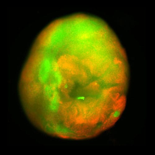
Under a fluorescence microscope at Northwestern University, a sphere of human spinal cord tissue — about 3 mm across, roughly the diameter of a mouse spinal cord — erupted with green light. It had been stained with a calcium-sensitive dye that lights up living neurons and their extending fibers. This green light provided unmistakable visual proof of neurites streaming outward from the tissue’s injured surface and into a synthetic gel, where they were growing in organized, parallel structures.

photo of Fluorescence microscopy reveals axons (green) extending from an injured spinal cord organoid
Fluorescence microscopy reveals axons (green) extending from an injured spinal cord organoid after treatment with the supramolecular therapy.

That sphere of spinal cord tissue was a spinal cord organoid — a three-dimensional, miniaturized tissue model grown in a dish from human induced pluripotent stem cells (iPSCs) to recapitulate key features of an actual organ. This one had been differentiated over 24 weeks into a range of spinal cord cell types: neurons, astrocytes, oligodendrocyte progenitor cells, and Schwann cells.

The Northwestern team also incorporated microglia — the brain and spinal cord’s resident immune cells — by coaxing iPSC-derived progenitors to infiltrate the organoid. The result was an immune-competent model capable of mounting an inflammatory response to injury.

photo of Contusion from a mechanical impactor
Contusion from a mechanical impactor produces diffuse cell death (red), simulating the blunt-force trauma behind real-world spinal cord injuries.

Then, to make the organoid useful, the team injured it — either slicing it with a scalpel to model surgical hemisection or crushing it with a mechanical impactor to deliver the kind of compressive contusion that causes most real-world spinal cord injuries.

Wednesday, February 18, 2026

Lab-Grown Human Spinal Cord Shows Healing After Injury in Major Breakthrough: Study

 Do you really think your stroke medical 'professionals' will look at this and say; 'Maybe something will come out of this for stroke'! I think nothing will happen. Their is NO leadership and NO strategy in stroke.


Lab-Grown Human Spinal Cord Shows Healing After Injury in Major Breakthrough: Study

Saturday, May 3, 2025

Industrial-grade collaborative robots for motor rehabilitation after stroke and spinal cord injury: a systematic narrative review

 You're fired for delivering nothing that get patients recovered! But you did get published, so congratulations on that useless win.

Industrial-grade collaborative robots for motor rehabilitation after stroke and spinal cord injury: a systematic narrative review

Abstract

Background

There is a growing interest in exploring industrial-grade collaborative robots (cobots) for rehabilitation. This review explores their application for motor rehabilitation of the upper and lower extremities after a stroke and spinal cord injury (SCI). The article highlights the inherent safety features of cobots, emphasizing their design advantages over custom-built or traditional rehabilitation robots in terms of potential safety and time efficiency.

Methods

Database searches and reference list screening were conducted to identify studies relating to the use of cobots for upper and lower extremity rehabilitation among individuals with stroke and SCI. These articles were then reviewed and summarized.

Results

Thirty-three studies were included in this review. The findings suggest that the use of cobots in motor rehabilitation is still in the early stages. Some of the cobots used were equipped with sensors to detect and respond to the movement of the extremities and minimize the risk of injury. This safety aspect is crucial for patients with motor impairments. Most training protocols implemented with the cobots engaged users in repetitive task-based exercises with an overall positive user experience. Thus far, these devices have been primarily evaluated in individuals with stroke and SCI that affect the lower extremities, with no study addressing upper extremity impairments. This initial focus serves as a preliminary step toward assessing their applicability for individuals with stroke and SCI.

Conclusions

Cobots may(What a weasel word, saying nothing!) have the capacity to transform therapy and support healthcare professionals in delivering more personalized and effective rehabilitation. However, there is limited evidence on their use to support upper and lower extremity rehabilitation among individuals with stroke and SCI. Further research and development are needed to refine these technologies and broaden their applications in rehabilitation settings to enhance functional recovery and overall quality of life for individuals with stroke and SCI.

Tuesday, January 7, 2025

Unlocking Spinal Cord Regeneration: Astrocytes Lead the Way

 Do our stroke medical 'professionals' have enough brains to see the possible use of this for stroke recovery?  NO? So, we have NO stroke medical professionals at all?

Unlocking Spinal Cord Regeneration: Astrocytes Lead the Way

Summary: Spinal cord injuries (SCI) are challenging to treat due to the limited regenerative capacity of the central nervous system. A new study reveals that while ependymal cells in the spinal cord show minimal repair potential, astrocytes can transdifferentiate into oligodendrocytes, aiding in remyelination.

Researchers also found that modifying the injury microenvironment with functional material transplantation enhances this regenerative process. These findings highlight astrocytes’ critical role and offer a promising approach to advancing SCI therapies.

Key Facts:

  • Ependymal Limitations: Ependymal cells show minimal proliferation and restricted repair potential post-SCI, especially in primates.
  • Astrocyte Potential: Some astrocytes transdifferentiate into oligodendrocytes under injury conditions, contributing to remyelination.
  • Therapeutic Boost: Functional material transplantation enhances astrocyte transdifferentiation and promotes spinal cord repair.

Source: Chinese Academy of Sciences

Spinal cord injury (SCI) remains one of the most devastating medical conditions, severely impacting quality of life and often leading to permanent disability.

The central nervous system (CNS) has a limited capacity for regeneration, which poses a significant challenge in treating SCI, since recovery becomes increasingly difficult once the spinal cord is damaged.

This shows astrocytes.
Their cross-species examination of primate and rodent spinal cords revealed new insights into the behaviors of ependymal and astrocyte cells following SCI, highlighting their dynamic roles in spinal cord repair. Credit: Neuroscience News

During development, neural stem cells in the spinal cord differentiate into various neural cells that form complex circuits.

As the spinal cord matures, these progenitor cells lose their regenerative potential, making adult spinal cord tissue less capable of recovery after injury. Identifying endogenous stem cells with diverse lineage potentials in the adult spinal cord is crucial for advancing repair strategies for SCI.

In a recent study published in PNAS, researchers led by Profs. DAI Jianwu and ZHAO Yannan from the Institute of Genetics and Developmental Biology of the Chinese Academy of Sciences explored the regenerative mechanisms behind SCI repair.

By constructing single-cell transcriptomic databases for both human spinal cord development and rhesus monkey SCI models, the researchers established a foundation for comprehensively analyzing spinal cord cell behavior.

Their cross-species examination of primate and rodent spinal cords revealed new insights into the behaviors of ependymal and astrocyte cells following SCI, highlighting their dynamic roles in spinal cord repair.

The research team discovered that, during spinal cord development, ependymal cells mature and gradually lose their neural progenitor cell properties, retaining only limited proliferative capacity.

After injury, ependymal cells displayed minimal activation and demonstrated no significant proliferation or cross-lineage differentiation.

Importantly, the study revealed that the reactivity of ependymal cells post-SCI is significantly lower in primates than in rodents, indicating more restricted regenerative potential in primates.

In contrast, astrocytes in the injured spinal cord exhibited significant activation. Through single-cell and lineage tracing analyses, the researchers revealed that some astrocytes could transdifferentiate into oligodendrocytes under injury-induced conditions, thereby contributing to the remyelination process.

Further investigation identified an intermediate population among astrocytes where key transcription factors such as SOX10 promoted their conversion into oligodendrocyte lineage cells.

To enhance the regenerative process, the team introduced functional material transplantation into the injury microenvironment.

This intervention significantly boosted the efficiency of astrocyte transdifferentiation into oligodendrocytes, suggesting that material transplantation not only mitigates the inhibitory effects of the injury site but also creates favorable conditions for promoting remyelination.

This study provides compelling evidence of the limited regenerative capacity of ependymal cells in adult primate spinal cord injury and underscores the transdifferentiation potential of astrocytes.

Furthermore, the research highlights how microenvironmental modulation can enhance the efficiency of astrocyte-mediated repair, offering a promising approach for future SCI therapies.

About this SCI and neuroscience research news

Author: Na Chen
Source: Chinese Academy of Sciences
Contact: Na Chen – Chinese Academy of Sciences
Image: The image is credited to Neuroscience News
Original Research: Closed access.
“Characterizing progenitor cells in developing and injured spinal cord: Insights from single-nucleus transcriptomics and lineage tracing” by DAI Jianwu et al. PNAS

Saturday, July 27, 2024

Dental pulp stem cells regenerate neural tissue in degenerative disorders and stroke rehabilitation: A scope systematic review

I still prefer handing your competent? doctor a pee cup and asking for stem cells in return.  It has only been 11+ years, where the fuck is your doctor's protocol on turning urine into stem cells?

Turning urine into brain cells could help fight Alzheimer’s, Parkinson’s

December 2012

The latest here:

Dental pulp stem cells regenerate neural tissue in degenerative disorders and stroke rehabilitation: A scope systematic review

Open AccessPublished:July 24, 2024DOI:https://doi.org/10.1016/j.heliyon.2024.e35080
>

Abstract

Background

Dental Pulp Stem Cells (DPSCs) possess a remarkable ability for tissue differentiation, making them highly efficient in tissue regeneration and inflammation regulation. This systematic study proposes to find an answer to the question, "Do DPSCs have the ability to regenerate and rehabilitate nerve tissue?"

Methods

This systematic review was conducted based on Preferred Reporting Items for Systematic Reviews and Meta-Analyses (PRISMA) criteria, and the principle of non-bias was respected. All the articles from 2014-2024 were extracted from the Web of Science, PubMed, and Scopus databases. This study extracted the antigens and pro-inflammatory factors associated with DPSCs' involvement and how they affect the CNS's neural tissue regeneration.

Results

Two persons of researchers searched the database. After screening the full texts, they included 11 articles in their study. DPSCs control the following antigens: CD73, CD34, CD90, CD105, CD14, CD45, CD19Oct-4, CD73, CD31, CD34CD29CD44. Even though hematopoietic markers did not change much, OCT-4 and CD-73 were increased by DPSCs. DPSC-derived exosomes suppressed the expression of IL-6, IL-1β, TNF-α, and TGF, key mediators of nerve tissue inflammation. Additionally, DPSCs show high Vascular Endothelial Growth Factor (VEGF) expression in mice brain tissue cultures. DPSCs reduce Subarachnoid Hemorrhage (SAH), a condition in which blood collects in the subarachnoid space and causes ischemia.

Discussion

DPSCs showed the ability to regenerate nerve tissue and brain ganglia, stimulating angiogenesis by expressing cell markers and controlling growth factors in mice, and high therapeutic potential in neurodegenerative disorders. The present study invites further research in neurological disorders, specifically strokes, to prescribe these stem cells to the human population.

Wednesday, April 10, 2024

A portable system to measure knee extensor spasticity after spinal cord injury

Even if this would work for stroke, it won't do a damn bit of good since there IS NOTHING OUT THERE TO CURE SPASTICITY!

A portable system to measure knee extensor spasticity after spinal cord injury

Abstract

Background

The pendulum test is a quantitative method used to assess knee extensor spasticity in humans with spinal cord injury (SCI). Yet, the clinical implementation of this method remains limited. The goal of our study was to develop an objective and portable system to assess knee extensor spasticity during the pendulum test using inertial measurement units (IMU).

Methods

Spasticity was quantified by measuring the first swing angle (FSA) using a 3-dimensional optical tracking system (with external markers over the iliotibial band, lateral knee epicondyle, and lateral malleolus) and two wireless IMUs (positioned over the iliotibial band and mid-part of the lower leg) as well as a clinical exam (Modified Ashworth Scale, MAS).

Results

Measurements were taken on separate days to assess test–retest reliability and device agreement in humans with and without SCI. We found no differences between FSA values obtained with the optical tracking system and the IMU-based system in control subjects and individuals with SCI. FSA values from the IMU-based system showed excellent agreement with the optical tracking system in individuals with SCI (ICC > 0.98) and good agreement in controls (ICC > 0.82), excellent test–retest reliability across days in SCI (ICC = 0.93) and good in controls (ICC = 0.87). Notably, FSA values measured by both systems showed a strong association with MAS scores (

 ~ −0.8) being decreased in individuals with SCI with higher MAS scores, reflecting the presence of spasticity.

Conclusions

These findings suggest that our new portable IMU-based system provides a robust and flexible alternative to a camera-based optical tracking system to quantify knee extensor spasticity following SCI.

Introduction

Spasticity is a common symptom present in a large number of individuals with spinal cord injury (SCI) [1, 2]. Despite having a considerable impact on independence and quality of life after SCI [3, 4], its quantification remains limited. Clinical exams, such as the Modified Ashworth Scale (MAS) [5] and the Tardieu scale [6], use nominal scales for the quantifications of spasticity and have limited validity and reliability [7,8,9,10,11]. Mechanical devices used to quantify resistance to a passive stretch at controlled amplitudes and velocities need expensive and bulky equipment and therefore are less suitable for routine use in clinical environments [12]. Thus, there is a pressing need for developing objective and portable systems to measure spasticity [13,14,15, 2, 16].(I see measuring spasticity as completely useless until interventions that cure spasticity work!)

The goal of our study was to develop a system to assess knee extensor spasticity, which is commonly observed in individuals with SCI [1, 4]. At present, the pendulum test is a widely used biomechanical test for evaluating knee extensor spasticity using kinematic analysis [17]. The test quantifies the effect of a gravity-induced stretch of the knee extensor muscle on the leg kinematics and is performed with the participant sitting or supine with the legs hanging over the edge of a table. The operator first brings the leg to full extension and then suddenly releases it letting the limb swing under the action of gravity. A reduction of the first swing motion of the leg (first swing angle, FSA) has been associated with increased stretch reflex activity [18,19,20,21]. The pendulum test has been validated in controls [22] and has a high test–retest reliability and sensitivity to detect variations in spasticity in humans with SCI [20, 23,24,25], correlating with clinical scores [26,27,28]. Although, the pendulum test has been widely used alongside clinical scales barriers remain to implementing this exam in the clinic [13, 16, 29, 30]. Over the years, the pendulum test has been instrumented using video recordings [22], electrogoniometers [31, 32], gyroscopes [28], and accelerometers [33] making comparisons and standardization across outcomes difficult. Video recordings and systems based on optical markers are considered the gold standard for administering the pendulum test in laboratory environments but the need for markers to be visible at all times complicates physical examinations and measurements are sensitive to soft tissue artifacts and errors in markers positioning [34]. Electrogoniometers require positioning on two segments of a joint and are sensitive to improper alignment with the joint axes [35]. Gyroscopes can lead to overestimation of joint angles at higher angular speeds and accuracy degrades with time, while estimations from accelerometers are affected by measurement noise and integration drift [36]. Inertial measurements units (IMUs) have been extensively adopted as a reliable and inexpensive alternative to video recordings and systems based on optical markers for estimating lower limb kinematics [37,38,39,40,41]. We hypothesized that a portable system using two wireless IMUs would have good reliability in assessing knee extensor spasticity during the pendulum test as a 3-dimensional optical tracking system.

To address this question, we evaluated FSA values obtained with an optical tracking system and an IMU-based system in individuals with and without SCI on two different days. FSA values were compared with the MAS in individuals with SCI.