Changing stroke rehab and research worldwide now.Time is Brain! trillions and trillions of neurons that DIE each day because there are NO effective hyperacute therapies besides tPA(only 12% effective). I have 523 posts on hyperacute therapy, enough for researchers to spend decades proving them out. These are my personal ideas and blog on stroke rehabilitation and stroke research. Do not attempt any of these without checking with your medical provider. Unless you join me in agitating, when you need these therapies they won't be there.

What this blog is for:

My blog is not to help survivors recover, it is to have the 10 million yearly stroke survivors light fires underneath their doctors, stroke hospitals and stroke researchers to get stroke solved. 100% recovery. The stroke medical world is completely failing at that goal, they don't even have it as a goal. Shortly after getting out of the hospital and getting NO information on the process or protocols of stroke rehabilitation and recovery I started searching on the internet and found that no other survivor received useful information. This is an attempt to cover all stroke rehabilitation information that should be readily available to survivors so they can talk with informed knowledge to their medical staff. It lays out what needs to be done to get stroke survivors closer to 100% recovery. It's quite disgusting that this information is not available from every stroke association and doctors group.

Showing posts with label blood clotting. Show all posts

Thursday, June 6, 2024

Common sugar substitute linked to increased risk of heart attack and stroke

 Is your competent doctor going to IMMEDIATELY contact the dietician and get the hospital diet protocol changed so this is not involved?

Common sugar substitute linked to increased risk of heart attack and stroke


Is sugar the new cigarettes? Why you may see new warning labels
Scroll back up to restore default view.

The safety of sugar substitutes is once again being called into question.

Researchers led by the Cleveland Clinic linked the low-calorie sugar substitute xylitol to an increased risk of heart attack, stroke or cardiovascular-related deaths, according to a study published today in the European Heart Journal.

Xylitol is a sugar alcohol that is found in small amounts in fruit and vegetables, and the human body also produces it. As an additive, it looks and tastes like sugar but has 40% fewer calories. It is used, at much higher concentrations than found in nature, in sugar-free gum, candies, toothpaste and baked goods. It can also be found in products labeled "keto-friendly," particularly in Europe.

The same research team found a similar association last year to the popular sugar substitute erythritol. The use of sugar substitutes has increased significantly over the past decade as concerns about rising obesity rates mount.

“We’re throwing this stuff into our food pyramid, and the very people who are most likely to be consuming it are the ones who are most likely to be at risk” of heart attack and stroke, such as people with diabetes, said lead author Dr. Stanely Hazen, chair of cardiovascular and metabolic sciences at Cleveland Clinic’s Lerner Research Institute.

Many heart attacks and strokes occur in people who do not have known risk factors, like diabetes, high blood pressure or elevated cholesterol levels. The research team began studying sugar alcohols found naturally in the human body to see if the compounds might predict cardiovascular risk in these people.

In the study, the investigators measured the level of naturally occurring xylitol in the blood of more than 3,000 participants after overnight fasting. They found that people whose xylitol levels put them in the top 25% of the study group had approximately double the risk for heart attack, stroke or death over the next three years compared to people in the bottom quarter.

The researchers also wanted to understand the mechanism at work, so they fed xylitol to mice, added it to blood and plasma in a lab and gave a xylitol-containing drink to 10 healthy volunteers. In all these cases, xylitol seemed to activate platelets, which are the blood component that controls clotting, said Hazen. Blood clots are the leading cause of heart attack and stroke.

“All it takes is xylitol to interact with platelets alone for a very brief period of time, a matter of minutes, and the platelet becomes supercharged and much more prone to clot,” Hazen said.

The next question is what causes naturally-occurring xylitol to be elevated in some people and how do you lower it, said Dr. Sadiya Khan, a cardiologist at Northwestern Medicine Bluhm Cardiovascular Institute and a professor of cardiovascular epidemiology at Northwestern Feinberg School of Medicine who was not involved in the new study.

Much more research needs to be done, said Hazen. In the meantime, he is telling patients to avoid eating xylitol and other sugar alcohols, whose spelling all end in ‘itol.’ Instead, he recommends using modest amounts of sugar, honey or fruit to sweeten food, adding that toothpaste and one stick of gum are probably not a problem because so little xylitol is ingested.

The report had key limitations.

First, the study of naturally occurring xylitol in people’s blood was observational and can show only an association between the sugar alcohol and heart risk. It does not show that xylitol caused the higher incidence of heart attack, stroke or death.

Nevertheless, given the totality of the evidence presented in the paper, “it’s probably reasonable to limit intake of artificial sweeteners,” said Khan. “Perhaps the answer isn’t replacing sugar with artificial sweeteners but thinking about more high quality dietary components, like vegetables and fruits, as natural sugars.”

Artificial sweeteners shouldn’t be difficult to avoid, said Joanne Slavin, PhD, RDN, a professor of food science and nutrition at the University of Minnesota-Twin Cities. They are listed on the ingredient list of packaged goods.

“Would I say never eat xylitol?” asked Slavin, who had no connection to the study. For some people who struggle to reduce sugar in their diet, sugar substitutes are one tool, and it comes down to personal choice, she said.

While Slavin found the study interesting and cause for some concern, she noted that sugar alcohols are expensive and are generally used in very small amounts in gum and sugar-free candies.

Another limitation of the study is that the participants whose xylitol levels in the blood were measured were at high risk for or had documented heart disease, and so the results may not apply to healthy individuals.

Still, many people in the general public share the characteristics of the study participants, said Hazen.

“In middle-aged or older America, it’s common to have obesity and diabetes or high cholesterol or high blood pressure,” he said.

This article was originally published on NBCNews.com

Tuesday, February 28, 2023

Zero-calorie sweetener linked to heart attack and stroke, study finds

Is this enough for your doctor to instruct the nutritionist to update the diet protocols in the hospital and what is provided for discharged survivors? Or do you have an incompetent doctor that doesn't keep up-to-date on stroke research?

Zero-calorie sweetener linked to heart attack and stroke, study finds

A sugar replacement called erythritol – used to add bulk or sweeten stevia, monkfruit and keto reduced-sugar products – has been linked to blood clotting, stroke, heart attack and death, according to a new study.

“The degree of risk was not modest,” said lead study author Dr. Stanley Hazen, director of the Center for Cardiovascular Diagnostics and Prevention at the Cleveland Clinic Lerner Research Institute.

People with existing risk factors for heart disease, such as diabetes, were twice as likely to experience a heart attack or stroke if they had the highest levels of erythritol in their blood, according to the study, published Monday in the journal Nature Medicine.

“If your blood level of erythritol was in the top 25% compared to the bottom 25%, there was about a two-fold higher risk for heart attack and stroke. It’s on par with the strongest of cardiac risk factors, like diabetes,” Hazen said.

Additional lab and animal research presented in the paper revealed that erythritol appeared to be causing blood platelets to clot more readily. Clots can break off and travel to the heart, triggering a heart attack, or to the brain, triggering a stroke.

“This certainly sounds an alarm,” said Dr. Andrew Freeman, director of cardiovascular prevention and wellness at National Jewish Health, a hospital in Denver, who was not involved in the research.

“There appears to be a clotting risk from using erythritol,” Freeman said. “Obviously, more research is needed, but in an abundance of caution, it might make sense to limit erythritol in your diet for now.”

In response to the study, the Calorie Control Council, an industry association, told CNN that “the results of this study are contrary to decades of scientific research showing reduced-calorie sweeteners like erythritol are safe, as evidenced by global regulatory permissions for their use in foods and beverages,” said Robert Rankin, the council’s executive director, in an email.

The results “should not be extrapolated to the general population, as the participants in the intervention were already at increased risk for cardiovascular events,” Rankin said.

The European Association of Polyol Producers declined to comment, saying it had not reviewed the study.

What is erythritol?

Like sorbitol and xylitol, erythritol is a sugar alcohol, a carb found naturally in many fruits and vegetables. It has about 70% of the sweetness of sugar and is considered zero-calorie, according to experts.

Artificially manufactured in massive quantities, erythritol has no lingering aftertaste, doesn’t spike blood sugar and has less of a laxative effect than some other sugar alcohols.

“Erythritol looks like sugar, it tastes like sugar, and you can bake with it,” said Hazen, who also directs the Cleveland Clinic’s Center for Microbiome and Human Health.

“It’s become the sweetheart of the food industry, an extremely popular additive to keto and other low-carb products and foods marketed to people with diabetes,” he added. “Some of the diabetes-labeled foods we looked at had more erythritol than any other item by weight.”

Erythritol is also the largest ingredient by weight in many “natural” stevia and monkfruit products, Hazen said. Because stevia and monkfruit are about 200 to 400 times sweeter than sugar, just a small amount is needed in any product. The bulk of the product is erythritol, which adds the sugar-like crystalline appearance and texture consumers expect.

An unexpected discovery

The discovery of the connection between erythritol and cardiovascular issues was purely accidental, Hazen said: “We never expected this. We weren’t even looking for it.”

Hazen’s research had a simple goal: find unknown chemicals or compounds in a person’s blood that might predict their risk for a heart attack, stroke or death in the next three years. To do so, the team began analyzing 1,157 blood samples in people at risk for heart disease collected between 2004 and 2011.

“We found this substance that seemed to play a big role, but we didn’t know what it was,” Hazen said. “Then we discovered it was erythritol, a sweetener.”

The human body naturally creates erythritol but in very low amounts that would not account for the levels they measured, he said.

To confirm the findings, Hazen’s team tested another batch of blood samples from over 2,100 people in the United States and an additional 833 samples gathered by colleagues in Europe through 2018. About three-quarters of the participants in all three populations had coronary disease or high blood pressure, and about a fifth had diabetes, Hazen said. Over half were male and in their 60s and 70s.

In all three populations, researchers found that higher levels of erythritol were connected to a greater risk of heart attack, stroke or death within three years.

But why? To find out, researchers did further animal and lab tests and discovered that erythritol was “provoking enhanced thrombosis,” or clotting in the blood, Hazen said.

Clotting is necessary in the human body, or we would bleed to death from cuts and injuries. The same process is constantly happening internally, as well.

“Our blood vessels are always under pressure, and we spring leaks, and blood platelets are constantly plugging these holes all the time,” Hazen said.

However, the size of the clot made by platelets depends on the size of the trigger that stimulates the cells, he explained. For example, if the trigger is only 10%, then you only get 10% of a clot.

“But what we’re seeing with erythritol is the platelets become super responsive: A mere 10% stimulant produces 90% to 100% of a clot formation,” Hazen said.

“For people who are at risk for clotting, heart attack and stroke – like people with existing cardiac disease or people with diabetes – I think that there’s sufficient data here to say stay away from erythritol until more studies are done,” Hazen said.

Oliver Jones, a professor of chemistry at RMIT University in Victoria, Australia, noted that the study had revealed only a correlation, not causation.

“As the authors themselves note, they found an association between erythritol and clotting risk, not definitive proof such a link exists,” Jones, who was not involved in the research, said in a statement.

“Any possible (and, as yet unproven) risks of excess erythritol would also need to be balanced against the very real health risks of excess glucose consumption,” Jones said.

Healthy volunteers

In a final part of the study, eight healthy volunteers drank a beverage that contained 30 grams of erythritol, the amount many people in the US consume, Hazen said, according to the National Health and Nutrition Examination Survey, which examines American nutrition each year.

Blood tests over the next three days tracked erythritol levels and clotting risk.

“Thirty grams was enough to make blood levels of erythritol go up a thousandfold,” Hazen said. “It remained elevated above the threshold necessary to trigger and heighten clotting risk for the following two to three days.”

Just how much is 30 grams of erythritol? The equivalent of eating a pint of keto ice cream, Hazen said.

“If you look at nutrition labels on many keto ice creams, you’ll see ‘reducing sugar’ or ‘sugar alcohol,’ which are terms for erythritol. You’ll find a typical pint has somewhere between 26 and 45 grams in it,” he said.

“My co-author and I have been going to grocery stores and looking at labels,” Hazen said. “He found a ‘confectionery’ marketed to people with diabetes that had about 75 grams of erythritol.”

There is no firm “accepted daily intake,” or ADI, set by the European Food Safety Authority or the US Food and Drug Administration, which considers erythritol generally recognized as safe (GRAS).

“Science needs to take a deeper dive into erythritol and in a hurry, because this substance is widely available right now. If it’s harmful, we should know about it,” National Jewish Health’s Freeman said.

Hazen agreed: “I normally don’t get up on a pedestal and sound the alarm,” he said. “But this is something that I think we need to be looking at carefully.”

Thursday, May 12, 2022

Individuals with long COVID may be at greater risk for abnormal blood clotting

 You don't want this risk so get vaccinated and boosted to reduce the severity of any COVID-19 you do get.  Which is why at the start of COVID-19 I was going to demand heparin.

Heparin:

Why I'm getting heparin.  Heparin binds to cells at a site adjacent to ACE2, the portal for SARS-CoV-2 infection, and "potently" blocks the virus, which could open up therapy options.

Anticoagulation Again Shown to Improve Survival in COVID-19 Patients;-Mortality risk about 50% lower

The latest here:

Individuals with long COVID may be at greater risk for abnormal blood clotting

Individuals with long COVID may have a higher risk for abnormal blood clotting, especially those who have difficulties with basic exercise for more than 12 weeks after infection, according to a study published in Blood Advances.

The findings highlight a potential role for antithrombotic therapy in the management of these patients, researchers noted.

Source: Adobe Stock.

Rationale and methods

Long COVID, characterized by persistent fatigue, reduced exercise tolerance, chest pain, shortness of breath and cognitive slowing, is an increasingly recognized complication of acute COVID-19 infection, according to Nithya Prasannan, researcher in the department of hematology at University College London Hospitals NHS Foundation Trust in the U.K., and colleagues.

“Acute COVID-19 is strongly linked with increased risk [for] thrombosis and a prothrombotic state, quantified by elevated von Willebrand factor antigen:ADAMTS13 ratio, and is associated with severity of acute COVID-19 infection,” they wrote.

Investigators sought to examine whether patients with long COVID referred to a dedicated post-COVID-19 clinic experienced a prothrombotic state associated with symptom severity. The analysis included 330 patients (median age, 46 years; range, 18-88; 60% female), 97% of whom had symptoms for 3 months or more after acute COVID-19 infection.

Researchers assessed thrombotic risk and characterized patients as being in a prothrombotic state if they had significantly more von Willebrand factor antigen than ADAMTS13 in the bloodstream.

Eighty-four percent of participants (n = 276) completed exercise tests that included walking on a flat surface and/or repeatedly going from a sitting to standing position from a chair while wearing oxygen monitors. Researchers measured oxygen levels and tested participants’ blood before and after exercise to measure lactate levels.

Key findings

Results showed 28% of the entire cohort had a von Willebrand factor antigen:ADAMTS13 ratio of 1.5 or higher, which researchers considered to be raised. This was four times more common among patients with impaired exercise capacity, characterized by desaturation levels 3% or more and/or an increase in lactate level more than 1 from baseline on 1-minute sit to stand test and/or 6-minute walk test (P < .0001). Among those 56 patients, more than half (55%) had had a raised a von Willebrand factor antigen:ADAMTS13 ratio.

About one-quarter of all patients with long COVID had high factor VIII levels that ranged from 2.1 IU/mL to 5.1 IU/mL, and 18% had elevated von Willebrand factor antigen levels of 1.7 IU/mL to 3.3 IU/mL.

Implications

Looking ahead, Prasannan and colleagues plan to assess patients’ bloodwork using different research platforms during the course of their long COVID to assess how risk for thrombosis may change with progression of symptoms, according to a press release.

“I hope that people will view this research as a step forward in understanding what causes long COVID, which will hopefully help us guide future treatment options,” Prasannan said in the release. “I encourage people experiencing long COVID to participate in clinical trials when available because the more data we have, the better we can understand this condition.”

References:

Long COVID-19 exercise capacity linked to abnormal blood clotting markers (press release). www.hematology.org/newsroom/press-releases/2022/long-covid-19-exercise-capacity-linked-to-abnormal-blood-clotting-markers. Published May 11, 2022. Accessed May 11, 2022.
Prasannan N, et al. Blood Adv. 2022;doi:10.1182/bloodadvances.2021006944
.


Wednesday, July 21, 2021

'New mechanism for stroke?': Blood clotting in coronavirus patients has experts looking at role of viruses in causing stroke

WHOM do we talk to to make sure this is followed up on? Nothing will happen since we have NO STROKE LEADERSHIP OR STRATEGY. You are screwed along with your children and grandchildren when they have strokes.

'New mechanism for stroke?': Blood clotting in coronavirus patients has experts looking at role of viruses in causing stroke

Posted 
A patient in bed surrounded by doctors.
Clotting may be a major factor in organ damage from COVID-19, according to new findings.(

AP: Zhang Yuwei via Xinhua

)
Share

A growing body of evidence is painting a grim picture of how coronavirus can wreak havoc from one end of the body to the other.

It's well established that COVID-19 affects the respiratory system and targets the lungs. But several studies and anecdotal evidence suggest the disease may also cause super-charged blood clotting that damages vital organs, including the kidneys, heart and brain.

The New England Journal of Medicine recently published a case report on a small cluster of strokes in New York that involved young, healthy people, who had the virus but no known risk factors for stroke.

But strokes, where a clot blocks blood flow to the brain, appear to be just one way damage from clotting can play out in COVID-19 patients.

There are also reports of 'COVID toes' - painful red inflammations of the feet linked to blood clots.

Then there is the widely reported story of 41-year-old Broadway actor Nick Cordero, whose right leg was amputated after he developed severe blood clots while he was in hospital being treated for coronavirus.

'New mechanism for stroke'

Some experts suggest that what's going on may represent an entirely new way in which blood clots can form.

It might be the case that viruses generally have a more direct role in causing strokes than has been previously thought, says an Australian doctor working on the frontline against COVID-19 in New York.

"We are asking ourselves... have we just discovered a new mechanism of stroke?" says Associate Professor Thomas Oxley, Interventional Neurologist at New York's Mt Sinai Hospital, where thousands of COVID-19 patients have been treated.

Strokes kill brain cells and are a major cause of death and disability. They can cause changes in language, mood, vision and movement. In up to 30 per cent of strokes, the cause is unknown.

Dr Oxley says there is some evidence the clotting leading to strokes and other issues might be being triggered directly by an interaction between the virus and the blood vessel wall.

"I'm a stroke neurologist but we don't usually think about viral infection as a cause for stroke. That [idea] is something very new in medicine."

The role of blood clots

Dr Oxley says the links coming to light between the virus and blood clotting show the virus is affecting the body's blood system, as well as the respiratory system.

"Up to this point, we've been focusing on pneumonia as the primary problem caused by COVID-19," Dr Oxley says. Pneumonia is when the tiny air sacs in the lungs become full of fluid, making it hard to breathe.

"We are now learning there is another major cause of disease by COVID and it's got to do with the formation of blood clots."

Among the problems doctors around the world have been reporting are an increase in clots in the deep veins of the legs (known as deep vein thrombosis or DVT) and the lungs (pulmonary embolisms).

Around one in four people with coronavirus who end up in intensive care will develop a pulmonary embolism, while the incidence of clots in the deep veins of the legs or in the lungs is 30 to 70 per cent.


Saturday, December 19, 2020

'New mechanism for stroke?': Blood clotting in coronavirus patients has experts looking at role of viruses in causing stroke

Now if we had ANY STROKE LEADERSHIP AND STRATEGY we could be assured that  this will be followed up and protocols created to prevent clotting.

'New mechanism for stroke?': Blood clotting in coronavirus patients has experts looking at role of viruses in causing stroke

A growing body of evidence is painting a grim picture of how coronavirus can wreak havoc from one end of the body to the other.

It's well established that COVID-19 affects the respiratory system and targets the lungs. But several studies and anecdotal evidence suggest the disease may also cause super-charged blood clotting that damages vital organs, including the kidneys, heart and brain.

The New England Journal of Medicine recently published a case report on a small cluster of strokes in New York that involved young, healthy people, who had the virus but no known risk factors for stroke.

But strokes, where a clot blocks blood flow to the brain, appear to be just one way damage from clotting can play out in COVID-19 patients.

There are also reports of 'COVID toes' - painful red inflammations of the feet linked to blood clots.

Then there is the widely reported story of 41-year-old Broadway actor Nick Cordero, whose right leg was amputated after he developed severe blood clots while he was in hospital being treated for coronavirus.

'New mechanism for stroke'

Some experts suggest that what's going on may represent an entirely new way in which blood clots can form.

It might be the case that viruses generally have a more direct role in causing strokes than has been previously thought, says an Australian doctor working on the frontline against COVID-19 in New York.

"We are asking ourselves... have we just discovered a new mechanism of stroke?" says Associate Professor Thomas Oxley, Interventional Neurologist at New York's Mt Sinai Hospital, where thousands of COVID-19 patients have been treated.

Strokes kill brain cells and are a major cause of death and disability. They can cause changes in language, mood, vision and movement. In up to 30 per cent of strokes, the cause is unknown.

Dr Oxley says there is some evidence the clotting leading to strokes and other issues might be being triggered directly by an interaction between the virus and the blood vessel wall.

"I'm a stroke neurologist but we don't usually think about viral infection as a cause for stroke. That [idea] is something very new in medicine."

The role of blood clots

Dr Oxley says the links coming to light between the virus and blood clotting show the virus is affecting the body's blood system, as well as the respiratory system.

"Up to this point, we've been focusing on pneumonia as the primary problem caused by COVID-19," Dr Oxley says. Pneumonia is when the tiny air sacs in the lungs become full of fluid, making it hard to breathe.

"We are now learning there is another major cause of disease by COVID and it's got to do with the formation of blood clots."

Among the problems doctors around the world have been reporting are an increase in clots in the deep veins of the legs (known as deep vein thrombosis or DVT) and the lungs (pulmonary embolisms).

Around one in four people with coronavirus who end up in intensive care will develop a pulmonary embolism, while the incidence of clots in the deep veins of the legs or in the lungs is 30 to 70 per cent.


Monday, July 13, 2020

Autopsies indicate blood clots are lethal in COVID-19

I don't know how you are supposed to self treat this if you get it but not bad enough to go to a hospital. I'm hoping my 325 aspirin is enough. Or may be I just  demand a Lovenox shot. Not sure if walk in clinics would have anticoagulation therapies immediately available.

Autopsies indicate blood clots are lethal in COVID-19

In mid-March, pathologist Sigurd Lax pulled on a water-repellant surgery coat, a surgical cap, special booties to cover his shoes, a respiratory mask, and two pairs of gloves, then walked into an operating room at Hospital Graz II in Austria. There before him lay a patient who died of COVID-19 only 48 hours earlier.
“COVID-19 is a new disease, and we really want to know what’s underlying it,” Lax says. There’s only one method to determine what causes illness and death. “It’s doing an autopsy.”
Methodically, Lax sliced into the patient, taking care not to spray any bodily fluids into the air. He’d waited two days to perform the autopsy to reduce the risk of transmitting SARS-CoV-2, the coronavirus that causes COVID-19. With little known about the illness at the time, he and his colleagues wanted to take extra precautions to avoid getting sick. Waiting 48 hours after death to perform the autopsy, however, didn’t rid the body of SARS-CoV-2 RNA.

“We don’t know if the virus is still infectious,” Lax says, but finding its genetic material lingering in the patient’s tissues prompted him to be even more cautious. This autopsy was the first of 11 he and his colleagues have performed on patients who died from COVID-19. In May, the pathologists’ published a paper in the Annals of Internal Medicine reporting what they’d found.
Lax, a professor of pathology at the Johannes Kepler University Linz, spoke with The Scientist about the results and what they reveal about possible therapeutics for COVID-19.
The Scientist: As you started performing these autopsies, what was most striking in terms of damage done to the body?
Sigurd Lax:Both lungs were symmetrically damaged, with damage to the alveoli, the tiny air sacs of the lungs. The pleurae, the double-lined membranes, surrounding the lungs showed very little inflammatory changes, a little fluid. Most cases had a strongly dilated heart, but the abdomen was not really involved. That was what we saw at first view.
TS: In your paper, you write that patients had blood clots in their small to mid-sized pulmonary arteries. What’s the significance of this finding?
SL: We saw an occlusion of multiple pulmonary arteries, usually in the periphery. There were some clots in the larger branches, but we think seeing more of them in the periphery means the clots are thrombotic rather than embolic. [Editor’s note: thrombotic clots develop in a blood vessel where there is damage, in this case in the lungs, while embolic clots form elsewhere, in a blood vessel in the leg, for example, and travel through body and become lodged in blood vessels of the lung.] And this concurs also with findings in pediatric cases, where you have occlusion of arteries in the fingers, for example, or necrosis in back, so in the skin on their back. So it’s not the pulmonary embolism which is the major cause of severe illness but the inflammation of the blood vessels and a change in the clotting system of the blood. And this causes occlusion of the pulmonary arteries, which increases the pressure of pulmonary circulation and then leads to an insufficiency of the heart to pump blood through the body properly.
TS: Does the virus cause the blood clots to form or is it the immune response to the virus that causes them?
SL:So far that’s not clear. I think one can speculate that either one or the other or both may be the reason, but I think it might be more the immune response, that inflammatory mechanisms lead to the thrombosis.
TS: When doing the autopsies, did you focus mainly on the lungs or were there other body regions that you also examined, such as the brain?
SL:In the first cases we only looked at the brain in one case, but we’ve done more autopsies since the paper came out and from those, the central nervous system doesn’t seem to be really specifically affected, at least grossly. I don’t have so far any microscopic data, but the analysis is ongoing.

TS:There have been reports that infection with SARS-CoV-2 could cause diabetes by leading to damage of insulin-producing cells. Was there any indication of that in the autopsies?
SL:Not really. I think there’s a coincidence with diabetes. I think that has to do with the vascular changes caused by diabetes. . . . These patients due to their diabetes have atherosclerosis, then they have heart disease, and this aggravates the course of these COVID-19–associated pulmonary changes because, you know, when you have a strong heart, you will be able to overcome the infection, but if you have some associated disease, particularly heart diseases, you will undergo cardiac failure.
TS: What do your results say, right now, about treating COVID-19 patients with a severe form of the disease?
SL:Right after being admitted to the hospital, ten out of our eleven patients received an anticoagulant at a prophylactic level to prevent clotting in the blood.(You talked about doing 11 autopsies, so even with immediate anticoagulation those 10 still died? What is the solution then?) We did not find thrombosis in the deep arteries of the pelvis and the deep blood vessels of the pelvis and upper legs, but the treatment was not enough to prevent thrombosis in the pulmonary arteries.
TS:Given the autopsy data and all of the information researchers are gathering on COVID-19, what are the biggest questions that have yet to be answered?
SL:What we really don’t know at the moment is how the virus goes through the body, what are the sequence of changes caused by the virus. That’s not completely solved.
Editor’s note: This interview has been edited for brevity.
To read more, click here

Saturday, June 6, 2020

Blood Clotting and Alzheimer’s


You'll have to ask your doctors how they will use this to prevent your likely chance of Alzheimers/dementia.  

Your chances of getting dementia.

1. A documented 33% dementia chance post-stroke from an Australian study?   May 2012.

2. Then this study came out and seems to have a range from 17-66%. December 2013.

3. A 20% chance in this research.   July 2013.

4. Dementia Risk Doubled in Patients Following Stroke September 2018 

5. Parkinson’s Disease May Have Link to Stroke March 2017

 The latest here:

Blood Clotting and Alzheimer’s


Alzheimer’s disease (AD) is the most common form of dementia, a condition in which the individual suffers the loss of cognitive faculties such as thinking, logic, judgment, language, problem-solving, and memory.
Image Credit: Lightspring/Shutterstock.com
It is a progressive condition and is thought to be linked to a reduced number of interneuronal connections in the brain.

Fibrinogen and AD

The brain in a patient with AD is the presence of abnormal proteins within and around the neurons, called beta-amyloid plaques and tau neurofibrillary plaques. The accumulation of these proteins is correlated with synaptic degeneration and cognitive decline.
Earlier studies have demonstrated abnormalities in the complex vascular network of the brain, but scientists have not been able to clarify if the vascular deficit caused the synaptic breakdown or if it only led to inflammation in the nervous system. Both of these conditions end in cognitive deterioration.
A study from the Gladstone Institutes demonstrated that fibrinogen, a key blood coagulation component, is involved in a cascade of events at both molecular and cellular events which lead to the loss of memory storage neurons within the brain. This is the pathophysiological basis of memory loss, according to the researchers.
The fibrinogen comes from the blood within the brain’s blood vessels. It leaks out of the blood, leading to activation of the microglia, which are the brain’s immune cells. This leads to their attack on the neurons at the synapses, causing their destruction.
The study employed cutting-edge technology to generate the first 3D volume images of AD, from humans and mouse experiments. The researchers then manipulated the mice, preventing the leakage of fibrinogen, which in turn kept the brain’s immune cells quiescent. This protected the brain against memory loss.
In a healthy brain, too, the presence of fibrinogen induced the same type of changes as in AD, but without any evidence of amyloid plaques. This was surprising, as amyloid has been the target of many research efforts to find a therapy that works for AD.
These findings lend support to the hypothesis that dementia is caused by two separate mechanisms. Or in other words, the symptoms of AD and vascular dementia could be due to independently operating underlying disease processes.

Vascular and Amyloid Hypotheses

On the one hand, vascular insufficiency causes dementia, as is seen in elderly people following a stroke. On the other hand, AD occurs in people whose brains show the appearance of amyloid plaques.
Alternatively, both could be part of a linked process. Both sets of people show the deterioration of cognitive abilities and when both pathologies are present, the decline in cognition is more profound than for either alone.

How are Amyloid-Beta and Fibrinogen Linked?

The additive effect of fibrinogen in AD could be partly explained by the fact that when amyloid interacts with fibrinogen or its successor, fibrin, increased amounts of fibrin are deposited within the blood vessels of the brain.
The amyloid-fibrinogen interaction leads to the formation of clots that are resistant to the thrombolytic action of tissue plasminogen activator (tPA) when they are formed directly from thrombin.
This resistance to breakdown could be due to the formation of a tighter fibrin network when associated with amyloid, or because amyloid interferes with the binding of plasminogen to fibrin.
In short, the interaction of fibrinogen or fibrin with amyloid could initiate or aggravate the hypoperfusion and inflammation in the brain.
Fibrin build-up interferes with the normal cerebral perfusion and induces microinfarcts, which lead to neuroinflammation, disruption of the blood-brain barrier, and dysfunction of the neurons.
Simultaneously, the amyloid activates fibrinogen, causing more fibrin to be generated via the intrinsic coagulation pathway. Amyloid also activates factor XII which triggers thrombosis, besides increasing the level of inflammation and vascular permeability through other soluble inflammatory mediators like kallikrein and bradykinin.

Fibrinogen-Linked Prothrombotic State in AD

AD patients may have a higher risk of thrombosis, as shown by higher levels of activated factors VII and V, factor V Leiden (a mutant and more degradation-resistant form of factor V), von Willebrand factor, and prothrombin fragments.
Patients with AD were tested for the risk of spontaneous emboli within the cerebral circulation, compared to healthy age-matched controls. A full 40% of them showed such events, vs only 15% in the controls, and this subset showed a faster cognitive decline over the next 6 months.
Again, microbleeds from cerebral blood vessels occur more frequently in AD than with non-AD controls, perhaps due to ischemia-induced hemorrhage.
Research in mice shows that by reducing the fibrinogen levels, the incidence of cerebral amyloid angiopathy (CAA) declines. With CAA, amyloid is deposited around cerebral blood vessels, causing narrowing and ischemia, as well as blood-brain barrier damage. CAA is present in up to 90% of AD patients.
Reduction in the level of fibrinogen also reduces microglial activation and improves cognitive performance in mouse models. On the other hand, AD is associated with a prothrombotic state: there is a higher tendency to form clots, reduced fibrin degradation, increased platelet activation, and higher levels of clotting factors.
Platelet activation might deliver fibrin directly to forming clots, contributing to the formation of resistant clots.

Directions for Future Therapy in AD

The presence of fibrin in cerebrospinal fluid helps distinguish a patient with AD from mild dementia, or non-dementia patients, and is linked with a more severe disease course. These findings could change the way the condition is perceived and treated.
The use of coagulation data in AD patients could be a novel biomarker, and research suggests that the inhibition of fibrinogen effects in AD could be a new therapeutic target.
Since anticoagulant therapy is a challenge in elderly patients, and AD increases the risk of major cerebral bleeds due to the presence of CAA, novel inhibitors that act on the amyloid-fibrinogen interaction could be useful in the therapy of this devastating condition.
Image Credit: Juan Gaertner/Shutterstock.com

Source

  • Strickland, S. (2017). Impact of the Coagulation System on the Pathogenesis of Alzheimer's Disease. Blood (2017) 130 (Supplement 1): SCI-3. https://doi.org/10.1182/blood.V130.Suppl_1.SCI-3.SCI-3. ashpublications.org/.../Impact-of-the-Coagulation-System-on-the
  • Suidan, G. L., et al. (2018). Abnormal Clotting of the Intrinsic/Contact Pathway in Alzheimer Disease Patients Is Related to Cognitive Ability. Blood Advances; 2(9): 954–963. doi: 10.1182/bloodadvances.2018017798. https://www.ncbi.nlm.nih.gov/pmc/articles/PMC5942003/
  • Cortes-Cantelli, et al. (2012). Fibrinogen and Altered Hemostasis in Alzheimer's Disease. Journal of Alzheimer's disease: JAD 32(3). DOI: 10.3233/JAD-2012-120820.
  • Merlini, M. et al. (2019). Fibrinogen Induces Microglia-Mediated Spine Elimination and Cognitive Impairment in an Alzheimer’s Disease Model. Neuron. DOI: https://doi.org/10.1016/j.neuron.2019.01.014

Thursday, May 28, 2020

Coronavirus seems to reach the brain. What could this mean for us?

My god, can no one think at all? Autopsies show this:

Translation: The lungs of the patients who died secondary to Covid-19 had wide-spread clotting, nine times more blood clots and blockages in the thousands of small capillaries and blood vessels in their lungs, compared with the lungs of patients who died from influenza.  

Micro clots could easily cause all these widespread brain damage reports.

Coronavirus seems to reach the brain. What could this mean for us?

JENNIFER FRONTERA has been treating people in intensive care for years. But she has never experienced anything like covid-19 before. “These patients are absolutely among the sickest any of us have ever encountered,” says the New York-based doctor. But the strange thing is, Frontera isn’t a lung disease specialist or a virologist, she is a neurologist. And it is the possible impact of the coronavirus on our brains that is worrying her.
It was early in the outbreak in New York that Frontera and her colleagues began to notice neurological symptoms in those with covid-19. People were passing out before they were hospitalised. Once in hospital, some of them started having unusual movements. Some had seizures and others had strokes.
Similar reports are coming in from hospitals around the world. Some neurological symptoms appear to be mild, such as the loss of smell and taste. At the other end of the spectrum, a few people have developed encephalitis – a potentially fatal inflammation of the brain.
It is a surprising discovery in a disease that was generally considered to attack the airways, and one of pressing concern. One big question is how the new coronavirus is causing these kinds of symptoms. Growing evidence suggests that the virus may work its way into the brain, directly attacking neurons. If that is the case, we may need to reconsider some of the treatments being developed for covid-19. And we must also prepare for potential long-term and chronic neurological conditions in some survivors.
Millions of people globally have now been infected with the new coronavirus, SARS-CoV-2, but we are still …

Continue reading
Subscribe now for unlimited access


Tuesday, November 7, 2017

Blood Leaking Fibrinogen Is the Cause of Multiple Neurological Diseases

We  need to know how much damage there is to the myelin sheaths post-stroke so we can determine if the solution to this is required for stroke recovery.
https://www.labroots.com/trending/cardiology/7243/blood-leaking-fibrinogen-cause-multiple-neurological-diseases

WRITTEN BY: Kara Marker
3 10 412

The body has built-in mechanisms to handle brain injury, but when something disrupts the repair process, neurological diseases like multiple sclerosis (MS), stroke, and Alzheimer’s develop. With a new study published in the journal Neuron, scientists from the Gladstone Institutes discovered the role of a blood protein in blocking repair processes in the brain.
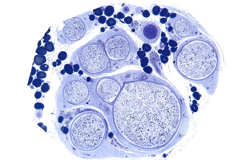
Low magnification micrograph of a vasculitic neuropathy. Credit: Wikimedia User Nephron
Without the myelin sheath, nerve fibers experience a plethora of problems centered around impaired signal transmission: cognition, sensation, movement, and more. When Gladstone researchers realized that it was the blood-clotting protein fibrinogen that was causing the repair problems, they set out to revolutionize the way scientists approach treating neurological disorders.
Fibrinogen, made in the liver, is one of 13 coagulation factors that promote blood clotting. In the right context, blood clotting can save lives by preventing excessive bleeding. In the wrong context, blood clotting can prevent the flow of blood, carrying oxygen and nutrients, to important bodily organs like the brain and heart.
They found that fibrinogen “leaks” into the central nervous system and disrupts the way nerve cells produce myelin, preventing repair. Normally, adult stem cells travel to damaged nerve sites and transform into new myelin-producing cells. However, fibrinogen prevents adult stem cells from making the reparative transition into myelin-producing cells.
"We thought it might be important to look instead at the toxic environment outside the cell, where blood proteins accumulate," explained senior investigator Katerina Akassoglou, PhD. "We realized that targeting the blood protein fibrinogen could open up the possibility for new types of therapies to promote brain repair."
Now that they know what’s stopping repair, scientists are hopeful that they can find a way to better approach treating neurological diseases like MS and Alzheimer’s.
“Repairing myelin by eliminating the toxic effects of vascular damage in the brain is a new frontier in disease therapeutics," said Gladstone’s Lennart Mucke, MD. "This study could change the way we think about how to repair the brain."
Sources: Gladstone Institutes, Healthline

Thursday, July 14, 2016

Blood Coagulation Detector May Help in Monitoring Stroke Risk

For your doctor to explain to you. 

Blood Coagulation Detector May Help in Monitoring Stroke Risk


An analyser recently developed to measure blood coagulability has the sensitivity to detect hypercoagulatibility associated with stroke risk in those without atrial fibrillation (AF), according to research published in PLOS ONE.
To estimate the risk of stroke among patients with AF and determine the requirement for anticoagulation therapy, the CHADS2 predictive score is used. Because some parts of this score are also associated with atherosclerosis risk and increased blood coagulability, a high score has been proposed as linked to hypercoagulability in both patients with and without AF. However, this association has not been fully investigated, partly owing to the lack of a sensitive means of detection.
Researchers from the Tokyo Medical and Dental University (TMDU), Tokyo, Japan, used a highly sensitive technique to measure small changes in blood coagulation, and found hypercoagulability in patients without AF with high CHADS2 scores.
Several physical and chemical factors affect blood clotting, some of which can be measured over time to determine blood coagulability and the likelihood of clot formation. Dielectric blood coagulometry (DBCM) is a recently developed test that measures changes in the dielectric permittivity of whole blood, representing clumping of red blood cells.
The researchers used DBCM to detect changes in the dielectric permittivity of whole blood at 10 MHz. Comparisons between untreated blood and that with added heparin or tissue factor enabled derivation of a coagulability index.
“We calculated the end of acceleration time as an index of coagulability from temporal changes in dielectric permittivity,” said Satomi Hamada, TMDU. “This value reduced when tissue factor was added, and increased with heparin present. It was also sensitive enough to detect small changes in coagulability, particularly in hypercoagulability.”
End of acceleration time (EAT) also boasts high reproducibility and reliability. The researchers found that patients receiving warfarin had a significantly longer EAT than those without, confirming the anticoagulation effect. They also showed that patients with a high CHADS2 score had a significantly shorter EAT that represented hypercoagulability compared with patients with lower CHADS2 scores.
“"Intriguingly, EAT varied widely in patients with CHADS2 scores of 0 or 1,” said lead author Yuki Hasegawa, TMDU. “This suggests that DBCM can identify high risk of thrombosis even in patients with low CHADS2 scores.”
SOURCE: Tokyo Medical and Dental University