Changing stroke rehab and research worldwide now.Time is Brain! trillions and trillions of neurons that DIE each day because there are NO effective hyperacute therapies besides tPA(only 12% effective). I have 523 posts on hyperacute therapy, enough for researchers to spend decades proving them out. These are my personal ideas and blog on stroke rehabilitation and stroke research. Do not attempt any of these without checking with your medical provider. Unless you join me in agitating, when you need these therapies they won't be there.

What this blog is for:

My blog is not to help survivors recover, it is to have the 10 million yearly stroke survivors light fires underneath their doctors, stroke hospitals and stroke researchers to get stroke solved. 100% recovery. The stroke medical world is completely failing at that goal, they don't even have it as a goal. Shortly after getting out of the hospital and getting NO information on the process or protocols of stroke rehabilitation and recovery I started searching on the internet and found that no other survivor received useful information. This is an attempt to cover all stroke rehabilitation information that should be readily available to survivors so they can talk with informed knowledge to their medical staff. It lays out what needs to be done to get stroke survivors closer to 100% recovery. It's quite disgusting that this information is not available from every stroke association and doctors group.

Showing posts with label brain in a dish. Show all posts

Friday, November 23, 2018

Scientists Have Grown 'Mini Brains' Resembling Those of Premature Babies, Inciting Controversy

  So whom is putting all these mini brains together into one version so our stroke researchers can all use the same starting point to solve all the problems in stroke?

Scientists Have Grown 'Mini Brains' Resembling Those of Premature Babies, Inciting Controversy

MICHELLE STARR
20 NOV 2018
For the first time, brain tissue grown in a lab has spontaneously exhibited electrical activity, and it looks startlingly similar to human brain activity. More specifically, it resembles the brain activity of premature babies.
Now, the report of this startling development is yet to be peer-reviewed, but if confirmed, it could be a huge discovery bringing on possibilities for studying the early development of brain disorders.
It also has left some scientists feeling hesitant.
These lab-grown brains are known as organoids - three-dimensional, miniature, simplified versions of organs grown in a lab for research purposes, such as testing drug responses, or cell development under certain adverse conditions.
Neuroscientist Alysson Muotri has been developing brain organoids in his lab at the University of California, San Diego for some years, but this is the first time he and his team have seen anything like human brain activity.
They grew the organoids in question from human pluripotent stem cells, or stem cells that can become any other kind of cell. They induced these 'blanks' to develop into cells that make up the cerebral cortex, the region of the brain responsible for really important things like memory, perception, cognition, thought, and sensory processing.
Hundreds of these tiny brains were grown in culture over a period of 10 months, with testing to ensure the right genes for brain development were being expressed. They also continuously monitored the organs with electroencephalography (EEG).
By six months, Nature reports, the brainlets were showing very energetic brain activity - much more than any the team had noted before.
On analysis, the activity wasn't much like the organised, predictable brain activity of an adult. But it did bear a semblance to a different kind of brain activity. It had patterns in common with the chaotic bursts of synchronised brain activity seen in preterm infants.
"While network activity from organoids does not exhibit the full temporal complexity seen in adults, the pattern of alternating periods of quiescence and network-synchronised events is similar to electrophysiological signatures present in preterm human infant EEG," the researchers wrote in their paper.
It wasn't exactly the same. But a machine learning model trained on preterm EEG signatures was able to identify many features in common with a normal developmental timeline. Past 28 weeks, the organoids seemed to be on a similar developmental trajectory as a preterm baby of the same age.
The brain organoids are not quite like the parts of actual human brains - not only are they scaled down and simplified, the also don't have other brain regions to connect to. They were also engineered to be deficient in a protein that's essential for the normal function of neurons.
But they could represent a step towards a better understanding of brain development, since the brains of premature babies aren't exactly easy to come by, and adult brains are notoriously complicated.
"While we do not claim functional equivalence between the organoids and a full neonatal cortex," the researchers wrote, "the current results represent the first step towards an in vitro model that captures some of the complex spatiotemporal oscillatory dynamics of the human brain."
The researchers are going to continue to try to develop the brainlets further to see if they continue to mature.
But others are genuinely concerned at the proximity of developing consciousness in a tub of culture in a lab. So far, none of the brains show any signs of consciousness, but as the experiment continues, it could be a possibility.
"The closer they get to the preterm infant, the more they should worry," neuroscientist Christof Koch of the Allen Institute for Brain Science in Seattle told Nature.
Given the careful engineering of the organoids to inhibit normal function, the researchers are not worried at this stage. However, if any start to show signs of consciousness, they will consider shutting the project down.
The research was presented at the Society for Neuroscience annual meeting in early November, and has been published on preprint resource bioRxiv.

Wednesday, November 14, 2018

The Human Mini Heart-in-a-Jar That Could One Day Replace Animal Testing

So whom is putting all these mini brains together into one version so our stroke researchers can all use the same starting point to solve all the problems in stroke?


The Human Mini Heart-in-a-Jar That Could One Day Replace Animal Testing


An interview with Dr. Kevin Costa, discussing the miniaturized heart-in-a-jar model developed by Novoheart, and its applications in personalized medicine.

What is the mini heart-in-a-jar?

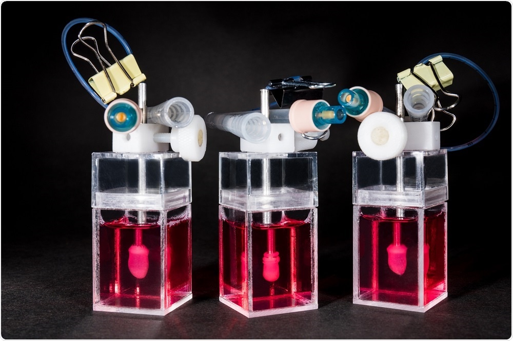
The mini-heart is a 3D engineered, living miniature human heart chamber designed to mimic the pumping function of the human heart. It's a single-chamber organoid model that’s about a centimeter in diameter, representing the ventricle which is the key chamber in the heart for pumping blood through the body, and also the one that’s most susceptible to arrhythmias and diseases that adversely affect heart function.
Mini-hearts - side view - Novoheart
We start with human pluripotent stem cells and differentiate these into ventricular-specific cardiomyocytes, which are characteristic of the ventricles. We then embed these cells into a hydrogel matrix made of collagen and other supporting factors that are poured into a custom developed mold. The cells attach to the matrix over a couple of days and grow into a hollow, spherical shape, following the mold.
With time in culture, the cells compact the collagen around the mold and become organized into a three-dimensional tissue. Part of the mold is similar to a rubber balloon – we deflate this to leave a hollow organoid chamber that pumps and beats like a miniature human heart.
We can control the heart rate using an electrical pacemaker, meaning we can simulate the heart under different conditions such as exercise, stress or sleep, and monitor the mini-heart using sensors to measure things like pressure and volume.
This allows us to make measurements that cardiologists are interested in, such as stroke volume, ejection fraction, and cardiac output. By combining the pressure and volume measurements, we can generate pressure/volume (PV) loops, which describe the pumping efficiency of the heart. This capability is rather unique for an in vitro model of the heart.

How easy is it to keep these mini-hearts alive?

One of the other aspects that we’re focused on is developing the technology to support the biologics. It's not just about engineering and innovating tissues but also the development of bioreactors. These are machines which are used to grow and monitor the mini-heart.
We designed these bioreactors so that there is minimal user manipulation of the mini-heart, with the idea being that it will grow on its own. There’s a little bit of intervention needed to withdraw the ‘balloon’, but the measurements are pretty hands-off. This helps to maximize throughput and minimize variability between users.
We’ve also developed software and analytics that help cardiologists and scientists discover the full capabilities of the mini-heart. They’re pretty sophisticated models and can be used to generate very complicated datasets. It would be a shame to just analyze the amplitude of the pressure wave, for example.
If you start integrating pressure measurements with volume and flow, you can start to build up a more complex picture of the organoid function, which contains important information about how the system responds to drugs, or how it reproduces aspects of a particular heart disease.
To fully utilize the valuable datasets we get from studying these mini-hearts, we’ve now started to integrate machine learning technologies to help analyze all of this data in an unbiased way. For example, the machine can assess whether a drug is having a positive or a negative effect based on how the chamber is working, in a highly efficient and effective manner.
This is only the basics of what machine learning can do, in reality, there are a lot more subtleties. Just one of the many possibilities would be to develop algorithms that can be trained to automatically classify new compounds into pre-existing drug categories based on how they work. They can identify whether a drug is a sodium channel blocker or calcium channel blocker, for example.
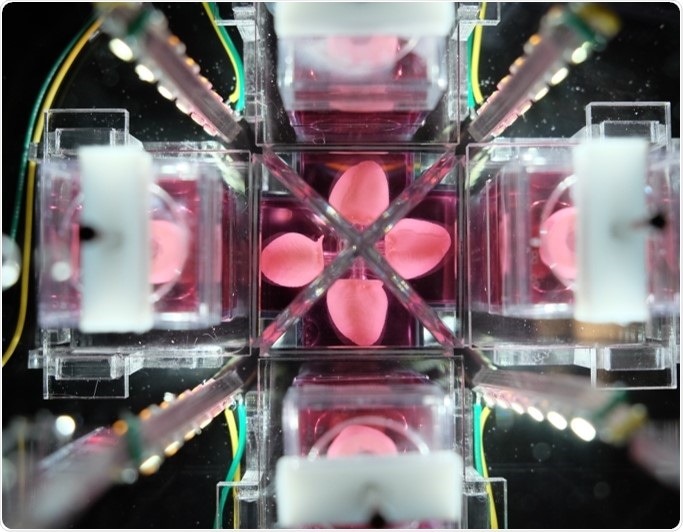
Multichamber of mini-hearts

How long does it take to create one mini-heart?

Once we have the stem cells, it takes a minimum of two weeks to turn those into beating cardiomyocytes. We then make the organoid chamber by placing the cells into the custom mold. The next day, we add some additional media to help culture the cells, and after 24 hours, we remove the outside of the mold, giving the cells full access to the culture media.
Within a couple of days, the cells will start automatically beating and after 7-10 days, we remove the silicone balloon from inside the structure and are left with a hollow, autonomous beating human mini-heart chamber.
An exciting thing about this technology is that it uses induced pluripotent stem (iPS) cells. This means that if you're interested in looking at how a drug might have toxic effects on the heart, you can test it on human cells immediately, with limited ethical implications.
We can also test a drug that is expected to cure heart disease on a model that’s been made with stem cells from a patient with the disease of interest. This offers a powerful tool in drug discovery, as it can give you information about both drug efficacy using diseased mini-hearts and cardiotoxicity using healthy mini-hearts.

What makes the heart-in-a-jar so revolutionary compared to previous tools in drug development?

Traditional tools used in drug development are single-cell cultures and small animal models, which have been tested for decades. Unfortunately, history tells us that those models are not especially predictive of how a drug will ultimately behave when it's delivered to human patients.
We're exceptionally good at curing heart disease and cancer in mice and rats, but a lot of those same therapies are not effective or even toxic when delivered to patients.
Novoheart’s mini-heart technology offers a human-based system using human heart cells. It’s all part of the stem cell revolution that occurred when iPS cells were developed.
We now have access to human tissues that we just didn’t have access to in the past. Human hearts are almost impossible to come by as there are so many critically ill patients in need of a transplant. It would be unethical to ship needed organs off to a company for drug testing.
When we present this to researchers, they recognize that it is really quite unique and potentially disruptive, in the sense that for the first time, you can actually have a miniature human heart in your laboratory for several weeks.
It's not something that's going to die after a couple of hours, and it gives you human-specific measures that should better predict how therapies will behave in actual patients. Ultimately this will make therapies safer and more effective for patients, while also reducing the wasted investment of resources required to develop a drug that is destined to fail in the clinic.

Could the model be used routinely in drug development?

Yes, that's what we are striving for. A key question we are working to answer right now is the most appropriate time in the development pipeline to implement the mini-hearts.
Of course, one idea is the earlier, the better. If something's not going to work, why bother spending time developing it?
On the other hand, pharmaceutical companies have libraries containing thousands of compounds that they're interested in screening, so developing a mini-heart for each one isn’t very practical with such a system. For early stage screening, you need an assay with much higher throughput.
We imagine the right time point for using Novoheart technology is mid-stage in the drug development pipeline, when you've already identified a few lead compounds, maybe a couple of dozen or so, that you are starting to seriously invest in and think about moving to animal studies. Before doing this, you would use the organoid chambers as a screen to inform how to move forward.
For Novoheart, mini-hearts are a first step. We're heart specialists so it made sense for us to start there.  Moreover, cardiotoxicity is one of the key reasons drugs fail in clinical trials. Ultimately we want to integrate heart organoids with liver organoids and lung organoids etc., to build up a whole system.
The bioreactor technology development has been designed with that plan in mind. We expect that at some stage we’ll have several organoids all interacting and being monitored by the same bioreactor, and hope that this might eventually be able to replace animal models altogether.

Saturday, December 16, 2017

Snapshots of Life: Growing Mini-Brains in a Dish

With all this earlier research our researchers should be able to put this all together and create real human brains that can be damaged by stroke and show how to stop and reverse such damage. But that is way too pie in the sky for our stroke medical professionals to understand and implement. 

Multiregional brain on a chip  Jan 2017


Draper Laboratory developing “Brain-on-a-Chip”  October 2012


"Alzheimer's-in-a-Dish" Docs Win Top Smithsonian Ingenuity Award Nov. 2015 


A patient’s budding cortex — in a dish?  June 2015 


Cell cultures in petri dishes open new doors to brain research  April 2017


 The latest here:

Snapshots of Life: Growing Mini-Brains in a Dish


Brain grown in a lab dish
Credit: Collin Edington and Iris Lee, Department of Biomedical Engineering, MIT
Something pretty incredible happens—both visually and scientifically—when researchers spread neural stem cells onto a gel-like matrix in a lab dish and wait to see what happens. Gradually, the cells differentiate and self-assemble to form cohesive organoids that resemble miniature brains!
In this image of a mini-brain organoid, the center consists of a clump of neuronal bodies (magenta), surrounded by an intricate network of branching extensions (green) through which these cells relay information. Scattered throughout the mini-brain are star-shaped astrocytes (red) that serve as support cells.
Collin Edington and Iris Lee created this striking image in Linda Griffith’s lab at Massachusetts Institute of Technology (MIT), Cambridge. While it looks like a single image, the picture actually took about 12 hours to produce, using a confocal laser scanning microscope to zoom in and capture the microscopic details, pixel by pixel, before digitally stitching the series of images all together. For their painstaking efforts, the team was one of 10 winners in the 2017 Koch Institute Image Awards at MIT, established to recognize and publicly display extraordinary works of art and science.
One of the things that fascinated Edington and Lee about this particular mini-brain was its surprisingly organized structure, which emerged over the course of about two weeks. In fact, one of the most interesting aspects of growing organoids in the lab is that by simply changing the chemical makeup and physical consistency of the hydrogel on which the stem cells are placed, it is possible to influence the ways in which they differentiate and assemble. This enables researchers to tinker with and fine-tune the gel’s characteristics to encourage cells to take on different forms. It also reveals a critical component of the development of complex organs—the cellular environment really matters.
In the experiment captured in this image, the researchers randomly seeded thousands of neural stem cells from human embryonic stem cell lines into a dime-sized well on a standard laboratory plate. Those cells divided and differentiated into hundreds of thousands of cells to form the mini-brain organoid shown above. By studying these mini-brains in the lab, researchers can explore the way a “typical” organoid functions, as well as learn what goes wrong in organoids generated from induced pluripotent stem cells derived from people affected by Alzheimer’s disease or other brain conditions.
Griffith’s lab isn’t stopping with the brain. In fact, this mini-brain is part of a much larger project, funded by NIH and Defense Advanced Research Projects Agency, to produce many different types of “mini-organs”—representing the liver, gut, lung, heart, and more. The goal is to link the individual organoids together to form a “human on a chip.” That integrated approach is important because organs don’t act in isolation in either health or sickness.
Alzheimer’s disease is a good example. While it’s primarily a disease of the brain, it has become increasingly clear that the gut and liver play a role, too. The “human on a chip” platform will make it possible to study such complex interactions in Alzheimer’s disease and many other conditions in the lab, with the ultimate goal to accelerate development of safe and effective new treatments.
Links:
Griffith Lab (MIT, Cambridge, MA)
Meet Chip (National Center for Advancing Translational Sciences/NIH)
NIH Support: National Center for Advancing Translational Sciences


Friday, October 20, 2017

New procedure enables cultivation of human brain sections in the petri dish

With this and earlier 6 posts on brains in a dish 
and

Nearly complete human brain grown in US lab: scientist.

Your stroke researchers and doctors should be able to come up with experiments to figure out how to stop the neuronal cascade of death by these 5 causes in the first week. But that won't occur since we don't have two functioning neurons to rub together in our stroke medical leadership. 


https://www.alphagalileo.org/ViewItem.aspx?ItemId=180119&CultureCode=en
19 October 2017 Universitaet Tübingen
Researchers at the University of Tübingen have become the first to keep human brain tissue alive outside the body for several weeks. The researchers, headed by Dr. Niklas Schwarz, Dr. Henner Koch and Dr. Thomas Wuttke at the Hertie Institute for Clinical Brain Research, published their findings in the latest edition of Scientific Reports. The reason for this success is apparently the use of human cerebrospinal fluid to cultivate the tissue in the laboratory. The tissue cultures were in good shape and able to function even after three weeks in a petri dish. Previous attempts to keep brain sections alive used a standard culture solution – which does not work well for human tissue. That is one reason why researchers often fall back on animal experiments. The scientists say the new procedure expands the range of options open for carrying out tests on human brain cells. For instance, it simplifies tests to find out whether brain tissue can tolerate new drugs.
“The human brain appears to have a very low tolerance for cultivation outside the human body,” says Dr. Henner Koch, who lead the study. It is not yet clear which substances in human cerebrospinal fluid are key to the survival of nerve tissue. That will be a goal for future analyses.
But it is clear that the new method answers some of the questions about human brain tissue which used to require animal experiments. In the future, researchers will be able to test the effects of new pharmaceuticals on human brain tissue using cell cultures in a petri dish. And the results of animal experiments are not always fully applicable to humans – leaving a margin of risk for voluntary subjects in the testing of new drugs.
The new technique will also make it easier to research genetic mutations associated with human nervous system disorders. “The method enables us to introduce a mutation into the brain cells and to investigate its effect on the tissue as a whole,” says lead author Dr. Niklas Schwarz. “Even though many neurological disorders can be studied using animal models – it is often uncertain that the results can be transposed to human brain cells.” The Tübingen researchers hope that their process will reduce the number on animals used in experiments.
Yet there will not be mass experiments on human brain sections in the future. The researchers can only use tissue which has been removed as part of necessary brain surgery. That may be the case when epilepsy can no longer be treated using drugs and the damaged area of the brain has to be removed. “Of course, we only use material from patients who have consented to its scientific use in writing,” says neurosurgeon and co-author, Dr. Thomas Wuttke.

Attached files

  • Human nerve cells cultivated in cerebrospinal fluid retain their structure and function after two weeks (left). Cells in the standard fluid (right) die early. Copyright: Niklas Schwarz & Henner Koch, 2017

Wednesday, July 26, 2017

Scientists create 3D-printed brain-like tissue from stem cells

With all this earlier research our researchers should be able to put this all together and create real human brains that can be damaged by stroke and show how to stop and reverse such damage. But that is way too pie in the sky for our stroke medical professionals to understand and implement. 

Multiregional brain on a chip  Jan 2017


Draper Laboratory developing “Brain-on-a-Chip”  October 2012


"Alzheimer's-in-a-Dish" Docs Win Top Smithsonian Ingenuity Award Nov. 2015 


A patient’s budding cortex — in a dish?  June 2015 


Cell cultures in petri dishes open new doors to brain research  April 2017


 The latest here:

Scientists create 3D-printed brain-like tissue from stem cells

Scientists in Australia have used a 3D printer to create nerve cells found in the brain using a special bio-ink made from stem cells.

Key points:

  • Stem cells from adult cells used to make "bio-ink"
  • Bio-ink printed into 3D scaffold and then stem cells turned into nerve cells found in the brain
  • Process could be used in the future to make replacement brain tissue from patient's own skin cells
The research takes us a step closer to making replacement brain tissue derived from a patient's own skin or blood cells to help treat conditions such as brain injury, Parkinson's disease, epilepsy and schizophrenia.
The bio-ink is made of human induced pluripotent stem cells (iPSC), which have the same power as embryonic stem cells to turn into any cell in the body, and possibly form replacement body tissues and even whole organs.
Jeremy Crook, who led the research, said the ability to customise brain tissue from a person's own body tissue was better for transplantation.
"That circumvents issues of immune rejection, which is common in organ transplantation," said Dr Crook, from the University of Wollongong and ARC Centre of Excellence for Electromaterials Science.


Correcting chemical imbalances

Dr Crook said many neuropsychiatric disorders result from an imbalance of key chemicals called neurotransmitters, which are produced by specific nerve cells in the brain.
For example, he said, defective serotonin and GABA-producing nerve cells are implicated in schizophrenia and epilepsy while defective dopamine-producing cells are implicated in Parkinson's disease.
The team used 3D printing to make neurones involved in producing GABA and serotonin, as well as support cells called neuroglia, they reported in the journal Advanced Healthcare Material.
In the future, they plan to print neurones that produce dopamine.


"That's absolutely achievable."
To make the neurones, Dr Crook and colleagues used their bio-ink to print layers of a hatched pattern to create a 5 millimetre-sized cube.
They then "crosslinked" the cube into a firm jelly-like substance.
Growth factors and nutrients were then fed into the holes of this spongey "scaffold", encouraging the stem cells to grow and turn into neurons and support cells, linking up to form tissue.
Waste was also removed via the holes in the scaffold.
Dr Crook said once scaled up, blood vessels would be needed, but small transplants could be theoretically possible using the tissue developed so far.

Impressive but risky too

Tissue engineer Makoto Nakamura from Toyama University in Japan said the study was "very impressive".
"This article indicates the good feasibility of 3D bioprinting with human iPS cells to engineer neural tissues," said Professor Nakamura, who recently wrote an overview on the use of 3D bioprinting in the journal Tissue Engineering.
But he said there were also risks with the technology.
One of the challenges of using iPSCs is that, like embryonic stem cells, they have the potential to develop into teratomas — disturbing looking tumours that contain more than one type of tissue type (think toenails growing in brain tissue, or teeth growing in ovary tissue).
According to Professor Nakamura, it would be important to ensure all the stem cells had turned into nerve cells in the final transplanted material.
"Undesired tissue may grow if even only one immature [stem] cell contaminates [the tissue to be transplanted]," he said.
Dr Crook said the team was currently carrying out animal experiments to test if teratomas developed from the 3D printed nerve cells.

3D brains?

While this is a first step towards 3D printing of whole organs, Dr Crook said a whole functioning brain would be a much more complex task.
"That's a whole different scale. The tissue we print is uniform, and not made up of different regions like a brain," said Dr Crook.
Still, it is a goal the researchers are heading towards.


Apart from providing customised transplants, 3D printed tissue could be useful for medical research.
For example, tissue from a patient with epilepsy or schizophrenia could be created, specifically to study their particular version of the condition.
"You can compare how neuronal networks form differently compared to healthy patient," said Dr Crook.
And the tissue could also be used to screen for effective drugs or electrical stimulation treatments.

Wednesday, April 19, 2017

Cell cultures in petri dishes open new doors to brain research

This has been out there for years and I bet our stroke researchers are doing nothing with this. Because we have NO STROKE LEADERSHIP OR STRATEGY.

"Alzheimer's-in-a-Dish" Docs Win Top Smithsonian Ingenuity Award Nov. 2015 

A patient’s budding cortex — in a dish?  June 2015 

The latest here:

Cell cultures in petri dishes open new doors to brain research

The most complex organ in humans is the brain. Due to its complexity and, of course, for ethical reasons, it is extremely difficult to do scientific experiments on it - ones that could help us to understand neurodegenerative diseases like Parkinson's, for example. Scientists at the Luxembourg Centre for Systems Biomedicine (LCSB) of the University of Luxembourg have now succeeded in turning human stem cells derived from skin samples into tiny, three-dimensional, brain-like cultures that behave very similarly to cells in the human midbrain. In the researchers' petri dishes, different cell types develop, connect into a network, exchange signals and produce metabolic products typical of the active brain. "Our cell cultures open new doors to brain research," says Prof. Dr. Jens Schwamborn, in whose LCSB research group Developmental & Cellular Biology the research work was done. "We can now use them to study the causes of Parkinson's disease and how it could possibly be effectively treated." The team publishes its results today in the prestigious scientific journal "Stem Cell Reports" (DOI: 10.1016/j.stemcr.2017.03.010).
The human midbrain is of particular interest to Parkinson's researchers: it is the seat of the tissue structure known medically as the substantia nigra. Here, nerve cells - specifically dopaminergic neurons - produce the messenger dopamine. Dopamine is needed to maintain smooth body movements. If the dopaminergic neurons die off, then the person affected develops tremors and muscle rigidity, the distinctive symptoms of Parkinson's disease. For ethical reasons, researchers cannot take cells from the substantia nigra to study them. Research groups around the world are therefore working on cultivating three-dimensional structures of the midbrain in petri dishes. The LCSB team led by stem cell researcher Jens Schwamborn is one such group.
The LCSB scientists worked with so-called induced pluripotent stem cells - stem cells that cannot produce a complete organism, but which can be transformed into all cell types of the human body. The procedures required for converting the stem cells into brain cells were developed by Anna Monzel as part of her doctoral thesis, which she is doing in Schwamborn's group. "I had to develop a special, precisely defined cocktail of growth factors and a certain treatment method for the stem cells, so that they would differentiate in the desired direction," Monzel describes her approach. To do this, she was able to draw on extensive preparatory work that had been done in Schwamborn's team the years before. The pluripotent stem cells in the petri dishes multiplied and spread out into a three-dimensional supporting structure - producing tissue-like cell cultures.
"Our subsequent examination of these artificial tissue samples revealed that various cell types characteristic of the midbrain had developed," says Jens Schwamborn. "The cells can transmit and process signals. We were even able to detect dopaminergic cells - just like in the midbrain." This fact makes the LCSB scientists' results of extraordinary interest to Parkinson's researchers worldwide, as Schwamborn stresses: "On our new cell cultures, we can study the mechanisms that lead to Parkinson's much better than was ever the case before. We can test what effects environmental impacts such as pollutants have on the onset of the disease, whether there are new active agents that could possibly relieve the symptoms of Parkinson's - or whether the disease could even be cured from its very cause. We will be performing such investigations next."
The development of the brain-like tissue cultures not only opens doors to new research approaches. It can also help to reduce the amount of animal testing in brain research. The cell cultures in the petri dishes are of human origin, and in some aspects resemble human brains more than the brains of lab animals such as rats or mice do. Therefore, the structures of human brains and its modes of function can be modelled in different ways than it is possible in animals. "There are also attractive economic opportunities in our approach," Jens Schwamborn explains: "The production of tissue cultures is highly elaborate. In the scope of our spin-off Braingineering Technologies Sarl, we will be developing technologies by which we can provide the cultures for a fee to other labs or the pharmaceutical industry for their research."

Wednesday, June 15, 2016

Brain in a Dish Provides New Way to Learn About Alzheimer’s

I'm sure we have enough genius stroke researchers out there that can create brain in a dish to study stroke.  At least if we had some leadership from our fucking failures of stroke associations.
http://neurosciencenews.com/alzheimers-brain-in-dish-4465/
Summary: Researchers report they have discovered a new method to replicate one of the earliest changes in Alzheimer’s disease.
Source: Babraham Institute.
Researchers at the Institute have found a way to replicate one of the earliest changes in Alzheimer’s disease in a dish. This means that it should now be possible to find out a lot more about why it happens – and how to stop it. The new findings are published in the journal Molecular Neurodegeneration and supported by the charity Alzheimer’s Research UK.
Currently, there are no effective treatments to slow or stop Alzheimer’s disease, only ones that aim to manage the disease’s symptoms. This is due in part to difficulty in identifying the first physical signs of the disease in its early stages. Early treatment is an important goal as it could prevent the irretrievable loss of nerve cells seen as the disease progresses.
Being able to properly understand what is happening in the brain as a result of Alzheimer’s disease requires an accurate and accessible system. Until now, much research has relied on post-mortem brain samples, either from people who have passed away with the disease or from mice bred to show features of Alzheimer’s.
Researchers at the Institute have used a method to sustain brain slices from mice in the laboratory. The advantage of these brain slices is that they retain a 3D structure, containing the many different types of cell found in the living brain. The brain slices can survive for several weeks, allowing longer-term studies into what happens to a brain affected by Alzheimer’s, and they can be imaged in real time, sampled and manipulated by the addition of drugs.
The team used the technique to study one of the earliest and most clinically relevant changes in human Alzheimer’s disease which is the loss of synapses – the connection points between nerve cells that allow them to communicate with each other. The brain slices the team studied were taken from young mice bred to develop features of Alzheimer’s disease and contained the hippocampus, a key part of the brain that is important for memory and is affected by the disease. The researchers were able to observe a build-up of the hallmark Alzheimer’s protein amyloid in the slices over time, gaining important insights into how this increase is associated with a loss of proteins in the synapses responsible for transmitting signals between nerve cells. They were also able to treat the slices with a drug to effectively modify the amount of amyloid protein they produced, highlighting the future value of this approach for testing potential new Alzheimer’s treatments.
Mice brain clices maintained in a petri dish to allow study of the earliest stages of Alzheimer’s disease. Credit: The researchers.
Claire Harwell, a PhD student at the Babraham Institute and first author on the paper, said: “This technique will help us to observe the earliest, usually hidden, stages of Alzheimer’s disease whilst providing an accessible system for the identification of promising treatments. For effective therapeutics to be developed, we must understand and target the disease at its earliest stages.”
The method reduces the numbers of animals required to undertake this type of research and means that researchers can study important stages in the disease process without the mice themselves experiencing disease symptoms. It also allows researchers to study live brain tissue and learn how it reacts to drugs in a way that isn’t possible with living humans or mice.
Professor Michael Coleman, research group leader at the Babraham Institute and Professor of Neuroscience at the John van Geest Centre for Brain Repair, explained: “The value of the system is that candidate drugs can be readily delivered and the effects monitored, thus filling a vital gap between cell culture and animal studies. We expect that this approach will be extremely useful in research into Alzheimer’s disease and potential treatments.”
This image compares brain slices from a healthy mouse (left panel) and a mouse with Alzheimer’s disease mutations (right panel). The brain sections have been labelled with fluorescent staining to identify synapses (shown as red). Comparing the images shows that synapses are reduced in mice brains that contain Alzheimer’s disease mutations, modelling what is known to happen in a human brain affected by Alzheimer’s. These images show brain slices cultured for six weeks. Image details: x60 magnification, red= synapses (synaptophysin), green= nerve cell axons (tau). NeuroscienceNews.com image is credited to the researchers.
Dr Simon Ridley, Director of Research at Alzheimer’s Research UK, said: “To develop new treatments for people with Alzheimer’s we need a better understanding of the disease and how it affects the brain. The brain is a very inaccessible organ, presenting researchers with a unique challenge, particularly when studying the very early stages of diseases like Alzheimer’s. It’s important for scientists to use a range of approaches to understand different aspects of Alzheimer’s and this new research provides a window into some of the early stages of the disease process. Research such as this has the power to open new doors to studies in humans and to help in the search for new treatments for this devastating disease.”
About this Alzheimer’s disease research article
Funding: This work was supported by Alzheimer’s Research UK. The Babraham Institute is strategically funded by the Biotechnology and Biological Sciences Research Council.
Animal research statement: As a publicly funded research institute, the Babraham Institute is committed to engagement and transparency in all aspects of its research. The research presented here used brains from mice in order to study the changes associated with the onset of Alzheimer’s disease.
All animal work was approved by the Babraham Institute Animal Welfare and Ethical Review Body and UK Home Office, and carried out in accordance with the Animals (Scientific Procedures) Act, 1986, under a Project Licence. In evaluating any research using animals, the Institute applies the principles of reduction, refinement and replacement. One of the benefits of this study is to provide new methods for this.
Source: Laurie Callahan – Babraham Institute
Image Source: This NeuroscienceNews.com images are credited to the researchers.

Monday, June 1, 2015

A patient’s budding cortex — in a dish?

If this could possibly be used to correct brain circuit functioning in a mentally ill patient then we should be able to use this for stroke repair. At least if we assign some genius researchers to the problem. Ask the ASA, NSA and WSO when they are going to sponsor such research. If not, their boards of directors and CEO need to be replaced with stroke survivors willing to take on the most difficult challenges.
http://loonylabs.org/2015/05/31/cortex-in-a-dish/

Wednesday, October 15, 2014

Breakthrough Replicates Human Brain Cells for Use in Alzheimer’s Research

Our stroke associations should be able to copy this work and support scientists that can create 'Stroke in a dish'. With that they could see what works to save neurons from the neuronal cascade of death. And listen to signals between neurons so we can find out exactly how damaged neurons can be repaired. Yet I doubt any one will look at this and see how it can be reused for stroke.  We must have complete idiots in charge of stroke strategy. Or worse yet, no strategy at all.
http://www.nytimes.com/2014/10/13/science/researchers-replicate-alzheimers-brain-cells-in-a-petri-dish.html?
For the first time, and to the astonishment of many of their colleagues, researchers created what they call Alzheimer’s in a Dish — a petri dish with human brain cells that develop the telltale structures of Alzheimer’s disease. In doing so, they resolved a longstanding problem of how to study Alzheimer’s and search for drugs to treat it; the best they had until now were mice that developed an imperfect form of the disease.
The key to their success, said the lead researcher, Rudolph E. Tanzi of Massachusetts General Hospital in Boston, was a suggestion by his colleague Doo Yeon Kim to grow human brain cells in a gel, where they formed networks as in an actual brain. They gave the neurons genes for Alzheimer’s disease. Within weeks they saw the hard Brillo-like clumps known as plaques and then the twisted spaghetti-like coils known as tangles — the defining features of Alzheimer’s disease.
The work, which also offers strong support for an old idea about how the disease progresses, was published in Nature on Sunday. Leading researchers said it should have a big effect.

More at link.