Emerging
research involving over 900 patients suggests the use of deep learning
on brain magnetic resonance imaging (MRI) scans may obviate the need for
T2WI MRI sequences in diagnosing acute ischemic stroke.
For the retrospective study, recently published in Academic Radiology,
researchers evaluated the capability of a deep learning research
application (Neuro Triage Application, version 1.2.5, Siemens
Healthineers) in detecting acute ischemic stroke (AIS) in a cohort of
947 people (mean sage of 64) who had diffusion-weighted imaging (DWI)
and fluid-attenuated inversion recovery sequences with brain MRI.
At
an average processing time of 24 seconds, the deep learning application
offered a 90 percent sensitivity rate, and 89 percent specificity rate
and a 95 percent area under the receiver operating characteristic curve
(AUROC) for diagnosing AIS on brain MRI, according to the study authors.
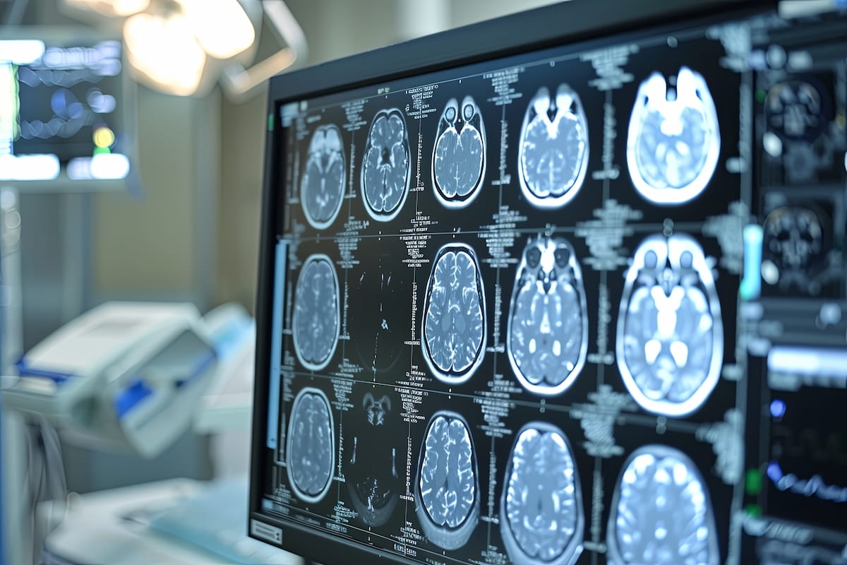 |
In
a new study, researchers found that a deep learning application for
brain MRI offered a 90 percent sensitivity rate and a 95 percent area
AUROC for diagnosing acute ischemic stroke at an average processing time
of 24 seconds. (AI-generated image courtesy of Adobe Stock.) |
In
a subgroup of 71 people who had additional T2WI sequences, the
researchers found equivalent sensitivity (88 percent) and AUROC rates
(88 percent) between the deep learning application (with no T2WI
sequences) and the use of T2WI sequences. They also noted comparable
specificity (81 percent for deep learning vs. 84 percent for T2WI) and
accuracy rates (85 percent for deep learning vs. 83 percent for T2WI).
However,
the study authors pointed out that use of T2WI sequences resulted in a
48-second processing time in comparison to 34 seconds with the deep
learning application.
“ … The addition of T2WI to DWI
and FLAIR does not provide any advantage when (the deep learning
application) is employed to detect AIS. Given that the golden hour for
stroke intervention is within 180 to 270 min, our findings suggest that
T2WI can be omitted to shorten the detection time for AIS because any
potential time delays caused by T2WI cannot be overlooked or regarded as
minor,” wrote lead study author Jimin Kim, M.D., who is affiliated with
the Department of Radiology at Eunpyeong St. Mary’s Hospital and the
Catholic University of Korea College of Medicine in Seoul, Korea, and
colleagues.
Assessing the performance of the deep
learning application for differentiating between lacunar and non-lacunar
AIS in a sub-analysis of 239 participants from the cohort, the
researchers noted equivalent specificity (89 percent) as well as
comparable accuracy rates (89 and 90 percent respectively) and
processing time (22 seconds and 23 seconds respectively).
Three Key Takeaways
1. Deep learning efficiency. The
use of a deep learning application significantly improves the
efficiency of diagnosing acute ischemic stroke (AIS) on brain MRI,
offering a high sensitivity (90 percent) and specificity (89 percent)
with an average processing time of 24 seconds, which is faster than the
traditional T2WI sequences.
2. Comparable diagnostic accuracy. The
deep learning application shows comparable sensitivity, specificity,
and accuracy rates to traditional T2WI sequences, suggesting that T2WI
can be omitted without compromising diagnostic accuracy for AIS.
3. Time-savings in acute care settings. Omitting
T2WI sequences in favor of the deep learning application may shorten
the detection time for AIS, which could be crucial given the critical
time window for stroke intervention. This is particularly important
because time delays caused by T2WI may negatively impact patient
outcomes.
However, the study
authors also found that for non-lacunar AIS, the deep learning
application had higher sensitivity (92 percent vs. 85 percent for
lacunar AIS) and AUROC rates (96 percent vs. 92 percent). They noted
prior research had revealed a slightly declining performance of the deep
learning algorithm with respect to decreasing infarct size.
“This
is likely attributed to the challenges that emerged when the (the deep
learning application) attempted to detect very minor abnormalities. …
However, the difference was marginal, so further evaluation is needed to
confirm the significance of this difference,” added Kim and colleagues.
(Editor’s note: For related content, see “Can Deep Learning Automate Amyloid Positivity Assessment on Brain PET Imaging?,” “FDA Clears New Version of AI Segmentation Software for Brain MRI” and “Can AI Enhance MRI Detection of Amyloid-Related Abnormalities in Patients with Alzheimer’s Disease?”)
Beyond
the inherent limitations of a single-center retrospective study, the
authors acknowledged the small cohort (71 patients) utilized to assess
the impact of T2WI MRI. They also noted that broader extrapolation of
the study findings may be limited as all MRI scans were obtained with 3T
devices from a single vendor.


